Symposium 17th Australia-Japan Colloids
←
→
Page content transcription
If your browser does not render page correctly, please read the page content below
th
    17 Australia-Japan Colloids
          Symposium

Version 1.0
Welcome Message

On behalf of the organising committee I am very pleased to welcome your participation in
the 17th Australia-Japan Colloids Symposium. These meetings have a history stretching back
to 1992 and have provided a great opportunity for Japanese and Australian Colloid and
Interface Scientists to engage both intellectually and socially. Many fruitful collaborations
and deep friendships have resulted from these meetings.

The COVID-19 pandemic has had a profound influence on the lives of people all over the
world and this event is not immune from its influence. The pandemic means that we are
unable to meet in person and hence this event is being held virtually. Whilst this brings
many challenges it also has provided us with a number of opportunities. With the support of
the Australasian Colloids and Interface Society we have been able to make the event free
and this has enabled us to bring together a record number of participants (over 180 at this
point). Additionally, we are trialling a number of new approaches in how our scientific
presentations are being delivered including pre-recorded talks, breakout virtual rooms for
poster presentations, graphical abstracts for social media promotion and 1 minute pre-
recorded lightning talks for poster presenters giving them the opportunity to get you excited
about their presentation.

I very much hope that in the near future we are able to resume meeting in person I think
that this event will also show us a new way that we can augment our traditional meetings.

I must acknowledge the outstanding work of the organising committee, Saffron Bryant,
George Franks, Stuart Prescott, Anna Wang and Catherine Whitby in getting this event off
the ground in a very short period of time. You all have been wonderful to work with. Thank
you.

I must acknowledge the valuable guidance of Professor Takanori Takiue as our international
adviser and the strong support of Shigeru Deguchi and Naoyuki Ishida when the idea of this
meeting was first raised. This event would never have taken place if it wasn’t for the initial
efforts of Greg Warr and Ben Boyd, who first championed the idea.

Thank you for your attendance. I think the exciting program we have assembled will reward
your participation well.

I wish you all a safe and healthy 2020. Sincerely,

                                              Vince Craig

                                               ii
The Organising Committee

  George Franks,
                                                  Stuart Prescott,
  University of
                                                  UNSW
  Melbourne

  Anna Wang,                                      Catherine Whitby,
  UNSW                                            Massey University

                                                  Saffron Bryant,
  Vince Craig, ANU
                                                  RMIT

                           Adviser to the local organising committee
                           Takanori Takiue, Kyushu University

                     iii
Important Information
For the latest updates and information, visit: https://complexfluids.net/aj/
If you’re having technical difficulties and need to contact the organizing committee,
please email: vince.craig@anu.edu.au, or saffron.bryant@rmit.edu.au
Courtesy on Zoom
Please keep yourself muted unless participating directly in the discussion.
Please turn off your video camera when you're not talking.
Please ask questions via the text chat and the session chair will collate them for the
speaker. During the question session, the raise hand feature can also be used, but
priority will go to the questions in the chat session.
If presenters are sharing video as well as their slides, side-by-side mode can be a nice
way of seeing both more easily.
Zoom can be accessed via the desktop client, mobile client or web browser.
Time Zones for Reference
Japan Eastern Western New United USA (New
Australia Australia Zealand Kingdom York)
GMT+9 GMT+10 GMT+8 GMT+12 GMT+1 GMT-4
iv
Keynote Speakers
Tam Greaves is an Associate Professor within Physics at RMIT University, and has
been at RMIT since 2014. She completed her Ph.D. in Experimental Physics in
2004 at Monash University, Australia. In 2005 she joined CSIRO as a Postdoctoral
Fellow, and worked there for nearly 10 years. She is a frequent user of the
Australian Synchrotron SAXS/WAXS beamline, and member of the advisory
committee for the BioSAXS beamline which is being built over the next 3 years.
Tam conducts research into understanding the fundamental physicochemical and
thermal properties of ionic liquids, their mixtures, liquid nanostructure and the
solvophobic effect. She is currently developing ionic liquid solvents for use with
biological molecules in a broad range of applications, including protein solubility,
stability and crystallization. A key focus of her research is the development of
high throughput methodologies for the experimental design, sample synthesis,
characterisation and data analysis.

Kiyoshi Kanie is a Professor in the Institute of Multidisciplinary Research for
Advanced Materials at Tohoku University in Sendai. He received his PhD from
Kyoto University in 2000. He held research associate positions at the University of
Tokyo and Tohokhu University before being appointed an Associate Professor at
Tohokhu University in 2008 and a Full Professor in 2019. His research interests
encompass, the design and synthesis of functional materials, liquid phase
synthesis of functional inorganic nanoparticles with controlled size and shape
and the development of organic-inorganic hybrid materials with dynamic
functions. He was the recipient of the Science Award of the division of colloid and
surface chemistry of the Chemical Society of Japan in 2010 and received the
Japan Institute of Metals and Materials meritorious award in 2014.

Naoyuki Ishida is Associate Professor in the Graduate School of Natural Science
and Technology at Okayama University. He received his Ph.D. degree form Kyoto
University in 2000. From 2000 to 2012, he worked at the National Institute of
Advanced Industrial Science and Technology (AIST) as a research scientist and
moved to Okayama University in 2012. He also spent one year at University of
Leeds in 2006 and eight months at the Australian National University in 2017 and
2018 as Visiting Fellow. His research is aimed at understanding of surface forces
by direct measurement and he has made significant contributions to our
understanding of the forces between hydrophobic surfaces. His research also
focuses on interaction forces in non-aqueous liquids, interactions of
biomolecules and surface functionalisation with stimuli-responsive polymers.

Catherine Whitby was awarded her PhD from the University of Melbourne in
2001. She held research associate positions at the University of Hull (United
Kingdom) and the University of Sydney (Aust.) before being appointed as an ARC
Future Fellow and Senior Research Fellow in the Ian Wark Research Institute at
the University of South Australia. In 2014 she was appointed as a Senior Lecturer
in Chemistry in the School of Fundamental Sciences at Massey University in New
Zealand. She is a physical chemist with expertise in colloid and surface chemistry.
She uses nanomaterials to modify the chemistry of drop and bubble surfaces.
This strategy enables her to control the structure, stability and flow of soft
materials. Her findings have been applied in food and pharmaceutical products
and in drilling fluids.

                         v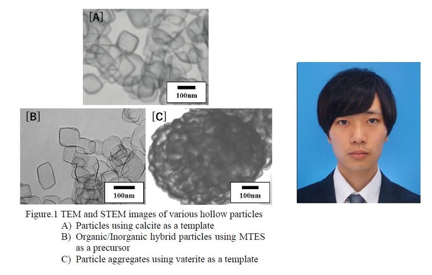
Program
       th
     17 September – DAY ONE
     Morning

Tokyo time       Sydney time     Speaker               Title                                         Page

                                       Chair: George Franks
8.45 – 9 am      9.45 – 10 am    Vince Craig           Welcome

9 – 9.15 am      10 – 10.15 am   Syuji Fuji            Shape-designable liquid marble                 1

9.15 – 9.30 am   10.15 – 10.30   Raymond               Mass transfer across nanometre thin films      2
                 am              Dagastine             between microbubbles

9.30 – 9.40 am   10.30 – 10.40   Joe Berry             Mechanics of Soft Materials                    3
                 am

9.40 – 9.50 am   10.40 – 10.50   Neha Sharma           Surface coating of Liposomal Membranes         4
                 am                                    with Gum Ghatti

9.50 – 10 am     10.50 – 11 am   Grant Webber          Specific-ion Effects in Complex Polymer        5
                                                       Brush Systems

10 – 10.30 am    11 – 11.30 am   Morning tea break

                                       Chair: Saffron Bryant
10.30 to 11 am   11.30 am - 12   Keynote: Prof.        Size- and shape-controlled liquid phase        6
                 pm              Kiyoshi Kanie         synthesis of inorganic nanoparticles and
                                                       application to organic-inorganic hybrid
11 to 11.15 am   12 – 12.15 pm   Hideya Kawasaki       materials
                                                       Ultrasonically activation of colloidal gold    7
                                                       nanoclusters toward sono-catalysis

11.15 to 11.30   12.15 – 12.30   Cathy McNamee         Change in the inter-surface forces between     8
am               pm                                    two charged surfaces containing salts by
                                                       the presence of a liquid flow
11.30 to 11.45   12.30 – 12.45   Srinivas Mettu        “Soft tip” Atomic Force Microscopy in          9
am               pm                                    Probing the Interactions of Different Oil
                                                       Components with Calcite Surface
11.45 to 12 pm   12.45 – 1 pm    Michael Higgins       Hydration Layer Structure of Biofouling        10
                                                       Resistant Nanoparticles

12 to 1 pm       1 to 2 pm       Lunch break

                                                  vi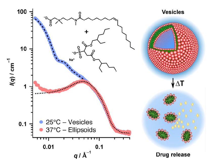
Afternoon

Tokyo time         Sydney time       Speaker                  Title                                            Page

12 to 1 pm         1 to 2 pm         Lunch break

                                          Chair: Catherine Whitby
1 to 1.30 pm       2 to 2.30 pm      Keynote A/Prof.          Solvent properties of protic ionic liquid-        11
                                     Tamar Greaves            water mixtures, and their application to
                                                              biological molecules
1.30 to 1.45 pm    2.30 – 2.45 pm    Chiho Watanabe           Delayed diffusion and unique phase                12
                                                              behaviour in polymer crowding micro-
                                                              emulsions
1.45 to 2 pm       2.45 – 3 pm       Stephen Holt             The adsorption of glucose oxidase on              13
                                                              mesoporous aluminium

2 to 2.10 pm       3 – 3.10 pm       Saffron Bryant           Selective Ion Transport across a Lipid Bilayer    14
                                                              in a Protic Ionic Liquid

2.10 to 2.20 pm    3.10 – 3.20 pm    Hank (Qi) Han            Conformational Changes of Protein with            15
                                                              Ionic Liquids: A Multi-technique Approach

2.20 to 2.30 pm    3.20 – 3.30 pm    Durga Dharmadana         Human neuropeptide substance P self-              16
                                                              assembles into semi-flexible nanotubes that
                                                              can be manipulated for nanotechnology
2.30 to 3 pm       3.30 to 4 pm      Afternoon tea break

                                               Chair: Ben Boyd
3 – 3.15 pm        4 – 4.15 pm       Anna Wang                Aggregation of highly negatively charged          17
                                                              vesicles

3.15 – 3.30 pm     4.15 – 4.30 pm    Rico Tabor               Precise molecular packing from designed           18
                                                              surfactants for vesicles and wormlike
                                                              micelles
3.30 – 3.45 pm     4.30 – 10.45 pm   Hiroki Matsubara         Common black film stability and synergetic        19
                                                              adsorption in ionic−nonionic mixed
                                                              surfactant systems
3.45 – 4 pm        4.45 – 5 pm       Thomas McCoy             Spontaneous self-assembly of                      20
                                                              thermoresponsive vesicles using a
                                                              zwitterionic and an anionic surfactant
4:10 pm onwards    5:10 pm onwards   Poster Session 1

                                                        vii
18th September – DAY TWO
     Morning

Tokyo time       Sydney time       Speaker                   Title                                        Page

                                             Chair: Ray Dagastine
8.35 – 8.50 am   9.35 – 9.50 am    Anthony Stickland         A model system for experimental studies       22
                                                             of gas generation in compressible
                                                             sediments
8.50 – 9 am      9.50 – 10 am      Ramzi Kutteh              Microhydrodynamics of arbitrary-shape         23
                                                             passive and active particles in linear
                                                             flow: daily life at the small scale
9 – 9.10 am      10 – 10.10 am     George Franks             Elastic plastic fracture mechanics            24
                                                             investigation of toughness of wet
                                                             colloidal particulate materials: influence
9.10 – 9.20 am   10.10 – 10.20     Ishihara Shingo           of saturation
                                                             Modelling   of interaction force between      25
                 am                                          particles and simulation of breakage
                                                             behaviour
9.20 – 9.35 am   10.20 – 10.35     YK Leong                  Thixotropic clay gels: microstructure and     26
                 am                                          role of colloidal forces

9.35 – 10 am     10.35 – 11 am     Morning tea break

                                              Chair: Rico Tabor
10 - 10.30 am    11 – 11.30 am     Keynote:                  Evaluation of Non-DLVO Forces between         27
                                   Prof. Naoyuki Ishida      Surfaces in Liquids by Atomic Force
                                                             Microscopy

10.30 - 10.45    11.30 - 11.45     Yuki Uematsu              Is the origin of the negative surface         28
am               am                                          charge of hydrophobic water surface OH
                                                             ion adsorption?
10.45 – 11.10    11.45 – 12.10     Duc Nguyen and Liwen      Soft-hard Janus nanoparticles for             29
am               pm                Zhu                       polymer encapsulation

11.10 - 11.20    12.10 - 12.20     Lee Shi Ting              LSPR-mediated high-resolution imaging         30
am               pm                                          for the study on cell adhesion and
                                                             spreading
11.20 - 11.30    12.20 - 12.30     Aaron Elbourne            Antibacterial liquid metals: biofilm          31
am               pm                                          treatment via magnetic activation

11.30 - 12.30    12.30 - 1.30 pm   Poster Session 2
pm

12.30 -1.30 pm   1.30 – 2.30 pm    Lunch break

                                                      viii
Afternoon

Tokyo time        Sydney time      Speaker                 Title                                          Page

12.30 - 1.30 pm   1.30 - 2.30 pm   Lunch break

                                        Chair: Arlene McDowell
1.30 - 2 pm       2.30 - 3 pm      Keynote: Dr.            Protein adsorption at the oil-water             32
                                   Catherine Whitby        interface: role of protein self-association

2 - 2.15 pm       3 - 3.15 pm      Leonie van ‘t Hag       SANS studies with lipid cubic phase to          33
                                                           investigate protein encapsulation and
                                                           conformation

2.15 - 2.30 pm    3.15 - 3.30 pm   Masumi Villeneuve       Hydrophobic seed proteins on starch             34
                                                           granule surface enables gluten-free rice
                                                           bread making
2.30 - 2.45 pm    3.30 - 3.45 pm   David Beattie           An ATR FTIR study of the interaction of         35
                                                           tio2 nanoparticle films with β-
                                                           lactoglobulin and bile salts
2.45 - 3 pm       3.45 - 4 pm      Andrew Clulow           Mimicking lipid self-assembly in digesting      36
                                                           milks

3 – 3.30 pm       4 – 4.30 pm      Afternoon tea break

                                           Chair: Anna Wang
3.30 – 3.45 pm    4.30 – 4.45 pm   Iijima Kazutoshi        Preparation of thin films and hydrogels of      37
                                                           polysaccharides via polyion complex
                                                           nanoparticles
3.45 – 4 pm       4.45 – 5 pm      Nick Reynolds           Self-healing hydrogels formed from              38
                                                           nanofibrillar assemblies of the amino acid
                                                           phenylalanine
4 – 4.15 pm       5 – 5.15 pm      Takuya Yanagimachi      Voltage driven characteristic instability of    39
                                                           nematic liquid crystal on chemically
                                                           patterned substrate
4.15 – 4.30 pm    5.15 – 5.30 pm   Nigel Kirby and         1. Small angle scattering at the Australian     40
                                   Susanne Seibt           Synchrotron. a fantastic tool for colloid       41
                                                           and interface science
                                                           2. The dynamics of gold nanorod growth
                                                           – a SAXS study
4.40 – 4.50 pm    5.40 – 5.50 pm   Vince Craig             Closing

                                                      ix
Thursday 17th September
Shape-Designable Liquid Marble
      Syuji Fujii1,2, Junya Fujiwara3, Florian Geyer4, Doris Vollmer4, Hans-Jürgen Butt4,
                          Tomoyasu Hirai1 and Yoshinobu Nakamura1,2
                1
                  Faculty of Engineering. Osaka Institute of Technology, Japan
    2
      Nanomaterials Microdevices Research Center, Osaka Institute of Technology, Japan
           3
             Graduate School of Engineering, Osaka Institute of Technology, Japan
                      4
                        Max Planck Institute for Polymer Research, Germany
                                       syuji.fujii@oit.ac.jp

   Liquid marbles (LMs) are liquid droplets
coated with solid particles adsorbed at the
liquid-gas interface and behave as non-wetting
soft, elastic objects [1,2]. A requirement for the
particles to work as an effective LM stabilizer
is that they are hydrophobic (intrinsic water
contact angle close to or larger than 90°). LMs
can be prepared using solid particles with
various chemical compositions including Fig. 1 Initials of the term polyhedral LM: PHLM,
organic, inorganic and composite particles. formed using transparent 2.35 mm-sized plates
Most of the literature is concerned with (near) and dyed water. LMs with different polyhedral
spherical particles and their flocs. However, morphologies. SEM image of a dried
there are no studies on LMs stabilized with cyanoacrylate-treated 3 µL polyhedral LM using
well-defined non-spherical particles or 0.34 mm-sized plates.
stabilizers. Thus, it is crucial to reveal and
understand the relationship between solid particle shape and LM structure formation to utilize
the LMs.
   In this study, a new type of armored droplets, so-called polyhedral LMs are introduced
(Figure 1) [3]. These LMs consist of liquid droplets stabilized by hexagonal plates made of
poly(ethylene terephthalate), which adsorb to the liquid-air interface. Depending on the
specific combination of plate size and droplet diameter, the plates self-assemble into highly
ordered hexagonally arranged domains. Even tetrahedral-, pentahedral-, and cube-shaped
LMs composed of only 4 to 6 plates are demonstrated. During evaporation of the internal
liquid, due to the high adsorption energy of the plates at the liquid-air interface, the overall
surface area stayed constant resulting in strongly deformed polyhedral LMs. In line with this,
highly asymmetric and super-long polyhedral LMs and letters are obtained due to the strong
interfacial jamming exerted by the rigid hexagonal plates. This is particularly pronounced for
larger plate sizes leading to LMs with unusually sharp edges.

References
[1] P. Aussillous, D. Quéré, Proc. R. Soc. A 462 (2006) 973.
[2] S. Fujii, S. Yusa, Y. Nakamura, Adv. Funct. Mater. 26 (2016) 7206.
[3] F. Geyer, Y. Asaumi, D. Vollmer, H.-J. Butt, Y. Nakamura, S. Fujii, Adv. Funct. Mater.
29 (2019) 1808826.

                                               1
Mass transfer across nanometre thin films
between microbubbles
Raymond R. Dagastine1, Yuqi Yang2,3, Matthew D. Biviano1,
Jixiang Guo3, Joseph D. Berry1,*
1
Department of Chemical Engineering,
University of Melbourne, Parkville 3010, Australia
2
State Key Laboratory of Heavy Oil Processing at Karamay,
China University of Petroleum-Beijing at Karamay,
Karamay 834000, China
3
Institute of Unconventional Oil and Gas Science and Technology,
China University of Petroleum, Beijing 102249, China
Email: rrd@unimelb.edu.au
The role of interfacial coatings in gas transport dynamics in foam coarsening is often difficult
to quantify. The complexity of foam coarsening measurements or gas transport measurements
between bubbles requires assumptions about the liquid thin film thickness profile in order to
explore the effects of interfacial coatings on gas transport. It should be possible to
independently quantify the effects from changes in film thickness and interfacial permeability
by using both atomic force microscopy (AFM) and optical microscopy to obtain time
snapshots of this dynamic process. Further, it is expected that the surfactant and polymer
interfacial coatings will affect the mass transfer differently. We measure the mass transfer
between the same nitrogen microbubbles pairs in an aqueous solution using two methods
simultaneously. First, we quantify the bubble volume changes with time via microscopy and
second, we use AFM to measure the film thickness and mass transfer resistances using a
model for the gas transport. Modelling of the interface deformation, surface forces and mass
transfer across the thin film agrees with independent measurements of changes in bubble size.
We demonstrate that an anionic surfactant does not provide a barrier to mass transfer, but
does enhance mass transfer above the critical micelle concentration. In contrast, a polymer
monolayer at the interface does restrict mass transfer. This approach may also have potential
in providing a novel method to rapidly screen interfacial species with potential utility in the
areas of ultrasound contrast imaging agents, oxygen carriers and therapeutic gases [1].
[1] Y. Q. Yang, M. D. Biviano, J. X. Guo, J. D. Berry and R. R. Dagastine, Journal of
Colloid and Interface Science, 2020, 571, 253-259.
2Mechanics of Soft Materials
Joe Berry1, Matthew Biviano1,2, Ray Dagastine1
1. Department of Chemical Engineering, University of Melbourne, Australia
2. Department of Physics, Technical University of Denmark, Denmark
Email: berryj@unimelb.edu.au
Soft materials have critical application in paints, food processing, biology and medicine. The
function of these materials depends upon responses to external stimuli that may be orders of
magnitude different in length and time-scales. A change in something as simple as size or
material stiffness of a drug delivery particle can mean the difference between successful
transport & release of a drug to a specific disease site in the body or expulsion; a change in
porosity or viscoelastic response of a hydrogel used for cell culture can significantly impact
biological functions such as cell proliferation and differentiation; and a change in elastic
modulus can determine if capsules containing flavour, fragrance or paint survive processing
& handling. These widely varying but poorly understood characteristics are fundamental to
the future design of advanced materials.
There is currently a lack of appropriate models required to determine the material properties
of micro-capsules and micro-particles. Instead, measurements are typically carried out on
larger samples under the assumption that materials made from the same components have the
same properties regardless of size and shape. For example, interfacial rheology of complex
interfaces is commonly carried out on flat interfaces, and material property measurements of
hydrogels are undertaken on large, thick films. Here we show that size and shape do matter,
and present new models required to interpret data resulting from experiments directly
undertaken with micro-capsules and micro-particles. The new models open up the possibility
of testing the rheological and/or mechanical properties of capsules and particles directly at
the length-scale and geometry relevant to the required application.
3Surface coating of Liposomal Membranes with
Gum Ghatti
Neha Sharma, Shigeru Deguchi
Japan Agency for Marine Earth Science and Technology
Email: nehash@jamstec.go.jp
Liposomal membranes are self-assembled lipid vesicles which are extensively studied for
drug delivery processes. Polysaccharides such as amylopectin, dextran and pullulan were
shown to exhibit prominent interaction with liposomal membranes1. Gum Ghatti (GG) is a
proteoglycan derived from Anogeissus Latifolia, widely available in the Indian subcontinent.
It is widely used emulsifier and thickening agent in pharmaceuticals, paper industries and
food items. In this study, we investigated GG-induced morphological changes in the nano-
sized liposomal membranes made up of neutral phospholipids; 1,2-dioleoyl-sn-glycero-3-
phosphocholine (DOPC), 1-palmitoyl-2-oleoyl-sn-glycero-3-phosphocholine (POPC) and
positively charged lipid; 1,2-dioleoyl-3-trimethylammonium-propane (DOTAP). We aimed
to coat GG on the surface of different liposomal membranes via the aid of electrostatic
interactions between lipid molecules and negatively charged GG. The changes were
evaluated by difference in size and surface charge of the membranes before and after addition
of GG. Further, the morphological changes in the membranes were observed with optical
microscopy and transmission electron microscopy (TEM). Rise in size of the membranes was
found to vary with concentration of GG. Figure 1 represents TEM images of POPC and
POPC_GG liposomal membranes which show a coating of GG on the surface of POPC
membranes. Interestingly, introduction of GG in cationic liposomal suspension caused
aggregation which can be seen with turbidity in the solution, possibly attributed to fusion of
the individual membranes. It is in agreement with a previous study where polysaccharides
exhibit coating or bridging of
lipid vesicles depending on the
concentration2. Our study
demonstrates change in activity
of polysaccharide (GG) with the
composition of lipids, and
contributes to the present
outlook of GG.
Figure 1. TEM images of POPC (left) and POPC_GG lipid vesicles (right).
References
[1] Sumamoto et al., Biochem. Biophy. Res. Commun. 1980, 94, 136.
[2] Sumamoto et al., J. Biochem. 1980, 88, 1219.
4Specific-ion Effects in Complex Polymer Brush
Systems
Grant B. Webber,a Edwin C. Johnson,a Hayden Robertson,a Isaac J. Gresham,b Andrew
Nelson,c Stuart W. Prescott,b Erica J. Wanlessa
a
Priority Research Centre for Advanced Particle Processing and Transport, University of
Newcastle, Callaghan, NSW 2308, Australia
b
School of Chemical Engineering, UNSW Sydney, UNSW Sydney, NSW 2052, Australia
c
ANSTO, Locked bag 2001, Kirrawee DC, NSW, 2232, Australia.
Email: grant.webber@newcastle.edu.au
Physicochemical behaviour of soft-matter and colloidal systems can be impacted by the
identity of any electrolyte present.1 These specific-ion effects go beyond simplistic models
based on ion valency and concentration and have be known for well over a hundred years. In
spite of years of research there is currently no widely accepted theory capable of explaining
or, importantly, predicting specific-ion effects universally. The concept of an underlying
series that can be used to order the magnitude of effects for a subset of ions has recently
gained wider acceptance, with myriad experiments indicating other ions can alter their
position in this series. Series reversal is even possible depending on the solvent. While most
experimental and theoretical studies to-date have focused on single electrolytes in a pure
solvent, real systems that exhibit specific-ion effects are often more complex. In this
presentation we will discuss recent research on two complex systems; thermoresponsive
polymer brushes in aqueous solutions of mixed electrolytes and specific-ion effects on multi-
responsive polymer brushes.
Electrolytes can act to either “salt-in” (stabilise) or “salt-out” (destabilise) a polymer brush
immersed in a solvent. We have used brushes of poly(ethylene glycol) methyl ether
methacrylate (POEGMA) copolymer to study the effect of mixtures of two salting-in, two
salting-out and a salting-in plus salting-out electrolyte. The impact of two salts of the same
type is concentration dependent, where the net effect can be cooperative or antagonistic. For
mixtures of different salt type the overall behaviour is more temperature sensitive, with the
salting-out ion dominating at low temperature (below the polymer lower critical solution
temperature) and the salting-in ion dominating at high temperature.
A poly(2-(2-methoxyethoxy) ethyl methacrylate-co-2-(diethylamino)ethyl methacrylate)
[P(MEO2MA-co-DEA)] copolymer brush of 90:10 mol% composition was used to study
specific-ion effects on a multi-responsive brush; PMEO2MA is thermoresponsive, PDEA pH
responsive.3 Here the effect of a particular salt was pH dependent; for example, KCl salted-in
the brush at pH 4 but acted to destabilise the brush at pH 9. In this case, the order of the
specific-ion effect could be reversed by changing pH and thus the
charge state of the PDEA.
[1] T.J. Murdoch; B.A. Humphreys; E.C. Johnson; G.B. Webber; E.J.
Wanless, J. Colloid Interface Sci., 2018, 526, 429-450 DOI:
10.1016/j.jcis.2018.04.086.
[2] V. Mazzini; V.S.J. Craig, Chem. Sci., 2017, 8 (10), 7052-7065
DOI: 10.1039/C7SC02691A.
[3] E.C. Johnson; T.J. Murdoch; I.J. Gresham; B.A. Humphreys; S.W.
Prescott; A. Nelson; G.B. Webber; E.J. Wanless, Phys. Chem. Chem.
Phys., 2019, 21 (8), 4650-4662 DOI: 10.1039/C8CP06644B.
5KEYNOTE: Size- and Shape-controlled Liquid
Phase Synthesis of Inorganic Nanoparticles and
Application to Organic-inorganic Hybrid
Materials
Kiyoshi Kanie
Institute of Multidisciplinary Research for Advanced Materials, Tohoku University
Email: kanie@tohoku.ac.jp
Recent remarkable progress in inorganic nanoparticles (NPs) synthesis enables us to obtain
various types of monodisperse NPs with uniform size and shape.[1]-[6] Thus, new
breakthroughs could be expected in NP-based novel functional materials. Especially, NP-
based periodic structure formation has attracted a considerable attention in material
science.[7]-[8] Among organic soft materials, liquid-crystalline (LC) organic dendron is one of
the most representatives to form self-organized periodical structures. Thus, we focused our
attention on introduction of such self-organization ability into inorganic NPs. As dendrons,
we synthesized phenetyl ether-type dendrons with an amino-group at the apex. These
dendrons themselves show thermotropic LC phases. The dendrons are attached as the outer
corona, through amidation, to the carboxylic groups at the surface of the inner aliphatic
corona encapsulating the NP. CO2H-modified monodisperse Au, CdS, and Fe3O4 NPs were
synthesized for the purpose of the study. The dendron-modified gold NP showed an LC
hexagonal columnar phase at 130 C and formed a simple cubic (SC) LC phase at 150 C.
The result indicated that the dendron-modified gold NPs can be regarded as organic-inorganic
hybrid dendrimers with thermotropic LC behaviour.[9]-[10] The dendron-modified CdS NPs
also formed a thermotropic cubic LC phase with a novel, low-symmetry structure, space
group P213 by annealing at 150 °C for 15 h. In contrast, unannealed dendron-modified CdS
NPs formed an amorphous structure. In such a state, it showed strong photoluminescence
(PL) when UV irradiated at 365 nm. However, PL was quenched when the dendron-modified
CdS NPs formed the cubic phase. Such PL quenching behavior was totally reversible, and
appears to be associated with the periodic structure.[11] Such unusual PL behavior might be a
powerful tool to develop future functional devices.
[1] K. Kanie, T. Sugimoto, Chem. Commun., 2004, 1584-1585.
[2] K. Kanie, T. Sasaki, M. Nakaya, A. Muramatsu, Chem. Lett., 2013, 42, 738-740.
[3] K. Kanie, Y. Seino, M. Matsubara, M. Nakaya, A. Muramatsu, New J. Chem., 2014, 38, 3548-
3555.
[4] Y. Kamikoriyama, H. Imamura, A. Muramatsu, K. Kanie, Sci. Rep., 2019, 9,
899.
[5] R. Suzuki, Y. Nishi, M. Matsubara, A. Muramatsu, K. Kanie, ACS Appl.
Nano Mater., 2020, 3, 4870-4879.
[6] C. Shen, M. Matsubara, M. Yabushita, S. Maki, A. Muramatsu, K. Kanie,
Nanoscale Adv., 2020, 2, 814-822.
[7] K. Kanie, T. Sugimoto, J. Am. Chem. Soc., 2003, 125, 10518-10519
[8] K. Kanie, A. Muramatsu, J. Am. Chem. Soc., 2005, 127, 11578-11579.
[9] K. Kanie, M. Matsubara, X. Zeng, F. Liu, G. Ungar, H. Nakamura, A.
Muramatsu, J. Am. Chem. Soc., 2012, 134, 808-811.
[10] M. Matsubara, A. Miyazaki, X. Zeng, A. Muramatsu, G. Ungar, K. Kanie,
Mol. Cryst. Liq. Cryst., 2015, 617, 50-57.
[11] M. Matsubara, W. Stevenson, J. Yabuki, X. Zeng, H. Dong, K. Kojima, S.
F. Chichibu, K. Tamada, A. Muramatsu, G. Ungar, K. Kanie, Chem, 2017, 2,
860-876.
6Ultrasonically Activation of Colloidal Gold
Nanoclusters Toward Sono-Catalysis
Hideya Kawasaki1), Junichi Yagi1), Kouhei Kawamura1), Ayaka Inui2), Ken Yamamoto2)
1)
Department of Chemistry and Materials Engineering, Kansai University;
2)
Department of Pure and Applied Physics, Kansai University
Email:hkawa@kansai-u.ac.jp
Thiolate-protected Au NCs (AunSRm), where n and m denote the numbers of gold atoms and
thiolate ligands (SR), have ultrasmall sizes (typically less than 2 nm in the core) and contain
up to hundreds of gold atoms. Au NCs have discrete energy levels because of the quantum
size effect, which is significantly different from the larger plasmonic Au nanoparticles (>3
nm) with continuous energy levels. Our groups have focused on photo-functionality of Au
NCs, such as photoluminescence, photocatalytic activity, and photosensitizing ability [1-5].
Apart from light, ultrasound has several advantages, such as operational simplicity, safety,
and being environmentally benign. Recently, ultrasound excited nanomaterials (e.g., TiO2)
have been drawing intense research because of the potential applications such as ultrasound-
activated catalysis (sonocatalysis), ultrasound-activated sensitizers (sonodynamic therapy),
and ultrasound-induced hydrogen production from water (Sono-Hydro-Gen process)[6,7].
However, the ultrasound-activated mechanism remains unresolved, and there is no evidence
on ultrasound activation of nanomaterials. As a result, the strategy for high-efficient sono-
catalytic nanomaterials becomes obscure.
Herein, we demonstrate the ultrasound (1 MHz) can excite thiolate-protected Au NCs by
observation of sonoluminescence and excited oxygen (i.e., singlet oxygen) from Au NCs[8].
The acoustic wavelength of ultrasound in water ranges from about 75 to 0.15 mm for a
frequency between 20 kHz and 10 MHz. As this wavelength is significantly above the
dimensions of Au NCs, the direct
coupling of the ultrasonic acoustic field
with the Au NCs is unlikely. On the
other hand, microbubble cavitation (the
growth and rapid collapse of
microbubbles) occurs in water under
ultrasonication to emit light
(sonoluminescence). It is proposed that
the sonoluminescence from the water
could excite Au NCs when the
sonoluminescence spectrum overlaps
with the absorption spectrum of the Au NCs, as like resonance energy transfer process.
[1] H. Kawasaki, K. Hamaguchi, I. Osaka, and R. Arakawa, Adv. Funct. Mater.2011, 21,
3508.
[2] H. Kawasaki and R. Jin et al., Chem. Mater. 2014, 26, 2777.
[3] M. Yamamoto, K. Shitomi et al., J. Colloid. Interf. Sci. 2018, 510, 221 .
[4] D. Hikosou, S. Saita, et al., J. Phys. Chem. C 2019, 122, 12494.
[5] T. Kawawaki, Y. Negishi, H. Kawasaki, Nanoscale Adv. 2020, 2, 17.
[6] K. Kerboua,O. Hamdaoui, Current Opinion in Green and
Sustainable Chemistry 2019, 18,84.
[7] X. Lin, J. Song, X. Chen,H. Yang, Angew. Chem. Int. Ed. 2020, 59,
14212.
[8] K. Kawamura, D. Hikosou et al., J. Phys. Chem. C, 2019,123, 26644.
7Change in the Inter-Surface Forces Between
Two Charged Surfaces Containing Salts by the
Presence of a Liquid Flow
C.E. McNamee, H. Kawakami
Shinshu University, Japan
mcnamee@shinshu-u.ac.jp
The forces acting between particles in an aqueous solution determine whether those
particles will disperse or aggregate. The ability to control these forces allows the physical
properties of these systems to be controlled and therefore their applications to be improved.
In real systems of particles in aqueous solutions, the liquid often contains a flow. The effect
of a flow on the forces acting in such systems, however, is still not clear.
In this study, we used a combined Atomic Force Microscope-peristaltic pump system to
determine the effect of a flow in aqueous salt (NaCl or MgCl26H2O) solutions between a
negatively charged silica particle and a negatively charged silicon wafer on the forces acting
in the system [1]. A flow decreased the inter-surface repulsive forces, if the water contained
ions that showed specificity to the surfaces, i.e., Na+ or Mg2+ ions. The difference in the
repulsive forces measured in the absence and presence of a liquid flow became greater, when
the concentration of these ions were increased by increasing the bulk salt concentration.
Fitting of the force curves with the DLVO theory showed that an increased flow rate caused
the surface potentials and Debye lengths of the systems to decrease. The difference between
the values measured in the absence and presence of a liquid flow became larger for higher
bulk salt concentrations. The Debye length decrease was explained by a shrinkage of the
electrical double layer due to (1) the deformation of the electric double layer by the liquid
flow, and (2) an increase in the number of ions near the charged surfaces and the subsequent
electrostatic screening increase, due to ions being introduced into the electrical double layer
by the flowing liquid. The decreased surface potential was explained by (1) the increased
electrostatic screening resulting from the decreased electrical double layer thickness, and (2)
an increase in the number of specifically adsorbed ions to the silica or silicon wafer surfaces.
[1] Kawakami, H.; McNamee, C.E. Langmuir 2018, 34, 8464-8471.
8“Soft tip” Atomic Force Microscopy in Probing
the Interactions of Different Oil Components
with Calcite Surface
Srinivas Mettu a,b, Hongna Ding c, Sheik Rahman d
a
School of Chemistry and the Department of Chemical Engineering, University of
Melbourne, Parkville, Victoria 3010, Australia.
b
Chemical and Environmental Engineering, RMIT University, Melbourne, Victoria 3000,
Australia.
c
School of Petroleum Engineering, Northeast Petroleum University, Daqing, Heilongjiang
Province, China.
d
School of Minerals and Energy Resources Engineering, University of New South Wales,
Sydney, NSW 2052, Australia.
Email: srinivas.mettu@unimelb.edu.au
The interactions of oil with rocks are complex, which involve van der Waals,
electrostatic, hydration, cation-π, steric interactions, etc. These interactions have been the
forefront of diverse fields of chemistry, biology, and material science, especially they control
the adsorption and transport of oil molecules on rock surface in petroleum industry. [1] [2] On
the other hand, in recent years, there has been an emerging interest on the impacts of ionic
chemistry on calcite-oil interactions. Scholars have found that Ca2+, Mg2+, and SO42- ions
changed the binding strength of oil molecules on calcite surface by altering the surface
properties, e.g., lattice structure, charges, and wettability of calcite surface. [3] [4] A deep
understanding of calcite-ion-oil interactions help the oil industry smartly develop the oil
reserves. The force spectroscopy technique that measures the attractive and repulsive forces
between oil droplet and solid surface are crucial in developing this understanding.
We studied the ionic effects on calcite-oil interactions by immobilizing a droplet of
mineral oil or crude oil to a tipless cantilever and used this "soft tip" to probe the calcite
surface in different salt solutions (NaCl, Na2SO4, CaCl2, and MgCl2) with an atomic force
microscopy (AFM). It was observed that for mineral oil that contains only hydrocarbons and
aromatics, the total forces were repulsive in NaCl and MgCl2 solutions but were attractive in
Na2SO4 and CaCl2 solutions, and the adhesion results followed CaCl2 >Na2SO4 >NaCl
(reference)>MgCl2. While for crude oil that comprises of hydrocarbons, aromatics, resin, and
asphaltenes, it was observed that the total forces were repulsive only in Na2SO4 solution but
were attractive in NaCl, CaCl2, and MgCl2 solutions, and the adhesion results followed
MgCl2 >>NaCl (reference)>Na2SO4 >CaCl2. The force results firstly suggested that the
interactions of oil with calcite surface could be extremely different due to the differences in
oil chemical components, i.e., cation-π interactions [5] and cation bridging interactions [6] for
mineral oil and crude oil, respectively. Secondly, these two kinds of interactions responded
differently to Ca2+, Mg2+, and SO42- ions, that is, the cation-π interactions were mitigated by
Mg2+ but were enhanced by Ca2+ and SO42-, whereas the cation bridging interactions were
mitigated by Ca2+ and SO42- but were enhanced by Mg2+.
[1] G. Hirasaki, SPE Formation Evaluation 1991, 6, 217-226.
[2] E. E. Meyer, K. J. Rosenberg, J. Israelachvili, Proceedings of the National
Academy of Sciences 2006, 103, 15739-15746.
[3] T. Austad, Enhanced oil recovery Field case studies 2013, 301-335.
[4] S. Rashid, M. S. Mousapour, S. Ayatollahi, M. Vossoughi, A. H. Beigy,
Colloids and Surfaces A: Physicochemical and Engineering Aspects 2015,
487, 142-153.
[5] H. Wang, D. J. Grant, P. C. Burns, C. Na, Langmuir 2015, 31, 5820-5826.
[6] A. RezaeiDoust, T. Puntervold, S. Strand, T. Austad, Energy & fuels 2009,
23, 4479-4485.
9Hydration Layer Structure of Biofouling
Resistant Nanoparticles
Paul J. Molino^1,4, Dan Yang^1,4, Matthew Penna2,4, Keisuke Miyazawa3, Brianna R.
Knowles1,4, Takeshi Fukuma3, Irene Yarovsky2,4 and Michael J. Higgins1,4
1. ARC Industrial Transformation Research Hub for Australian Steel Manufacturing,
NSW 2522, Australia
2. School of Engineering, RMIT University, Melbourne, Victoria 3001, Australia
3. Division of Electronic Eng. and Computer Science, Kanazawa University, Kakuma-
machi, Kanazawa 920-1192, Japan
4. ARC Centre for Electromaterials Science (ACES), Intelligent Polymer Research
Institute/AIIM Faculty, Innovation Campus, Squires Way, University of Wollongong,
NSW 2522, Australia
Email: mhiggins@uow.edu.au
Hydrophilic surface chemistries can strongly bind water to produce surfaces that are highly
resistant to protein adsorption and fouling. The interfacial bound water and its unique
properties have intrigued researchers for decades yet the relationship between the water three-
dimensional structure and function in antifouling coatings remains elusive. Here, we use
hydrophilic, epoxy organosilane modified silica nanoparticles to demonstrate cheap, robust
and practically applied coatings that we discover have broad-ranging, ultra-low antifouling
properties when challenged by various proteins, bacteria and fungal spores. To understand
their remarkable antifouling properties, Frequency Modulation-Atomic Force Microscopy is
used to directly observe the interfacial water structure on single nanoparticles at sub-atomic
resolution (Figure 1), which we validate using all-atom MD simulations that strikingly
predict similar structures of water layers on the original and ultra-low fouling surfaces [1].
Unprecedented convergence of experimental and modelling data reveal that suitably spaced,
flexible chains with hydrophilic groups interact with water molecules to produce a confluent,
quasi-stable layer, consisting of dynamic interfacial water, provides an effective basis for
antifouling performance of ultrathin, hydrophilic surface chemistries
Figure 1: 3D FM-AFM cross-sectional image of interfacial
water structure on silica nanoparticle.
[1] Molino*, Yang* et al. "Hydration Layer Structure of Biofouling-
Resistant Nanoparticles." ACS Nano 12.11 (2018): 11610-11624.
10KEYNOTE: Solvent properties of protic ionic
liquid-water mixtures, and their application to
biological molecules
Tamar Greaves, Qi (Hank) Han, Dilek Yalcin, Radhika Arunkumar, Andrew Christofferson,
Calum Drummond
College of Science, Engineering and Health, RMIT University
Email: tamar.greaves@rmit.edu.au
Protic ionic liquids (PILs) are cost efficient “designer” solvents which can be tailored
to have properties suitable for a broad range of applications. PILs are also being combined
with molecular solvents to enable more control over the solvent environment, driven by a
need to reduce their cost and viscosity. However, there are relatively few structure-property
studies which look at these more complex mixtures. We have explored the solvation
properties of common PIL-molecular solvents using various techniques,1 including Kamlet-
Aboud-Taft parameters determined from solvatochromic dyes, and MD simulations.2-3 These
have identified many interesting solvent properties of these solutions and gives insight into
their interactions with solutes.
In the second part of this presentation I will discuss how we are using our
understanding of PIL-water solvent properties to design and characterise solvents for
biological molecules. In particular, we are targeting being able to control protein solubility
and stability, which are critical for applications in bioprocessing, biocatalysis, protein
crystallography and cryopreservation. We have explored lysozyme as a model protein in
various PIL-water systems, predominantly using spectroscopic techniques and small angle x-
ray scattering (SAXS).4-5 From this we have been able to identify which PILs are more
biocompatible, and to identify specific conformational changes of lysozyme due to the
presence of PILs. More recently, protein crystallography has been used to identify specific
binding sites of the PIL ions and water to lysozyme.
References
1. Yalcin, D.; Drummond, C. J.; Greaves, T. L., High throughput approach to
investigating ternary solvents of aqueous non-stoichiometric protic ionic liquids. Phys. Chem.
Chem. Phys. 2019, 21, 6810-6827.
2. Yalcin, D.; Christofferson, J.; Drummond, C. J.; Greaves, T. L., Solvation properties
of protic ionic liquid – molecular solvent mixtures. Phys. Chem. Chem. Phys. 2020, 22,
10995-11011.
3. Yalcin, D.; Drummond, C. J.; Greaves, T. L., Solvation properties of protic ionic
liquids and molecular solvents. Phys. Chem. Chem. Phys. 2020, 22, 114-128.
4. Wijaya, E. C.; Separovic, F.; Drummond, C. J.; Greaves, T. L., Activity and
conformation of lysozyme in molecular solvents, protic ionic liquids (PILs) and salt-water
systems. Phys. Chem. Chem. Phys. 2016, 18, 25926-25936.
5. Arunkumar, R.; Drummond, C. J.; Greaves, T. L., FTIR
Spectroscopic Study of the Secondary Structure of Globular Proteins in
Aqueous Protic Ionic Liquids. Frontiers in Chemistry 2019, 7, Article
74.
11Delayed diffusion and unique phase behaviour
in polymer crowding micro-emulsions
Chiho Watanabe, Miho Yanagisawa
1
Graduate School of Integrated Sciences for Life, Hiroshima University (Japan), 2Komaba
Institute for Science, The University of Tokyo (Japan)
Email: cwatan@hiroshima-u.ac.jp
Molecular behaviour in milliliter-scale bulk solutions and
small picoliter-scale microemulsions is often different[1–3].
This difference is attributed to as “size effect”. Although the
factors of the size effect are considered to be the small
volume and the membrane interface, few attempts have been
made to separate and quantify these factors. In this study, we
evaluated the size effect by measuring the molecular
diffusion in highly concentrated polymer crowding solutions
confined in micro-emulsions. First, molecular diffusion in
various sized spherical oil-in-water micro-emulsions of
polymer crowding solution covered by a lipid monolayer[4] Fig.1 FCS measurement in a
was investigated by fluorescence correlation spectroscopy micro-emulsion.
(FCS) (Fig.1). The results show that the molecular diffusion
of the globular protein bovine serum albumin (BSA) and the linear polymer poly(ethylene
glycol) (PEG) slowed down as the radius R of the spherical emulsion decreased below 20 µm
at polymer concentrations are less than one molecule of the inter-protein distance and above
the overlap concentration c*, respectively. Furthermore, the molecular diffusion in the disk-
shaped emulsions was measured in the same way and the diffusion coefficient D was found
to be delayed than that in the spherical emulsions under the same volume conditions. On the
other hand, the diffusion coefficient D was independent of the distance from the membrane
interface. These results suggest that the slow molecular diffusion is not due to the distance
from the membrane nor the small volume effect but is due to the increase in the membrane
area/volume ratio due to micrometric confinement. In addition to these results, we will
illustrate the liquid-liquid phase separation (LLPS) in such micro-emulsions and discuss the
origin of the size effect caused synergistically by polymer crowding and confinement in
microscopic spaces[5,6].
[1] C. Watanabe, M. Yanagisawa, Phys. Chem. Chem. Phys. 2018, 20, 8842–8847.
[2] A. Kato, M. Yanagisawa, Y. T. Sato, K. Fujiwara, K. Yoshikawa, Scientific Reports
2012, 2, 1–5.
[3] N. Biswas, M. Ichikawa, A. Datta, Y. T. Sato, M. Yanagisawa, K.
Yoshikawa, Chemical Physics Letters 2012, 539–540, 157–162.
[4] M. Yanagisawa, T. Sakaue, K. Yoshikawa, in International Review
of Cell and Molecular Biology (Eds.: R. Hancock, K.W. Jeon),
Academic Press, 2014, pp. 175–204.
[5] C. Watanabe, Y. Kobori, J. Yamamoto, M. Kinjo, M. Yanagisawa, J.
Phys. Chem. B 2020, 124, 1090–1098.
[6] C. Watanabe, M. Yanagisawa, Biophys Rev 2020, 12, 385–386.
12The Adsorption of Glucose Oxidase on
Mesoporous Aluminium Oxide Films
Stephen A. Holt, † Akira Yamaguchi,* Kazuya Katayama*
† Australian Centre for Neutron Scattering, ANSTO, Lucas Heights, NSW.
* Institute of Quantum Beam Science, Ibaraki University, 2-1-1 Bunkyo, Mito, Ibaraki 310-
8512, Japan
Email: sph@ansto.gov.au, akira.yamaguchi.sci@vc.ibaraki.ac.jp
Protein adsorption at an inorganic nanoporous material is a fundamental process in the
chromatographic separation of proteins and synthesis of an artificial biocatalytic system.1-3
The nanoporous material possesses both an outer surface and internal pore surfaces, and both
can be regarded as adsorption sites for protein molecules. The distribution of protein
molecules at both adsorption sites affects the chromatographic separation and performance of
the biocatalytic system. The purpose of this study is to clarify whether negatively-charged
glucose oxidase (GOD) can penetrate into pores in mesoporous aluminum oxide (MAO)
films. The MAO film used in this study was prepared by a surfactant-templated method,4 and
contained ordered pores with diameter of ca. 10 nm, much smaller than that of a conventional
nanoporous aluminum oxide.
The MAO film was deposited on a microscope cover glass slip for ex situ structural
characterization of the by X-ray reflectometry (XRR), neutron reflectometry (NR), and
scanning electron microscopy. The same deposition ‘recipe’ was then used for films
deposited on thick silicon wafers for in situ experiments, where the GOD containing solution
is in contact with the MAO film, using NR. The NR profiles before/after adsorption GOD
were continuously measured in a flow cell with varying H2O/D2O ratios applied to ‘highlight’
different aspects of the system. The results indicated almost exclusive outer surface
adsorption with the negatively charged GOD molecules unable to enter the positively charged
channels.
Fig. 1 Schematic illustrations of the distribution of GOD molecules at the MAO film in 10
wt% GOD solution and in water after removal of excess amount of GOD adsorbed.
References
[1] J. Nawrocki, J. Chromatogr. A, 1997, 779, 29-71.
[2] K. Ariga, A. Vinu, Y. Yamauchi, Q. Ji, and J. P. Hill, Bull.
Chem. Soc. Jpn, 2012, 85, 1-32.
[3] T. Itoh, A. Yamaguchi, T. Kyotani, T. Hanaoka, and F.
Mizukami, Adv. Porous Mater., 2016, 4, 157-165.
[4] Y. Shibuya, K. Katayama, K. Akutsu-Suyama, and A.
Yamaguchi, ACS Omega, 2019, 4, 17890-17893.
13Selective Ion Transport across a Lipid Bilayer
in a Protic Ionic Liquid
Saffron J. Bryant,a,b Alvaro Garcia,a,c Ronald J. Clarke,a Gregory G. Warra
a
School of Chemistry, The University of Sydney, NSW, Australia, 2006
b
School of Science, RMIT University, Melbourne, Victoria 3001, Australia
c
School of Life Sciences, University of Technology Sydney, NSW, Australia, 2007
Email: saffron.bryant@rmit.edu.au
Spontaneous bilayer formation and amphiphilic self-assembly in ionic liquids is well
established.1, 2 However, to date, this has been a largely uncontrolled process. Using a
tethaPodTM system (Figure 1), we demonstrate the formation of a tethered lipid bilayer in a
purely ionic liquid environment.
Furthermore, using electrical impedance spectroscopy, we show for the first time the function
of a membrane transport protein in a pure ionic liquid. The transporter in question;
valinomycin, not only retains full functionality in an ionic liquid, it also retains its selectivity
for potassium over sodium ions.
This paves the way for the development of new, non-aqueous systems such as microreactors,
and biosensors. Furthermore, stability in an ionic liquid could be used for long-term storage
of tethered membranes and related devices, or use at temperatures not accessible with an
aqueous environment.
Figure 1. Photograph of a tethaPodTM system from SDx Tethered Membranes Pty Ltd
(Sydney, Australia).
References
1. S. J. Bryant, K. Wood, R. Atkin and G. G. Warr, Soft Matter,
2017, 13, 1364-1370.
2. Z. Chen, T. L. Greaves, C. Fong, R. A. Caruso and C. J.
Drummond, Physical Chemistry Chemical Physics, 2012, 14,
3825-3836.
14Conformational Changes of Protein with Ionic
Liquids: A Multi-technique Approach
Hank (Qi) Han, Tamar Greaves
School of Science, College of Science, Engineering and Health, RMIT University, 124 La
Trobe Street, Melbourne, VIC 3000, Australia
Email: qi.han@rmit.edu.au
Ionic liquids (ILs) are liquids that are comprised entirely of ions. They are designer solvents
which can help stabilise some proteins. However, it is challenging to understand protein
structure in ILs. In this talk, we show a wide range of ILs used for lysozyme and GFP as
model proteins. We introduce a multi-technique approach including spectroscopies, small
angle x-ray scattering and crystallography to explore the conformational changes of proteins
and such approaches can improve our understanding on structure-property relationships for
future solvent design for proteins.
15Human neuropeptide substance P self-
assembles into semi-flexible nanotubes that can
be manipulated for nanotechnology
Durga Dharmadana1,2, Jozef Adamcik3, Timothy M Ryan4, Samuel Appiah Danso1,5,
Christopher J H Chong6, Charlotte E Conn2, Nicholas P Reynolds6, Raffaele Mezzenga3,
Céline Valéry1
1
School of Health and Biomedical Sciences, Translational Immunology and Nanotechnology
Program, RMIT University, Bundoora VIC 3083, Australia
2
School of Science, College of Science, Engineering and Health, RMIT University,
Melbourne VIC 3000, Australia
3
Laboratory of Food and Soft Materials, ETH-Zürich, CH-8092 Zürich, Switzerland.
4
The Australian Synchrotron, ANSTO Research facilities, Clayton VIC 3168, Australia.
5
CSIRO Manufacturing, Microscopy Group, Clayton VIC 3168, Australia.
6
Department of Physics and Chemistry, Institute for Molecular Science, La Trobe University,
Bundoora VIC 3086, Australia.
Email: durga.dharmadana@rmit.edu.au
Tailoring peptide self-assembled nanostructures for specific applications has been reported in
diverse areas, from nanoelectronics to water purification, to drug delivery [1]. Unlike proteins,
short peptides are accessible synthetically, with sequences that can be easily modified.
However, the majority of peptide nanostructures reported to date form polymorphic,
irreversible, insoluble “amyloid-like” nanofibrils [2] . Expanding the space of peptide
nanostructure morphologies and properties is hence desirable. Specifically, few sequences
were reported to form nanotubes, and these only include unnatural peptides, chimeric
molecules or truncated natural sequences [3]. Other than a few exceptions [4], these sequences
formed poly-disperse assemblies. Here we report, for the first time, the intrinsic, reversible
self-assembly of the human neuropeptide Substance P into high aspect-ratio, semi-flexible,
monodisperse nanotubes that can associate into tapes or films with nematic properties. We
show that substance P nanotubes are versatile materials for nanotechnology, as they can be
precipitated or mineralised while conserving their core-shell morphology and dimensions.
Our discovery will open new biomimetic approaches for innovative bio-nanotechnology
applications, beyond direct relevance for neuropeptide functional assemblies.
References
[1] F. Zhao, M. L. Ma, B. Xu, Chemical Society Reviews 2009, 38, 883-
891.
[2] J. Adamcik, R. Mezzenga, Angewandte Chemie International
Edition 2018, 57, 8370-8382.
[3] C. Valéry, F. Artzner, M. Paternostre, Soft Matter 2011, 7, 9583-
9594.
[4] C. Tarabout, S. Roux, F. Gobeaux, N. Fay, E. Pouget, C. Meriadec,
M. Ligeti, D. Thomas, M. IJsselstijn, F. Besselievre, Proceedings of
the National Academy of Sciences 2011, 108, 7679-7684.
16Precise molecular packing from designed
surfactants for vesicles and wormlike micelles
Rico F. Tabor
School of Chemistry, Monash University, Clayton, VIC 3800, Australia
Email: rico.tabor@monash.edu
Controlling molecular packing of surfactants results in their diverse bulk aggregation, from
small micelles to elongated worms, vesicles and liquid crystalline phases. We explore the
utility of two common approaches to achieving desired self-assembly structures: (a) additives
that locate between surfactant molecules, adjusting their interactions and packing; and (b)
using molecular design to achieve specific packing arrangements. The former is more easily
achieved, but is naturally more sensitive to other components in the system and suffers
limitations for formulation design. Nonetheless, we use betaines as a model class of
surfactant that favour formation of wormlike micelles, and demonstrate that by addition of
various different components, self-assembly can be directed towards formation of nano-discs,
microemulsion droplets, vesicles or networked worm structures [1].
Conversely, through molecular design of highly tailored carbohydrate surfactants, we
demonstrate that similar control over self-assembly can be achieved in a much more robust
fashion, rendering molecules that form non-ionic worms and vesicles [2]. This class of
molecules shows not only diverse self-assembly in dilute micellar systems, but also forms a
rich panel of liquid crystal structures at higher concentrations. Moreover, these molecules can
be synthesized from sustainably derived building blocks, giving them potential as ‘green’
molecules for industrial formulation. The design rules that we develop may aid formulation
of future products, as well as contributing to our fundamental understanding of how
surfactant architecture and interactions control self-assembly in bulk
solution.
[1] McCoy, T. M., Valiakhmetova, A., Pottage, M. J., Garvey, C. J.,
Campo, L. d., Rehm, C., Kuryashov, D. A., Tabor, R. F. Langmuir 32
(2016) 12423.
[2] Moore, J. E., McCoy, T. M., Sokolova, A. V., de Campo, L., Pearson,
G. R., Wilkinson, B. L., Tabor, R. F. Journal of Colloid and Interface
Science 547 (2019) 275.
17Aggregation of highly negatively charged
vesicles
Anna Wang
School of Chemistry, UNSW Sydney, NSW 2052, Australia
Email: anna.wang@unsw.edu.au
The adhesion of lipid bilayers is a critical process in membrane trafficking, multicellularity,
creation of tissues, and for fusion-based processes. However, while oppositely-charged lipid
bilayer membranes adhere and fuse readily, like-charged membranes typically only adhere in
the presence of divalent cations, fusogenic peptides, or depletants.
We use a recently reported method to generate solutions of giant vesicles that can relieve
membrane tension by elastic and extensive means [1]. We find that having a highly dynamic
bilayer system enables like-charged lipid vesicles to form assemblies ranging from pairs to
three-dimensional foams (shown below), without the addition of fusogens or adhesives.
Further analysis of the shape of the hemifused pairs reveals that they appear much like
bubbles. Indeed, with only monovalent salts and fatty acids in solution, our system draws
strong parallels with bubbles. Force balances along the junction between vesicles shows that
the hemifused pairs are at equilibrium.
Figure 1 Microscopy of vesicles reveal hemifused pairs (left) and foams (right)
[1] J.T. Kindt, J.W. Szostak, A. Wang ACS Nano 2020
18You can also read


