The Non-phosphorylating Glyceraldehyde-3-Phosphate Dehydrogenase GapN Is a Potential New Drug Target in Streptococcus pyogenes
←
→
Page content transcription
If your browser does not render page correctly, please read the page content below
ORIGINAL RESEARCH
                                                                                                                                            published: 15 February 2022
                                                                                                                                       doi: 10.3389/fmicb.2022.802427

                                             The Non-phosphorylating
                                             Glyceraldehyde-3-Phosphate
                                             Dehydrogenase GapN Is a Potential
                                             New Drug Target in Streptococcus
                            Edited by:       pyogenes
                     Haike Antelmann,
     Freie Universität Berlin, Germany       Philip Eisenberg 1 , Leon Albert 2 , Jonathan Teuffel 3 , Eric Zitzow 1 , Claudia Michaelis 1† ,
                        Reviewed by:         Jane Jarick 1 , Clemens Sehlke 1 , Lisa Große 1 , Nicole Bader 2 , Ariane Nunes-Alves 3,4† ,
                      Peter E. Nielsen,      Bernd Kreikemeyer 1 , Hermann Schindelin 2 , Rebecca C. Wade 3,4,5 and Tomas Fiedler 1*
 University of Copenhagen, Denmark
                                             1
                    Vladimir Muronetz,         Institute of Medical Microbiology, Virology, and Hygiene, Rostock University Medical Centre, Rostock, Germany, 2 Rudolf
Lomonosov Moscow State University,           Virchow Center for Integrative and Translational Bioimaging, University of Würzburg, Würzburg, Germany, 3 Molecular
                                Russia       and Cellular Modeling Group, Heidelberg Institute for Theoretical Studies, Heidelberg, Germany, 4 Center for Molecular
                       Chaaya Iyengar,       Biology (ZMBH), DKFZ-ZMBH Alliance, Heidelberg University, Heidelberg, Germany, 5 Interdisciplinary Center for Scientific
  National Institute of Pharmaceutical       Computing (IWR), Heidelberg University, Heidelberg, Germany
   Education and Research, Mohali,
                                  India
                                             The strict human pathogen Streptococcus pyogenes causes infections of varying
                 *Correspondence:
                      Tomas Fiedler
                                             severity, ranging from self-limiting suppurative infections to life-threatening diseases
   tomas.fiedler@med.uni-rostock.de          like necrotizing fasciitis or streptococcal toxic shock syndrome. Here, we show
                 † Present   addresses:      that the non-phosphorylating glyceraldehyde-3-phosphate dehydrogenase GapN is an
                     Claudia Michaelis,
                                             essential enzyme for S. pyogenes. GapN converts glyceraldehyde 3-phosphate into 3-
  Department V, Division Microbiology,
       Berliner Hochschule für Technik,      phosphoglycerate coupled to the reduction of NADP to NADPH. The knock-down of
                        Berlin, Germany      gapN by antisense peptide nucleic acids (asPNA) significantly reduces viable bacterial
                   Ariane Nunes-Alves,
Institute of Biotechnology, Technische
                                             counts of S. pyogenes laboratory and macrolide-resistant clinical strains in vitro. As
    Universität Berlin, Berlin, Germany      S. pyogenes lacks the oxidative part of the pentose phosphate pathway, GapN appears
                                             to be the major NADPH source for the bacterium. Accordingly, other streptococci
                   Specialty section:
         This article was submitted to
                                             that carry a complete pentose phosphate pathway are not prone to asPNA-based
Microbial Physiology and Metabolism,         gapN knock-down. Determination of the crystal structure of the S. pyogenes GapN
               a section of the journal
                                             apo-enzyme revealed an unusual cis-peptide in proximity to the catalytic binding site.
             Frontiers in Microbiology
                                             Furthermore, using a structural modeling approach, we correctly predicted competitive
         Received: 27 October 2021
         Accepted: 14 January 2022           inhibition of S. pyogenes GapN by erythrose 4-phosphate, indicating that our structural
        Published: 15 February 2022          model can be used for in silico screening of specific GapN inhibitors. In conclusion, the
                              Citation:      data provided here reveal that GapN is a potential target for antimicrobial substances
       Eisenberg P, Albert L, Teuffel J,
       Zitzow E, Michaelis C, Jarick J,
                                             that selectively kill S. pyogenes and other streptococci that lack the oxidative part of the
          Sehlke C, Große L, Bader N,        pentose phosphate pathway.
       Nunes-Alves A, Kreikemeyer B,
 Schindelin H, Wade RC and Fiedler T         Keywords: X-ray crystallography, homology modeling, computational docking, PNA (peptide nucleic acid),
     (2022) The Non-phosphorylating          NADPH, Streptococcus pyogenes, drug target, GapN
         Glyceraldehyde-3-Phosphate
  Dehydrogenase GapN Is a Potential
                                             Abbreviations: 3-PG, 3-phosphoglycerate; asPNA, antisense PNA; CDM-LAB, chemically defined medium for lactic
   New Drug Target in Streptococcus          acid bacteria; CPP, cell penetrating peptide; E4P, erythrose 4-phosphate; G3P, glyceraldehyde 3-phosphate; GapN,
                             pyogenes.       non-phosphorylating glyceraldehyde-3-phosphate dehydrogenase (EC 1.2.1.9); GapDH, glyceraldehyde-3-phosphate
          Front. Microbiol. 13:802427.       dehydrogenase (EC 1.2.1.12); oxPPP, oxidative part of the pentose phosphate pathway; PNA, peptide nucleic acid; RMSD,
    doi: 10.3389/fmicb.2022.802427           root mean square deviation; S7P, sedoheptulose 7-phosphate.

Frontiers in Microbiology | www.frontiersin.org                                     1                                        February 2022 | Volume 13 | Article 802427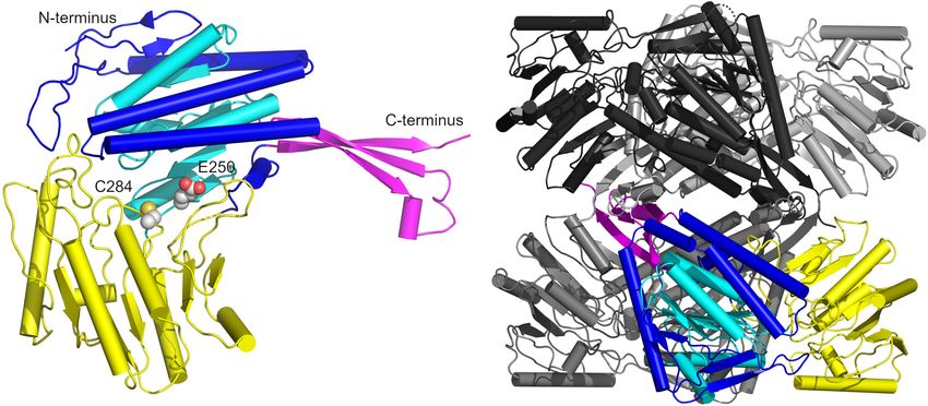
Eisenberg et al.                                                                                                      GapN—A Potential Drug Target

INTRODUCTION                                                               associated with the ability of the bacteria to enter human
                                                                           respiratory cells, thereby escaping extracellular antibiotics such
In the last decades, a rapid increase in antibiotic resistances            as penicillin (Facinelli et al., 2001).
of pathogenic bacteria has been observed (World Health                         Another unfavorable observation is the occurrence of
Organization [WHO], 2014). This alarming development is                    S. pyogenes isolates carrying a specific mutation in the penicillin-
exacerbated by the so-called discovery void of new antimicrobial           binding protein PBP2x, leading to a reduced β-lactam antibiotic
substances during the last 30 years (Silver, 2011). Infections             sensitivity in the last years (Musser et al., 2020; Southon et al.,
with antibiotic-resistant bacteria represent a severe problem in           2020; Vannice et al., 2020). Such mutations might be the starting
different ways. First, the prognosis for patients infected with            point for resistance development (Southon et al., 2020).
multiresistant bacteria is unfavorable. Second, infections with                Considering all of the above, it is of paramount importance to
multiresistant bacteria represent a high financial burden for the          search for new treatment strategies against S. pyogenes infections.
public healthcare systems due to long hospitalizations of patients             The central metabolism of bacteria is a potential target for
requiring complex therapeutic measures (Resch et al., 2009; Roca           the development of new antimicrobial substances. New targets
et al., 2015; Thabit et al., 2015).                                        for antimicrobial substances should be metabolic processes that
    Usually, antibiotics are not specific for a particular bacterial       (i) are not present or not essential in human cells, and (ii) are
species or even a genus. They normally not only kill the targeted          not present or not essential in the majority of the species of the
pathogenic bacteria, but also severely interfere with (part of)            physiological flora.
the commensal flora (Willing et al., 2011). Hence, there is an                 The     non-phosphorylating         glyceraldehyde-3-phosphate
urgent need for the development of new and efficient antibiotics           dehydrogenase GapN has been predicted to be an essential
targeting specifically those bacteria that need to be eradicated.          enzyme in S. pyogenes M49 based on gene essentiality analyses
    S. pyogenes (group A streptococcus, GAS) is one of the most            via transposon mutant libraries (Le Breton et al., 2015).
important gram-positive pathogens. It usually first colonizes              GapN catalyzes the irreversible oxidation of glyceraldehyde
the skin or mucosal membranes of the upper respiratory                     3-phosphate (G3P) to 3-phosphoglycerate (3-PG) while reducing
tract and induces local purulent infections (e.g., tonsillitis,            NADP+ to NADPH (Habenicht, 1997). Hence, it provides
pharyngitis, impetigo), however, toxin-mediated, invasive or               NADPH that is usually generated in the oxPPP, which is missing
systemic diseases (e.g., scarlet fever, toxic-shock-like syndrome,         in S. pyogenes and other streptococci (Crow and Wittenberger,
necrotizing fasciitis) as well as autoimmune-sequelae (rheumatic           1979). Furthermore, since this reaction does not consume
fever, rheumatic heart disease) can occur (Walker et al., 2014).           inorganic phosphate, it is hypothesized that GapN maintains
The estimated global annual burden of S. pyogenes diseases                 glycolysis under low-phosphate conditions (Levering et al.,
comprises about 616 million cases of tonsillitis/pharyngitis, 111          2012). Therefore, GapN is a promising target for specific
million cases of skin infections and 0.5 million deaths due to             inhibition of growth of S. pyogenes and other streptococci, since
severe infections (Carapetis et al., 2005).                                GapN-like enzymes are most likely not essential for bacteria
    Tonsillitis caused by S. pyogenes (strep throat) is a frequent         possessing a complete pentose phosphate pathway. Iddar et al.
reason for the prescription of antibiotics outside hospitals. These        (2003) previously investigated physicochemical and catalytic
infections are usually treated with penicillin for 7–10 days or with       properties of the S. pyogenes GapN and showed that (a) the
macrolides for 3 days (Bisno et al., 2002; Shulman et al., 2012).          enzyme is active in a pH range between 5 and 13 with its pH
According to recent studies, basic penicillins and macrolides are          optimum at 8.5, (b) the enzyme is stable at temperatures between
two of the most frequently prescribed antibiotics in Germany               15 and 40◦ C but loses activity at temperatures above 45◦ C, and
(20 and 15% of patients given antibiotic treatment, respectively;          (c) that the enzyme oxidizes D- and DL-G3P using NADP as a
Centers for Disease Control and Prevention [CDC], 2019b)                   cofactor with km values of 0.666 mM for G3P and 0.385 mM for
(Holstiege et al., 2020) and in the United States (23.4 and                NADP (Iddar et al., 2003).
15.9% of outpatients given antibiotic treatment, respectively).                Antisense peptide nucleic acids (asPNAs) are useful tools for
While penicillin resistance has not yet been observed in                   interfering with specific target molecules. PNAs have peptide
S. pyogenes, macrolide resistant strains are frequently isolated           backbones decorated with nucleobases capable of base pairing.
from patients. In the respective studies, up to 38% of the clinical        Because of their chemical composition, PNAs are inaccessible to
isolates were erythromycin-resistant (Rubio-Lopez et al., 2012).           nucleases and proteases. However, PNAs do not readily enter the
Furthermore, penicillin treatment frequently fails, which might            target cell. This problem can be overcome by cell penetrating
be associated with either intracellular persistence of the bacteria        peptides (CPPs) fused to the PNA. For S. pyogenes, the (RXR)4 XB
or the formation of impenetrable biofilms (Facinelli et al., 2001;         peptide has been shown to allow sufficient entry of PNAs
Baldassarri et al., 2006).                                                 (directed against the gyrase subunit A gene gyrA) into the bacteria
    In the “2019 Antibiotics Resistance Threat Report” of                  to accomplish growth inhibition (Herce et al., 2009; Patenge et al.,
the Centers for Disease Control and Prevention of the                      2013; Barkowsky et al., 2019).
United States the erythromycin-resistant S. pyogenes is classified             Here, we describe the application of (RXR)4 XB-fused gapN-
as a concerning threat that requires careful monitoring and                asPNAs for growth inhibition of S. pyogenes laboratory strains
preventative action as a result of the rising number of resistant          and macrolide-resistant clinical isolates. Our data support the
strains isolated (Centers for Disease Control and Prevention               hypothesis that the essentiality of GapN is due to the lack of
[CDC], 2019a). Erythromycin resistance in S. pyogenes is                   NADPH in the absence of this enzyme in streptococci lacking the

Frontiers in Microbiology | www.frontiersin.org                        2                                   February 2022 | Volume 13 | Article 802427
Eisenberg et al.                                                                                                                       GapN—A Potential Drug Target

oxPPP. Accordingly, GapN represents a promising target for new                          TABLE 2 | PNA sequences.
antibiotic strategies against S. pyogenes by cutting off the NADPH
                                                                                        Species                Target                 CPP            PNA sequence
supply of the bacteria.
                                                                                        S. dysgalactiae        gapN                 (RXR)4 XB         TTGTCAACGT
                                                                                        ssp. equisimilis
MATERIALS AND METHODS                                                                                              gyrA             (RXR)4 XB         TGCATTTAAG
                                                                                                            - (scr_gapN)            (RXR)4 XB         TCAGTCAGTT
Bacterial Strains and Culture Conditions                                                                     - (scr_gyrA)           (RXR)4 XB         ATTAGACTGT
S. pyogenes strains and their characteristics are listed in Table 1.                    S. pyogenes            gapN                 (RXR)4 XB         TTGCCAACGT

Furthermore, S. cristatus DSM8249 and S. dysgalactiae ssp.                                                         gyrA             (RXR)4 XB         TGCATTTAAG

equisimilis ATCC 12394 were used in this study. Generally,                                                  - (scr_gapN)            (RXR)4 XB         CATGTGCTAC

streptococci were grown in Todd-Hewitt medium supplemented                              S. cristatus           gapN                 (RXR)4 XB        CACGTGACAC

with 0.5% (w/v) yeast extract (THY medium) at 37◦ C under                                                          gyrA             (RXR)4 XB         CTTGCATTAA

a 5% CO2 enriched atmosphere, except for S. cristatus, which                                                - (scr_gapN)            (RXR)4 XB        CCCCGGAAAT
                                                                                                             - (scr_gyrA)           (RXR)4 XB        TAGTACTACT
was grown in Brain Heart Infusion (BHI) medium. For growth-
and phosphate-dependent GapN activity analyses, S. pyogenes
and S. cristatus were grown in chemically defined medium for
                                                                                        to make sure that the scrambled PNA are not complementary to
lactic acid bacteria (CDM-LAB) as previously described (Fiedler
                                                                                        the start codon region of any gene in the respective genome.
et al., 2011) but with 0.5 g/l arginine instead of 0.125 g/l.
While CDM-LAB contains 42 mM phosphate, in the CDM-
LAB low phosphate variant, the phosphate concentration was                              Kill Assays
reduced to 1 mM.                                                                        Bacterial susceptibility to PNA was tested by using the method
                                                                                        previously published by Barkowsky et al. (2019). The protocol
Peptide Nucleic Acids                                                                   was adopted for S. cristatus and S. dysgalactiae ssp. equisimilis.
Each PNA was fused to the cell penetrating peptide (RXR)4 XB                            THY or BHI overnight cultures of the respective strains were
(R—arginine, X—6-aminohexanoic acid, B—beta alanine),                                   dissolved in PBS containing either 20% (v/v) BHI (S. pyogenes),
which has previously been shown to work efficiently in                                  15% BHI (S. cristatus), 10% BHI (S. dysgalactiae ssp. equisimilis)
S. pyogenes M49 (Barkowsky et al., 2019). High-performance                              to 105 CFU/ml. Antisense PNA or the respective scrambled
liquid chromatography purified and matrix-assisted laser                                control PNA were mixed 1:9 with the bacterial suspension and
desorption/ionization mass spectrometry analyzed CPP-fused                              incubated for 6 h at 37◦ C in a rotator. As a control, bacteria
PNAs were obtained from Peps4LS GmbH (Heidelberg,                                       were incubated under the same conditions in the absence of PNA.
Germany). Results of the quality controls performed by                                  Subsequently, viable counts were determined by plating serial
Peps4LS GmbH for each PNA used in this study are shown as                               dilutions on THY or BHI agar plates.
Supplementary Table 1. Sequences and characteristics of the
PNAs used in this study are listed in Table 2. Antisense-PNA                            Growth Experiments
were designed to bind to the start codon region of their target                         To analyze the transcription and enzyme activity at different
gene. All scrambled controls were designed by shuffling the bases                       growth phases, CDM-LAB or CDM-LAB low phosphate was
of the specific PNA in random order. Each scrambled PNA was                             inoculated 1:20 with bacteria of THY or BHI overnight
blasted (short sequence algorithm) with the respective genome                           cultures suspended in 0.9% (w/v) NaCl and incubated at 37◦ C,
                                                                                        5% CO2 . The bacterial growth was controlled by measuring
                                                                                        the optical density at 600 nm. S. pyogenes samples were
TABLE 1 | Clinical S. pyogenes isolates and their characteristics                       collected in the exponential (OD600 nm = 0.35–0.45, 2.5 h
(Koller et al., 2010).                                                                  after inoculation), transition (OD600 nm = 0.9–1.1, 4 h after
Strain              Class of     Antibiotic       emm type Specific inhibition by
                                                                                        inoculation) and stationary phase (OD600 nm = 1.55–1.65, 7 h
                   infection    resistance                  gapN asPNA (µM)a            after inoculation), whereas S. cristatus samples were collected in
                                                                                        the exponential (OD600 nm = 0.35–0.5, 2.5 h after inoculation)
591                  Skin            –               49              ≥1
                                                                                        and stationary phase (OD600 nm = 2.55–2.65 (42 mM phosphate),
HRO-K-021           Throat          E, L             12              ≥3
                                                                                        OD600 nm = 1.65–1.9 (1 mM phosphate), 6.5 h after inoculation).
HRO-K-033           Throat           E                4              ≥3
                                                                                        Subsequently, the samples were spun down at 4,000 × g, 4◦ C for
HRO-K-071           Throat          E, T             77              ≥3
                                                                                        10 min. The pellets were flash frozen in liquid nitrogen and stored
HRO-K-075           Throat           E               81              ≥4
                                                                                        at –20◦ C.
HRO-K-094           Throat           E                1              ≥2
HRO-K-100           Throat          E, T             58              ≥2
HRO-K-177          Invasive         E, L             28              ≥3
                                                                                        Quantitative Real-Time PCR
                                                                                        For qPCR, RNA was isolated from respective samples via
E, erythromycin, L, levofloxacin, T, tetracycline.
a Minimum concentration with a significant reduction in colony-forming units            phenol-chloroform extraction. For that purpose, bacteria
compared to scrPNA-treated controls in the kill assay (p < 0.05, n = 4, Mann-           were suspended in 1 ml TRIZOL reagent and treated
                                                                                                                                       R

Whitney U-test).                                                                        in a Precellys 24 homogenizer (Bertin Technologies SAS,

Frontiers in Microbiology | www.frontiersin.org                                     3                                       February 2022 | Volume 13 | Article 802427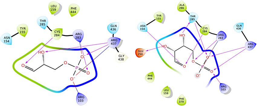
Eisenberg et al.                                                                                                   GapN—A Potential Drug Target

Montigny-le-Bretonneux, France) with two cycles of 30 s each at a         with acid-washed glass beads. Two cycles of 30 s each at a speed
speed of 6,500 rpm and cooling in between for 2 min on ice. After         of 6,500 rpm were applied. In between, samples were cooled on
centrifugation at 16,000 × g and 4◦ C for 5 min the supernatant           ice. Cell debris was removed by centrifugation at 16,000 × g and
was mixed with 600 µl acid phenol-chloroform-isoamylalcohol               4◦ C for 10 min. Clear supernatants were loaded on StrepTactin
(25:24:1) (APCI) and 20 µl 1 M sodium acetate in a Precellys              sepharose columns (2 ml volume) prewashed with buffer W
24 homogenizer with two cycles of 10 s at a speed of 4,000 rpm,           (2.5 column volumes). Unbound proteins were washed from
incubated for 5 min at room temperature and centrifuged for               the column with buffer W (15 column volumes). The protein
5 min at 5,300 × g and 4◦ C. With the upper, aqueous phase                carrying a StrepTag was eluted from the column in fractions of
this procedure was repeated three times. Finally, the aqueous             1 ml each in buffer E. Buffer E is buffer W supplemented with
phase was mixed with 1 ml pure ethanol at –20◦ C and incubated            10 mM desthiobiotin. The elution fractions were checked for
for 16-18 h at –20◦ C and then centrifuged for 60 min at 18,500           protein purity by SDS-PAGE. Fractions containing pure protein
× g and 4◦ C. The nucleic acid pellet was washed with 1 ml                were pooled and dialyzed overnight in PBS or 0.1 M citric acid
75% (v/v) ethanol in DEPC-treated water at –20◦ C, dried and              (pH 6.0) to prepare them for enzymatic assays.
suspended in 50 µl DEPC-treated water. The remaining DNA
in the samples was removed using the TURBO DNA-freeTM                     Measurement of GapN Activity
Kit (Invitrogen, Waltham, MA, United States) and the RNA                  For the measurement of GapN activity in S. pyogenes or
concentration was determined using a PicoDrop. Fifty nanogram             S. cristatus protein crude extracts, bacteria were subjected to
of total RNA were transcribed into cDNA using the First Strand            kill assays or growth experiments as described. After 6 h of
cDNA synthesis kit (Invitrogen, Waltham, MA, United States)               incubation in the presence of 2 µM gapN asPNA, 2 µM
following the manufacturer’s instructions. The real-time PCR              scrambled control PNA or in the absence of PNA bacteria were
was carried out with Maxima SYBR Green/ROX qPCR Master                    harvested by centrifugation at 16,000 × g and 4◦ C for 10 min.
Mix (Thermo Scientific, Waltham, MA, United States) on a                  The bacterial pellets were suspended in 500 µl PBS and total
LightCycler 480 II (Roche, Basel, Switzerland). The 5S rRNA               protein extracts were prepared in a Precellys 24 homogenizer
gene transcript was used for normalization. For that purpose              with two cycles of 30 s each at a speed of 6,500 rpm. Between
the primer pair 50 -TGAGTGTCATTGTGGCAAGAGC-30 /50 -                       the cycles the samples were cooled on ice for 2 min. Cell debris
AGAGAATACGACGATGCACAGG-30 was used. The gapN                              was removed by centrifugation at 16,000 × g and 4◦ C for
transcript was detected using the primers 50 -GAAGAAGGGCT                 10 min. Protein concentration in the supernatant was measured
TCGTATGG-30 and 50 -AGAACCTGCCAAGTTAACGG-30 .                             using a Qubit protein assay kit following the manufacturer’s
Primer efficiency was tested on genomic S. pyogenes M49 DNA               instructions. For the measurement of activity of purified GapN,
before use in reverse transcription reactions. The relative gene          the protein concentration was adjusted to 70 µg/ml. The specific
expression was determined by the 11CT method.                             GapN activity was measured as described in Iddar et al. (2003)
                                                                          with modification of the protocol. The assay mixture contained
Heterologous Expression and                                               50 mM Tricine buffer (pH 8.5), 3 mM 2-mercaptoethanol, 10 mM
Purification of GapN and GapDH                                            NADP+ , and 2 mM DL-G3P and 20 µl protein sample in a total
The chromosomal DNA of S. pyogenes M49 strain 591 served as               volume of 200 µl. The reaction was started by adding DL-G3P.
the template for PCR amplification of the gapN gene (primers              The reduction of NADP+ to NADPH was detected by measuring
50 -GAGATGAATTCTTGGCAAAACAATATAAAA-30                          and        the absorption at 340 nm in a spectrophotometer for up to
50 -CAATA-GTTGGATCCATCTGTATAGACTA-30 ) and the                            60 min. As controls, the assay was carried out without addition
gapDH gene (primers 50 -AGGAAATCAGGATC-CGTAGT                             of protein sample and without addition of G3P, respectively.
TAAAGTTGGT and 50 - CGTTATAACGTCGACTTTAGCAAT                              GapN activity was calculated with the Lambert-Beer equation
TTTTGC-30 ) with the PhusionTM High Fidelity PCR Kit                      and expressed in units per milligram of total protein (U/mg),
(Thermo Scientic, Waltham, MA, United States). The resulting              where one unit is defined as the formation of 1 µmol NADPH per
gapDH PCR fragment was ligated into the pASK-IBA2 vector                  minute. For determination of cofactor specificity, 10 mM NAD+
(IBA GmbH, Göttingen, Germany) system via the BamHI and                   was added to the assay mixture instead of NADP+ . Potential
SalI restriction sites. The gapN fragment was ligated into the            activating or inhibiting effects on GapN activity were tested by
pASK-IBA6 vector via the EcoRI and BamHI restriction sites.               adding 25 mM NaCl, 25 mM KCl, 25 mM NH4 Cl, 10 mM
E. coli DH5α cells were transformed with recombinant vectors.             D-glucose, 10 mM D-fructose, 10 mM sodium pyruvate, 10 mM
The correct insertion of the PCR product was confirmed by                 ATP, 2 mM erythrose 4-phosphate (E4P), 2 mM sedoheptulose
sequencing. For heterologous expression, the recombinant                  7-phosphate (S7P), 0.4 mM NADH or 0.4 mM NADPH to
E. coli strain was grown in 500 ml LB medium at 37◦ C under               the assay mixture.
vigorous shaking. At an optical density (600 nm) of about 0.4,
expression was induced by addition of anhydrotetracycline                 Measurement of GapDH Activity
(AHT, 0.2 µg/ml). Cells were harvested after overnight shaking            The specific GapDH activity was measured as described in Iddar
at 22◦ C and pellets were stored at –20◦ C. For purification of the       et al. (2003) with modification of the protocol. Purified GapDH
Strep-tagged proteins, cell pellets were thawed, suspended in             was adjusted to a protein concentration of 100 µg/ml. The assay
5 ml buffer W (100 mM Tris-HCl pH 8.0, 1 mM EDTA, 150 mM                  mixture contained 50 mM Tricine buffer (pH 8.5), 3 mM 2-
NaCl) and cell disruption was achieved by the FastPrep method             mercaptoethanol, 10 mM NAD+ , 10 mM PO4 3− , 1 mM DL-G3P

Frontiers in Microbiology | www.frontiersin.org                       4                                 February 2022 | Volume 13 | Article 802427
Eisenberg et al. GapN—A Potential Drug Target
and 5 µl protein sample in a total volume of 200 µl. The reaction Waals scaling on the receptor grid and 0.8 × scaling on the
was started by adding DL-G3P. The reduction of NAD+ to ligands. The docking was conducted using default parameters
NADH was detected by measuring the absorption at 340 nm in a and no additional restraints on the docking optimization. 5
spectrophotometer for up to 120 min. As controls, the assay was docking poses per ligand were generated and they were assessed
carried out without addition of either protein sample or without by the number of (un-) favorable protein-ligand interactions,
addition of G3P. Activity was calculated with the Lambert-Beer the docking scores, and by visual inspection. Docking with
equation and expressed in units per milligram of total protein Autodock4.2 was conducted using the Autodock Tools GUI
(U/mg), where one unit is defined as the formation of 1 µmol (Morris et al., 2008). A Lamarckian Genetic Algorithm was
NADH per minute. For determination of cofactor specificity, applied with the number of evaluations set to “long.” All other
10 mM NADP+ was added to the assay mixture instead of NAD+ . parameters were set to their default values. The resulting poses
were analyzed according to both visual inspection and by the
Multi-Angle Light Scattering Analysis of computed docking scores available in the Autodock Tools GUI.
GapN
Size-exclusion chromatography coupled to multi-angle light
Crystal Structure Determination of the
scattering (SEC-MALS) was used to determine the oligomeric Apo-Form of Streptococcus pyogenes
state of GapN in solution. The purified protein was separated at GapN
room temperature on a HiLoad 16/600 Superdex 200 pg column Crystals of the apo-enzyme (WT and C284S mutant) were grown
(Cytiva, Marlborough, MA, United States) in 1 × PBS (pH 7.4) at 20◦ C by the vapor diffusion method in hanging drops. The WT
buffer at a flow rate of 0.5 ml/min. The molecular weight of apo-enzyme crystals were grown by mixing 1 µl protein (WT)
the eluting proteins was determined using a Dawn Heleos 8 at a concentration of 15 mg/ml in 1 × PBS (pH 7.4) with 1 µl
light scattering detector and an Optilab T-rEX refractive index mother liquor containing 0.2 M Li2 SO4 , 0.1 M Bis-Tris pH 5.5
detector (Wyatt Technologies, Santa Barbara, CA, United States). and 25% PEG 3350. Crystals of the C284S variant were obtained
Data were analyzed using the ASTRA software (version 6.1.5.22, at a protein concentration of 2.5 mg/ml, by again mixing 1 µl of
Wyatt Technologies, Santa Barbara, CA, United States). the protein solution in 1 × PBS (pH 7.4) with 1 µl of reservoir
containing 0.2 M Li2 SO4 , 0.1 M Bis-Tris pH 6.5 and 18% PEG
Homology Modeling of Streptococcus 3350. Crystals grew either as plates or prisms in 4–5 days.
pyogenes GapN Diffraction data were collected at the ESRF (Grenoble, France)
Template-based homology modeling of the tetrameric holo- for the wild-type apo-enzyme on beam line ID30-B on a Dectris
enzyme was conducted using the SWISS-MODEL webserver Pilatus3 6 M detector at a wavelength of λ = 0.918401 Å and
(Waterhouse et al., 2018) and the sequence of S. pyogenes GapN for the C284S variant on beam line ID23-2 at a wavelength
obtained from resequencing (see “Results” section). A holo- of λ = 0.873128 Å on a Dectris Pilatus3 × 2 M detector. All
structure of GapN from S. mutans (PDB 1QI1) (Cobessi et al., diffraction data were collected at a temperature of 100 K after
2000) was chosen as the most promising template due to adding 25% glycerol as cryo-protectant. The collected data sets
its high sequence-identity of 86%. The molecular structures were indexed, integrated and scaled with XDS and the resulting
of the substrate, G3P, and the cofactor, NADP, were inserted scaling was analyzed with the program Aimless (CCP4). As
into the modeled protein structure by aligning it to the Aimless detected significant anisotropy in the data sets, the
template and subsequently transferring the ligands using PyMol data generated with XDS were reprocessed with Staraniso1 with
(DeLano, 2002). an < I/σI > cutoff of 1.2.
The apo-structure of wild-type GapN was solved by molecular
replacement using Phaser MR with the apo-enzyme from
Molecular Docking S. mutans (PDB entry: 1EUH) as a search model. Subsequent
Docking of the native substrate (G3P) and two known inhibitors molecular replacement calculations for the C284S GapN mutant
of the template protein from S. mutans, E4P and S7P, used a tetramer of the initial S. pyogenes apo-structure as a
was conducted using the GLIDE (Friesner et al., 2004) and search model. The structures were initially refined with Refmac
Autodock4.2 (Morris et al., 2008) molecular docking programs. 5 (CCP4) using NCS restraints, Babinet scaling and isotropic
The GLIDE docking calculations were carried out within the temperature factor refinement and extensive rebuilding in Coot.
Maestro (Schrödinger, LLC, New York, NY, United States, 2020) Subsequently, coordinates, B-factors, occupancies, and TLS-
environment for molecular modeling (Sastry et al., 2013). After parameters were refined with Buster after adding hydrogens
import into the Maestro environment, the protein structure was with Hydrogenate.
prepared with the “PrepWizard” utility to generate a protonation
state at pH 7.0 ± 3.0, to assign bond orders and to perform a Statistical Analysis
restrained energy minimization with the OPLS2005 force field. The number of biological replicates (n) and the tests used to
Similarly, all ligand structures were prepared using the “LigPrep” determine statistical significance for each data set are indicated in
program and protonated at a pH of 7.2 ± 0.4. The three- the respective figure captions. Statistical analyses were performed
dimensional structures of E4P and S7P were generated with using GraphPad Prism 8 software.
the “2D-sketcher” utility of Maestro (2020). The “standard-
precision” mode of GLIDE was used with 1 × van der 1
http://staraniso.globalphasing.org/cgi-bin/staraniso.cgi
Frontiers in Microbiology | www.frontiersin.org 5 February 2022 | Volume 13 | Article 802427Eisenberg et al. GapN—A Potential Drug Target
RESULTS importantly, the sequence derived in this study shows a
frameshift in comparison to the NCBI derived sequence, leading
GapN Is the Major NADPH Source in to an open reading frame extension of 24 bp and, consequently,
to a protein with 8 additional amino acid residues at its amino-
Streptococcus pyogenes
terminus. Apart from this, several other differences were found
We hypothesized that the GapN mediated reaction is the major
in the 30 -end of the gene, leading to differences in amino acid
source of NADPH in S. pyogenes. Since NADPH is needed in
residues at five positions near the C-terminus of the protein as
numerous anabolic reactions, e.g., fatty acid and amino acid
compared to the NCBI-derived sequence. In BLASTp analyses,
biosynthesis, it is an essential cofactor. In bacteria, NADPH is
the resulting protein shows a slightly higher similarity to GapN
usually derived from the oxidative part of the pentose phosphate
proteins of other sequenced S. pyogenes serotype strains (99.2–
pathway. Since the enzymes of the oxPPP are not encoded in the
99.7%) as compared to the one based on the NCBI derived
genome of S. pyogenes, the GapN mediated conversion of G3P to
sequence (98.6–99.1%). According to our analysis, there are no
3-PG might represent the major NADPH generating reaction in
differences between the gapN gene sequences of the NZ131
these bacteria. This hypothesis is supported by the observation
and 591 strains. The reference sequence meanwhile available for
that either GapN or the oxPPP are missing in the majority of
strain 591 is identical to the sequence we determined.
sequenced streptococci available at the KEGG database with only
To determine the state in which GapN can be found while
a subset of organisms containing both pathways (Table 3).
in solution, we used size-exclusion chromatography coupled
To further validate this hypothesis, we analyzed the cofactor
to multi-angle light scattering. GapN (496 aa) eluted at
specificity of GapN and the conventional GapDH of S. pyogenes
approximately 14 ml, revealing a calculated weight-average molar
M49 strain 591. For that purpose, both genes were ligated into
mass of 2.022 × 10⁵ Da (± 2.494%) (Supplementary Figure 1).
vectors of the pASK-IBA series for heterologous expression
Inserting our sequence in Expasy ProtParam revealed a molecular
and subsequent purification via StrepTactin based affinity
weight of 52937.55 Da per monomer or 2.118 × 10⁵ Da for a
chromatography. In this context, we re-sequenced the gapN
tetramer. The determined molar mass and the molecular weight
genes of the S. pyogenes M49 strains NZ131 (GeneBank:
of MALS and ProtParam are in very good agreement under the
OK337836) and 591. A BLASTnt analysis revealed significant
assumption that the enzyme is present in solution as a tetramer.
differences in our sequence data in comparison to the NCBI
The purified GapN and GapDH of S. pyogenes M49 strain
derived gapN sequence of the NZ131 strain (Figure 1). Most
591 were analyzed for their cofactor specificity. Therefore, the
enzymatic activity of both enzymes was measured in the presence
of NAD+ as well as NADP+ . Purified GapN showed a specific
TABLE 3 | Presence (+) or absence (−) of the oxPPP and GapN in activity of 33.6 ± 9.3 U/mg protein (n = 4) in the presence of
streptococcal species. NADP+ , while no activity was observed in the presence of NAD+ .
In contrast, GapDH was completely inactive in the presence of
Species GapN oxPPP NADP+ , while with NAD+ a specific activity of 21.1 ± 5.5 U/mg
S. oralis + + (n = 4) was detected. This indicates that GapN is highly specific
S. pneumoniae + + for NADP while GapDH cannot utilize NADP at all. The Km
S. sanguinis + + values of the purified GapN were determined to be 0.36 mM
S. cristatus + + (± 0.05 standard error) for NADP and 0.58 mM (± 0.19 standard
S. pyogenes + − error) for DL-G3P.
S. agalactiae + − Furthermore, we analyzed the impact of different salts, sugars
S. mutans + − and metabolic intermediates on the specific activity of the
S. thermophilus + − purified S. pyogenes GapN. All substances tested were previously
S. equi + − described to affect the activity of GapN in S. mutans (Crow and
S. uberis + − Wittenberger, 1979). The respective substances were added to
S. parauberis + − the standard GapN activity assay in the concentrations indicated
S. dysgalactiae + − in Figure 2. Of the 11 substances tested, only E4P and ATP
S. gallolyticus + − significantly reduced the activity of the S. pyogenes GapN by 90
S. salivarius + − and 50%, respectively, with the other substances displaying no
S. infantarius + − significant impact.
S. iniae + −
S. equinus + − GapN Is Essential in Streptococcus
S. anginosus − +
pyogenes and Other Streptococci
S. intermedius − +
S.macedonicus − + Lacking the Oxidative Part of the
S.gordonii − + Pentose Phosphate Pathway
S.pseudopneumoniae − + During growth of S. pyogenes M49 strain 591 in chemically
S.suis − + defined medium for lactic acid bacteria (CDM-LAB), the gapN
S.mitis − + transcript is present in all growth phases (Figure 3A). The
Frontiers in Microbiology | www.frontiersin.org 6 February 2022 | Volume 13 | Article 802427Eisenberg et al. GapN—A Potential Drug Target
FIGURE 1 | Resequencing of the gapN gene of S. pyogenes M49 strains NZ131 and 591. (A) 30 -end of the gapN gene sequence as determined in this work for the
NZ131 and 591 strains (lower lines) in comparison to the NCBI derived sequence of the NZ131 strain (upper lines). (B) Amino acid sequences of the N-terminus of
GapN for the NZ131 and 591 strains according to the resequencing in this work (lower line) in comparison to the NCBI derived sequence for NZ131 (upper line).
Stop codons are indicted in bold underlined letters (A) or asterisks (B). Discrepancies in sequences are indicated by gray backgrounds.
transcript abundance is highest during exponential growth, stays as scrambled (scr) PNA were used. Both asPNA and scrPNA
relatively stable in the transition phase but strongly drops in the were fused to the cell penetrating peptide (CPP) with the
stationary phase of the bacteria (Figure 3A). Correspondingly, sequence (RXR)4 XB (R—arginine, X—6-aminohexanoic acid,
specific GapN activity is detectable in all growth phases B—beta alanine), as it has previously been described that this
(Figure 3B). In contrast to the transcript abundance, however, CPP facilitates the uptake of PNA into S. pyogenes (Barkowsky
the specific activity does not decrease significantly toward the et al., 2019). For all eight strains a significant reduction of the
stationary phase, hinting at a high stability of the enzyme. viable counts was achieved upon treatment with gapN asPNA
Next, we analyzed whether a knock-down of GapN can affect (Figure 4 and Table 1). The scrambled control PNA also reduced
the viability of S. pyogenes. For this purpose, the M49 laboratory the viable counts, indicating that the cell penetrating peptide
strain 591 and seven macrolide-resistant clinical S. pyogenes exhibits cell toxicity. The effect of the asPNA was, however,
isolates (Table 1), together representing eight different emm types significantly higher than the effect of the scrambled controls in
and four different FCT-types, were subjected to kill assays with all tested strains. Upon treatment with 5 µM gapN asPNA the
gapN-specific asPNA. The gapN asPNA were complementary viable counts of all strains were between one and two orders
to the gapN start codon region. As a control, water as well of magnitude lower than upon treatment with the scrambled
control PNA. Already upon treatment with 2 µM of gapN specific
asPNAs for 6 h, the specific GapN activity in protein crude
extracts of S. pyogenes M49 strain 591 was significantly reduced
to about 25% as compared to the untreated control, whereas the
scrambled control PNA or PNA targeting the essential gyrase
subunit A encoding gyrA gene did not reduce GapN activity
significantly (Figure 5).
Additionally, we tested the effect of gapN asPNA treatment on
two further streptococcal species. S. dysgalactiae ssp. equisimilis
resembles S. pyogenes in as it also lacks the oxPPP but possesses
a GapN. S. cristatus possesses both GapN and the oxPPP. For
S. cristatus and S. dysgalactiae ssp. equisimilis, asPNA treatment
has not been described previously. However, using asPNA
targeting gyrA we could show that (RXR)4 XB is a suitable
CPP to mediate PNA uptake for both species as gyrA asPNA
treatment reduced the viable counts significantly in comparison
to the scrambled control PNA (Figure 6). In accordance with
the hypothesis that GapN is only essential in species lacking the
oxPPP, the viable counts of S. dysgalactiae ssp. equisimilis were
significantly reduced upon treatment with gapN asPNA, while
FIGURE 2 | Influence of different substances on the specific activity of purified this was not the case for S. cristatus (Figure 6).
S. pyogenes GapN. Specific activities are shown relative to the specific activity Consequently, we checked whether GapN is active in
in a standard assay mix. E4P, erythrose 4-phosphate; S7P, sedoheptulose S. cristatus at all. Therefore, the growth phase associated specific
7-phosphate, mean values and standard deviations; n = 4, one-way ANOVA,
***p < 0.001, ****p < 0.0001.
GapN activity of S. cristatus growing in CDM-LAB was analyzed
and compared to S. pyogenes 591. As can be seen in Figure 7A,
Frontiers in Microbiology | www.frontiersin.org 7 February 2022 | Volume 13 | Article 802427Eisenberg et al. GapN—A Potential Drug Target
FIGURE 3 | Growth phase associated gene expression and enzyme activity of GapN. (A) gapN transcript abundance and (B) specific GapN activity in crude extracts
of S. pyogenes during exponential (exp.), transition (trans.) and stationary (stat.) growth phases in CDM-LAB medium. Mean values and standard deviations,
***p < 0.001, n = 3, pairwise comparison with exponential phase using paired t-test.
GapN activity can be detected in crude extracts from S. cristatus, at a resolution of 2 Å against data derived from triclinic crystals
but the activity is far lower than the activity detected in crude containing two tetramers in the asymmetric unit (PDB: 7PKJ)
extracts of S. pyogenes in the exponential and stationary phases. (Table 4). The structure revealed a covalent modification of the
In contrast, the specific GapDH activity in the crude extracts was enzyme at the active site residue C284, which could be modeled
roughly the same in S. cristatus and S. pyogenes in both growth as β-mercaptoethanol although this compound was not present
phases (Figure 7B). during purification. Hence, the enzyme must have been modified
It has been proposed previously that in S. pyogenes GapN following heterologous expression in E. coli. To eliminate this
is needed to keep glycolysis running under low phosphate modification and generate a true apo-state of the enzyme, the
conditions, as the GapN-mediated conversion of G3P to 3-PG C284S variant was generated and crystallized. Its crystal structure
does not consume inorganic phosphate, in contrast to the GapDH was also determined by molecular replacement, with a tetramer
and phosphoglycerate kinase mediated reactions (Levering et al., of the wild-type protein as search model. The variant again
2012). Therefore, GapN activity in crude extracts of S. pyogenes crystallized in the P1 space group, but in this case the unit cell
and S. cristatus grown in CDM-LAB low phosphate conditions contained four tetramers and the structure was refined at 1.5 Å
was analyzed. However, the strongly reduced phosphate supply resolution (PDB entry 7PKC). Due to its higher resolution, the
(1 mM in comparison to 42 mM in the conventional CDM- structure of the C284S mutant is presented and discussed here.
LAB) did not lead to an increased GapN activity in either The GapN-monomer from S. pyogenes, like its counterparts
species (Figure 7). from other organisms can be subdivided into three domains
(Figure 8A; Cobessi et al., 1999). Starting at the N-terminus,
The High Resolution Crystal Structure of residues 2–118, 145–252, and 450–464 form the cofactor binding
domain containing a Rossmannoid fold (residues 145–252, cyan
GapN in the Apo-State Provides a with the remainder of the domain in blue) which harbors five
Starting Point for Structure-Based Drug β-strands and three α-helices and mediates binding to NADP
Design (Hanukoglu, 2015). The two other domains are the protruding
At present, no structures of GapN from S. pyogenes have been domain composed of residues 119–144 and 464–475 (magenta),
reported. The closest homolog for which crystal structures have which is critical for oligomerization, and the catalytic α/β domain
been determined is the enzyme from S. mutans, which shares encompassing residues 253–445 (yellow). The catalytically active
86% sequence identity with S. pyogenes GapN. This enzyme, as cysteine, which is responsible for the nucleophilic attack on the
well as more distantly related homologs from Bacillus halodurans carbonyl carbon of G3P, is located at position 284, in close
(58% identity), Methanocaldococcus janaschii (37% identity) and proximity to E250, the other key catalytic residue. E250 interacts
Escherichia coli (35% identity), is present as a homotetramer with with a water molecule, which deprotonates the cysteine in the
D2 symmetry in crystal structures. To further characterize the deacylation step of the NADPH generating mechanism (Cobessi
function of the S. pyogenes GapN and provide a starting point for et al., 2000). The monomers assemble into a tetramer displaying
future screening campaigns, we determined the crystal structure D2 symmetry which is formed by two interlacing dimers with
of the GapN by molecular replacement using the apo-form of an dimer-dimer contacts primarily mediated by the protruding
S. mutans tetramer as a search model. The structure was refined domain, while contacts within each dimer are formed by a
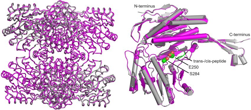
Frontiers in Microbiology | www.frontiersin.org 8 February 2022 | Volume 13 | Article 802427Eisenberg et al. GapN—A Potential Drug Target FIGURE 4 | Survival of different S. pyogenes strains in the presence of (RXR)4 XB-gapN-asPNA. Mean values and standard deviations, n = 5 (HRO-K021) or n = 4 (all other strains), *p < 0.05, **p < 0.01, pairwise comparison via Mann-Whitney U-test. β-sheet of the protruding domain interacting with a β-sheet of the respective A-chains were aligned (Figure 9A). The comparison catalytic α/β domain via salt bridges as well as α-helices (residues revealed an unexpected difference for residues G438 and T439 232–251) of the Rossmannoid fold of each chain (Figure 8B). (Figure 9B). In all chains of the wild-type and C284S variant a cis- A superimposition of our structure with that of GapN from peptide was observed while only half of the chains in the reference S. mutans (PDB: 1EUH) resulted in a root mean square deviation structure from S. mutans were modeled in the cis configuration. (RMSD) of 0.61 Å for the tetramer and 0.41 Å, if only the Considering that the formation of a cis-peptide outside an X-P Frontiers in Microbiology | www.frontiersin.org 9 February 2022 | Volume 13 | Article 802427
Eisenberg et al.                                                                                                                  GapN—A Potential Drug Target

                                                                                        have the necessary conformational changes to accommodate the
                                                                                        substrate and possible competitive inhibitors. Both the latter
                                                                                        structures have mutations in the active site: C284T (PDB:
                                                                                        1QI1) and E250A (PDB: 2ESD). We found that by using the
                                                                                        SWISS-MODEL webserver the conformations of the catalytic
                                                                                        residues were appropriately rebuilt when using the 1QI1 structure
                                                                                        as the template (Figure 10). On the other hand, when the
                                                                                        2ESD structure was used as the template, E250 adopted a
                                                                                        conformation which did not align to the reported “intermediate”
                                                                                        or “inside” conformations (Munoz-Clares et al., 2011). Hence,
                                                                                        we chose 1QI1 as the structural template. Assessment of the
                                                                                        resultant homology model using statistical criteria computed
                                                                                        by the SWISS-MODEL server indicated a high certainty in the
                                                                                        generated model (Supplementary Figure 2). Aligning the model
                                                                                        to the template structure in PyMol resulted in a remarkably low
                                                                                        RMSD of 0.081 Å.
                                                                                            From our models and the literature on NAD(P)-dependent
                                                                                        aldehyde dehydrogenases (Munoz-Clares et al., 2011), the
                                                                                        catalytic cysteine (C284) can either adopt a “resting”-
                                                                                        conformation, in which the thiol points away from the ligand,
                                                                                        or an “attacking”-conformation in which it points toward the
                                                                                        aldehyde moiety of the substrate. E250 is also involved in the
                                                                                        catalytic cycle in which it activates water molecules and can
                                                                                        either adopt an “inside” or “intermediate” conformation, in
                                                                                        which it points, respectively, toward the binding site or in the
                                                                                        opposite direction.
                                                                                            To assess the quality of the generated model and to provide
                                                                                        a benchmark for evaluating future inhibitor candidates, the
  FIGURE 5 | Influence of PNA treatment on the specific GapN activity. GapN
                                                                                        native ligand G3P was re-docked to GapN using both GLIDE
  activity in protein crude extracts of S. pyogenes M49 treated with either 2 µM        and Autodock4.2. Re-docking with GLIDE was successful in
  (RXR)4 XB-gapN-asPNA, (RXR)4 XB-gyrA-asPNA or (RXR)4 XB-gapN-scrPNA                   reproducing the binding pose observed in the 1QI1 structure
  (scr) in comparison to an untreated control. Mean values and standard                 after manually rotating C284 to the “resting” conformation for
  deviations, n = 3, ∗ p < 0.05, unpaired two-tailed t-test.
                                                                                        the docking assay. The best pose achieved a docking score of –
                                                                                        7.7 kcal/mol and featured mainly favorable interactions between
                                                                                        the ligand and the protein. Aligning the generated ligand pose to
(X denoting any amino acid) peptide is rather rare, the electron                        the structure of G3P from the crystal structure 1Q1 resulted in an
density maps of our structures and the published structures were                        RMSD of 1.08 Å. The only small difference between this pose and
critically examined (Jabs et al., 1999). This analysis confirmed the                    the template structure was in the position of the aldehyde oxygen.
formation of a cis-peptide bond in both S. pyogenes as well as in                       GLIDE did not reproduce its interaction with N154. Instead,
S. mutans. As this peptide bond is within 11.2 Å of the active                          the aldehyde oxygen formed a hydrogen-bond to T285 while
site, it might be relevant for the proper function of the enzyme.                       its interaction with C284 was successfully reproduced. It should
Furthermore, in the more distantly related Bacillus halodurans                          also be noted that the restrained energy-minimization routine in
GapN, a cis-peptide can be observed in all chains between G447                          Maestro leads to the loss of the hydrogen-bond of the aldehyde-
and P448 (PDB: 3PRL), further underscoring the importance                               oxygen of G3P to N154 present in the original crystal structure.
of this feature.                                                                        The best results from docking G3P to a structure with C284 in the
                                                                                        “attacking” conformation resulted in docking scores between –
Homology Modeling Facilitates                                                           7.6 and –7.3 kcal/mol but showed an alternative positioning of
Prediction of GapN Inhibitors                                                           the hydroxyl-group of G3P. In all of these slightly worse-scoring
To pave the way for future drug screening assays, we generated a                        poses, the hydroxyl-group interacted with T285 rather than with
structure of the tetrameric holo-enzyme by homology modeling                            R437 (as in 1QI1). Re-docking in Autodock resulted in a similar
before the crystal structures for S. pyogenes GapN were available.                      pose and a docking score of –7.3 kcal/mol.
Due to its high sequence-identity of 86%, GapN from S. mutans                               A comparison of the crystal structure of the apo-enzyme and
was chosen as the template protein for the model. There are four                        the homology model of the holo-enzyme (which, as stated before,
structures of this protein available in the RCSB protein data bank,                     was built before the crystal structure of the apo-enzyme was
two of which, 2ESD and 1QI1 (D’Ambrosio et al., 2006), consist                          determined) revealed a very high degree of similarity between
of the holo-enzyme with the physiological substrate, G3P, bound.                        the two and alignment in PyMol (DeLano, 2002) resulted in
Holo-enzymes were preferred, because the catalytic site would                           an RMSD of 0.345 Å (Figure 10A). The structures differed

Frontiers in Microbiology | www.frontiersin.org                                    10                                  February 2022 | Volume 13 | Article 802427
Eisenberg et al. GapN—A Potential Drug Target FIGURE 6 | Survival of different streptococcal species in the presence of (RXR)4 XB-PNA directed against gapN or gyrA. Mean values and standard deviations, n = 4 (all PNAs) or n = 8 (controls) *p < 0.05, pairwise comparison with corresponding scrPNA using Mann-Whitney U-test. FIGURE 7 | Phosphate concentration and growth phase associated specific GapN activity in S. pyogenes and S. cristatus. Activity of (A) GapN and (B) GapDH in protein crude extracts of S. pyogenes M49 and S. cristatus during exponential (exp.) and stationary (stat.) phase of growth in CDM-LAB (42 mM phosphate) resp. CDM-LAB low phosphate (1 mM phosphate; low Pi) media. Mean values and standard deviations, n = 3. mainly in the binding site, where the absence of G3P appeared and affinities of the two compounds for S. pyogenes GapN. While to result in an alternative conformation of R437 which is similar E4P was predicted by both GLIDE and Autodock to bind to the to that observed in apo-structures of GapN from S. mutans that enzyme with a binding affinity comparable to G3P: –7.6 kcal/mol lack a ligand: PDB 1EUH and 2ID2 (Figure 10B). However, (GLIDE; Figure 11), –7.2 kcal/mol (Autodock), both algorithms the cis-peptide bond between G438 and T439 observed in the predicted no inhibitory effect of S7P: ∼–5 kcal/mol (GLIDE); – crystal structure of the apo-enzyme was not present in the 0.19 kcal/mol (Autodock), whose binding was sterically hindered. homology model of the holo-enzyme or its structural template Docking G3P to the S. mutans GapN template structure 1QI1 from S. mutans. It is unclear whether this cis-peptide bond arises after manually mutating T284 back to the wild-type cysteine because of the missing ligands in the apo-form of the protein. resulted in a pose with the same score of –7.6 kcal/mol (GLIDE). Crow and Wittenberger (1979) purified the template protein E4P was docked with a slightly higher affinity score for the from S. mutans and identified both E4P and sedoheptulose- template protein of –8.1 kcal/mol while S7P was predicted to have 7-phosphate (S7P) to have significant inhibitory effects on its a similarly low affinity for the binding site of roughly –4 kcal/mol catalytic activity, with E4P having greater inhibitory activity than (GLIDE). Docking S7P to 1QI1 with Autodock resulted in a S7P. To assess the quality of our structural model, we used slightly higher affinity for the S. mutans GapN than for the computational docking algorithms to predict the binding modes modeled S. pyogenes GapN of roughly –1.4 kcal/mol. Frontiers in Microbiology | www.frontiersin.org 11 February 2022 | Volume 13 | Article 802427
Eisenberg et al.                                                                                                                                   GapN—A Potential Drug Target

TABLE 4 | Data collection and refinement statistics.

                                                        Apo-structure of wild-type S. pyogenes GapN                  Apo-structure of C284S mutant of S. pyogenes GapN

Data collection

Space group                                                                      P1                                                                P1

Cell dimensions
a, b, c (Å)                                                             97.75, 99.89, 106.18                                              97.67, 126.04, 148.39
α, β, γ (◦ )                                                            77.67, 75.19, 67.12                                                96.36, 104.81, 84.23
Resolution (highest shell) (Å)                                        48.15–1.99 (2.17–1.99)                                              47.63–1.50 (1.52–1.50)
Diffraction best/worst direction after cut-off (Å)                          1.989/3.378                                                        1.499/3.481
Anisotropic 1B (Å2 )                                                            17.23                                                             3.36
aR                                                                          0.304 (1.002)                                                     0.122 (0.705)
     sym
bR                                                                          0.213 (0.655)                                                     0.087 (0.497)
     pim
CC1/2                                                                       0.847 (0.396)                                                     0.992 (0.466)
c<    I/σI >                                                                3.195 (0.943)                                                     5.444 (1.569)
Completeness (sphere;%)                                                     0.629 (0.137)                                                     0.685 (0.149)
Completeness (ellipse;%)                                                    0.875 (0.707)                                                     0.900 (0.638)
Redundancy                                                                   2.91 (3.23)                                                       2.76 (2.89)
Refinement
Resolution (highest shell) (Å)                                        20.03–1.99 (2.08–1.99)                                              19.97–1.50 (1.58–1.50)
No. reflections (highest shell)                                           153,157 (2,918)                                                   744,550 (14,532)
dR          eR
     work /    free                                               0.1923/0.2319 (0.2930/0.3204)                                    0.1999/0.2316 (0.2734/0.2842)
No. of atoms                                                                   30,879                                                            63,495
   Protein                                                                     28,364                                                            56,829
   Water                                                                        2,243                                                             6,361
   Ions SO4 2− /PO4 3−                                                            90                                                               155
   Glycerol                                                                      182                                                               150
B-factors (Å2 )                                                                 24.99                                                             16.85
   Protein (best/worst chain)                                           21.97 (20.68/32.51)                                                15.02 (13.42/22.02)
   Water                                                                        27.39                                                             24.57
   Ions SO4 2− /PO4 3−                                                          72.48                                                             45.08
   Glycerol                                                                     49.47                                                             54.06
f Ramachandran        statistics (%)                                      96.24/3.31/0.45                                                    96.49/3.09/0.42
RMS deviations in
Bond lengths (Å)                                                                0.008                                                             0.008
Bond angles (◦ )                                                                 1.01                                                             1.01
Torsion angles (◦ )                                                             14.77                                                             14.13
Planar groups (Å)                                                              0.0121                                                            0.0135

  sym = 6 hkl 6 i | Ii —< I > | /6 hkl 6 i Ii where Ii is the measurement and < I > is the weighted mean of all measurements of I.
aR                                                     ith
   pim = 6 hkl 1/(N-1)
bR                      1/2 6 | I (hkl)—I (hkl) | /6 6 I(hkl), where N is the redundancy of the data and I (hkl) the average intensity.
                             i i                      hkl i
c < I/σI > indicates the average of the intensity divided by its standard deviation.
dR     = 6 hkl | | Fo | –| Fc | | /6 hkl | Fo | where Fo and Fc are the observed and calculated structure factor amplitudes.
     work
eR    same as R for 5% of the data randomly omitted from the refinement. The number of reflections includes the Rfree subset.
   free
f Ramachandran statistics were calculated with Coot.

Numbers in parentheses refer to the respective highest resolution data shell in each dataset.

DISCUSSION                                                                                   Xu et al., 2018). In bacteria, the major NADPH supply for most
                                                                                             species are the two oxidizing reactions in the pentose phosphate
GapN related enzymes occur not only in bacteria but also in                                  pathway (Spaans et al., 2015). This sugar degrading pathway,
archaea, fungi and plants (Habenicht, 1997; Iddar et al., 2005),                             however, is absent in some bacteria, e.g., in several streptococcal
however, the physiological importance of this enzyme is not                                  species. In such species, the GapN mediated reaction is likely to
entirely clear in most organisms. It is likely though that the                               be the major source for the production of NADPH.
GapN mediated reaction contributes to the NADP/NADPH                                             Essentiality of GapN has previously been proposed for
metabolism. NADPH is an essential cofactor in numerous                                       S. pyogenes on the basis of a high-throughput transposon mutant
anabolic reactions such as in the biosynthesis of fatty acids,                               library screening approach for the identification of essential
desoxyribonucleotides and, e.g., proline and glutamic acid, two                              genes (Le Breton et al., 2015). This is in line with our data,
amino acids S. pyogenes is prototrophic for (Levering et al., 2016;                          as survival of S. pyogenes laboratory and clinical strains was

Frontiers in Microbiology | www.frontiersin.org                                         12                                           February 2022 | Volume 13 | Article 802427
Eisenberg et al. GapN—A Potential Drug Target FIGURE 8 | Overview of the crystal structure of the S. pyogenes GapN C284S mutant. (A) Architecture of the monomer with the protruding domain colored in magenta, the cofactor binding domain in blue encompassing the Rossmannoid fold (cyan), and the catalytic α/β domain in yellow. The catalytically active C284 and E250 are shown in CPK representation. (B) The GapN tetramer with one monomer colored as in (A) and the other three monomers in black, light gray and dark gray. FIGURE 9 | Superimposition of structures of the S. pyogenes C284S GapN mutant and S. mutans GapN. (A) Comparison of the apo-forms of the GapN tetramers from S. mutans (PDB: 1EUH) in gray and S. pyogenes (C284S variant) in magenta. (B) Comparison of monomers with the cis-peptide bond between residues G438 and T439 indicated in green as well as the catalytic residues S284 (in lieu of C284) and E250 highlighted in CPK representation with C-atoms in green. significantly reduced upon asPNA-based inhibition of GapN growth at concentrations of 10 µM (KFF)3 K (Nekhotiaeva et al., production. We observed a significant specific reduction of 2004), and minimal inhibitory concentrations of 6.25–12.5 µM viable bacteria by gapN asPNA at concentrations above 1–4 µM, were described for PNAs against rpoD, respectively, depending depending on the S. pyogenes strain. These concentrations are on the CPP used for PNA translocation into the cells (Bai in the same range as previously published for asPNA treatment et al., 2012). Moreover, (RXR)4 XB-coupled anti rpoA PNA were against well-known essential genes in S. pyogenes and other effective in Listeria monocytogenes, as indicated by minimal gram-positive bacteria. (RXR)4 XB coupled gyrA-specific asPNA inhibitory concentrations of no more than 4 µM against clinical were reported to significantly reduce S. pyogenes viable counts isolates (Abushahba et al., 2016). Hence, the gapN-specific asPNA at concentration ≥ 4 µM in the same experimental setup as concentrations needed to significantly reduce viable counts of used in our study (Barkowsky et al., 2019). In Staphylococcus S. pyogenes are in the range reported for other essential genes of aureus, asPNA against fmhB and gyrA resulted in impaired gram-positive bacteria. Frontiers in Microbiology | www.frontiersin.org 13 February 2022 | Volume 13 | Article 802427
You can also read


