The role of the yolk syncytial layer in germ layer patterning in zebrafish
←
→
Page content transcription
If your browser does not render page correctly, please read the page content below
Development 127, 4681-4689 (2000)                                                                                                4681
Printed in Great Britain © The Company of Biologists Limited 2000
DEV3237

The role of the yolk syncytial layer in germ layer patterning in zebrafish

Shaw-Ree Chen and David Kimelman*
Department of Biochemistry and Center for Developmental Biology, Box 357350, University of Washington, Seattle,
WA 98195-7350, USA
*Author for correspondence (e-mail: kimelman@u.washington.edu)

Accepted 18 August; published on WWW 9 October 2000

SUMMARY

Formation of the three germ layers requires a series of             transcripts without affecting ubiquitously expressed genes
inductive events during early embryogenesis. Studies in             in the blastoderm. We also present data that indicate the
zebrafish indicate that the source of these inductive signals       stability of existing proteins in the YSL is unaffected by
may be the extra-embryonic yolk syncytial layer (YSL).              RNase injection. Using this technique, we show that RNA
The characterization of genes encoding the nodal-related            in the YSL is required for the formation of ventrolateral
factor, Squint, and homeodomain protein, Bozozok, both of           mesendoderm and induction of the nodal-related genes in
which are expressed in the YSL, suggested that the YSL              the ventrolateral marginal blastomeres, revealing the
has a role in mesendoderm induction. However, these                 presence of an unidentified inducing signal released from
genes, and a second nodal-related factor, cyclops, are also         the YSL. We also demonstrate that the dorsal mesoderm
expressed in the overlying marginal blastomeres, raising            can be induced independently of signals from the YSL and
the possibility that the marginal blastomeres can induce            present evidence that this is due to the stabilization of β-
mesendodermal genes independently of the YSL.                       catenin in the dorsal marginal blastomeres. Our results
  We have developed a novel technique to study signaling            demonstrate that germ layer formation and patterning
from the YSL in which we specifically eliminate RNAs in             in zebrafish uses a combination of YSL-dependent and
the YSL, thus addressing the in vivo requirement of RNA-            -independent inductive events.
derived signals from this region in mesendoderm induction.
We show that injection of RNase into the yolk cell after            Key words: Zebrafish, Squint, Yolk syncytial layer, Bozozok, Germ
the 1K cell stage (3 hours) effectively eliminates YSL              Layer

INTRODUCTION                                                        homeodomain protein Bozozok (Boz; Fekany et al., 1999;
                                                                    Koos and Ho, 1998, 1999; Yamanaka et al., 1998), both of
Induction and patterning of the three germ layers are two of        which have been shown to be necessary and sufficient for
the earliest and most crucial events of embryogenesis. Studies      dorsal mesoderm induction, are expressed in the dorsal YSL
in Xenopus have demonstrated that a vegetally localized             (Erter et al., 1998; Fekany et al., 1999; Feldman et al., 1998;
maternal T-box transcription factor, VegT, is required for the      Koos and Ho, 1998; 1999; Rebagliati et al., 1998; Yamanaka
transcription of inducing factors that position the endoderm at     et al., 1998). However, both of these genes are also expressed
the bottom of the embryo, and the mesoderm in the overlying         in the marginal blastomeres (Erter et al., 1998; Feldman et al.,
cells (reviewed in Kimelman and Griffin, 1998). In zebrafish,       1998; Koos and Ho, 1998; Rebagliati et al., 1998), suggesting
the mechanism of mesendoderm induction is less clear.               that these blastomeres contain sufficient information to induce
Transplantation experiments demonstrate that the extra-             dorsal mesoderm without a contribution from the YSL.
embryonic yolk cell, and an associated syncytial layer of           Endoderm formation also requires Sqt function, as shown by
cytoplasm and nuclei known as the yolk syncytial layer (YSL),       the double mutant of sqt and another nodal-related factor,
is capable of ectopically inducing mesoderm and endoderm            cyclops (cyc; Gritsman et al., 2000; Rebagliati et al., 1998;
when transplanted to the animal pole of a host embryo               Rodaway et al., 1999). cyc is expressed only in the marginal
(Mizuno, 1996; Ober and Schulte-Merker, 1999; Rodaway et            blastomeres (Gritsman et al., 2000; Rebagliati et al., 1998),
al., 1999). This suggests that the mesendoderm is induced by        again suggesting that the marginal blastomeres contain the
YSL-derived signals.                                                necessary factors to induce endoderm, and may not require
   Although it is clear that the YSL is sufficient to induce        additional signals from the YSL. sqt and boz mutants do not
mesoderm and endoderm, whether it is required to do so was          address whether or not the expression of each is required in the
not known. Consistent with the ability of the YSL to induce         YSL for the induction of dorsal mesoderm or endoderm, since
dorsal mesoderm, the nodal-related factor Squint (Sqt; Erter et     these mutants eliminate the function of each gene in both the
al., 1998; Feldman et al., 1998; Rebagliati et al., 1998) and the   YSL and marginal blastomeres. In order to address whether or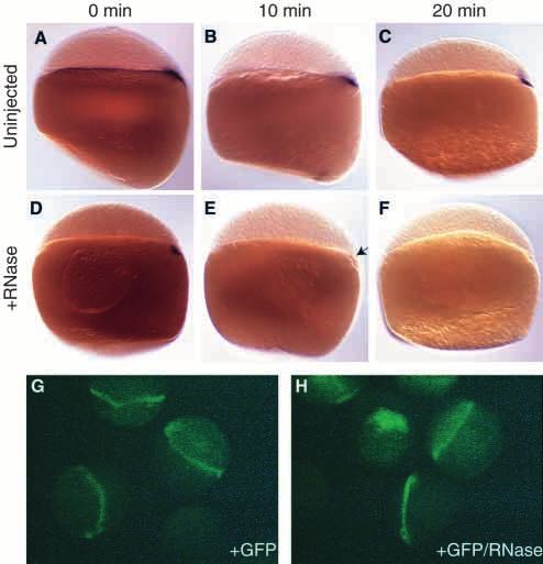
4682 S.-R. Chen and D. Kimelman

not the expression of sqt and boz in the YSL is required for            pyrocarbonate (DEPC; Sigma) was incubated in closed eppendorf
dorsal mesoderm and endoderm formation, it is necessary to              tubes for approximately 20 hours in a 37°C air incubator. The tubes
specifically eliminate the expression of these genes in the YSL.        were then transferred to a 37°C heat block and incubated for 10
   In order to determine whether or not the YSL is required for         minutes with the caps open to allow volatile DEPC to escape. The
establishing the mesendoderm, we chose to eliminate all RNA             overnight incubated RNase, with or without DEPC, was diluted 1:2
                                                                        in water with dextran such that the final concentration of RNase and
transcripts from the YSL by injecting RNase into the yolk cell          dextran was 10 µg/ml and 4 mg/ml. This mixture was then injected
at the time that the YSL forms. Formation of the YSL at the             into embryos. For in vitro analysis of RNase activity, the overnight
1K cell stage (3 hours) marks the time at which gap junctions           incubated RNase with or without DEPC was diluted 1:2 in water, and
between the yolk cell and the overlying marginal blastomeres            6 µl of this was added to 1 µg of capped RNA (synthesized using
are closed, resulting in the inability of large proteins to             the Ambion Message Machine System) and incubated at room
freely pass between the extra-embryonic and embryonic                   temperature for 1 hour.
compartments (Kimmel and Law, 1985). Therefore, injection
of RNase was expected to target transcripts in the YSL                  In situ hybridization
specifically, thus addressing whether or not the expression of          Whole-mount in situ hybridization was performed using digoxigenin-
sqt and boz in this region is required for dorsal mesoderm and          labeled antisense RNA and visualized using anti-digoxigenin Fab
                                                                        fragments conjugated with alkaline phosphatase (Roche Molecular
endoderm formation. As no factors have yet been identified              Biochemicals) as previously described (Griffin et al., 1995; Melby et
that are required for ventrolateral mesoderm induction,                 al., 1997). Double staining was carried out by detecting the
injection of RNase into the yolk cell should also indicate              flourescein-labeled probe first, using Fast Red (Sigma) as a substrate.
whether transcripts in the YSL are required for all mesoderm            The reaction was stopped by washing the embryo in PBST (PBS/0.1%
formation.                                                              Tween) several times, followed by two washes in 100 mM glycine,
   We show that injection of RNase specifically eliminated              pH 2.5, for 10 minutes each. Embryos were rinsed several times in
RNA in the YSL within 20 minutes, and we present evidence               PBST, and blocked for at least 1 h in 2% goat serum and 2 mg/ml
that protein stability is not affected. We demonstrate that the         BSA in PBST. The second probe was visualized using anti-
YSL is required for ventrolateral mesendoderm induction, and            digoxigenin Fab fragments.
that RNA in the YSL is required between the 1K cell stage                  In situ probes used: boz (Koos and Ho, 1998; Yamanaka et al.,
                                                                        1998), ntl (Schulte-Merker et al., 1992), gsc (Stachel et al., 1993),
(3 hours) and sphere stage (4 hours) for ventrolateral ntl              gta5 (Rodaway et al., 1999), sqt (Erter et al., 1998; Rebagliati et al.,
expression. We also show that the expression of sqt and cyc in          1998) and cyc (Rebagliati et al., 1998; Sampath et al., 1998). Embryos
the ventrolateral blastomeres depends on the YSL. In contrast,          were photographed in either Permount (Fisher) or 70% glycerol.
we find that dorsal mesendoderm induction occurs
independently of the YSL via stabilization of β-catenin in the          Dorsalization with LiCl
dorsal blastomeres. These results demonstrate a required role           Embryos were treated in their chorions with 0.3M LiCl for 9 minutes
for the YSL in establishing the germ layers, and indicate that          at the 64-cell stage (2 hours) (Stachel et al., 1993). The embryos were
an unidentified signal in the YSL is needed to induce                   then Pronase treated, washed and incubated at 28.5°C.
ventrolateral mesendoderm.

                                                                        RESULTS
MATERIALS AND METHODS
                                                                        To address the requirement of the YSL for mesendoderm
Embryos                                                                 induction, we wanted to specifically affect the ability of the
Adult AB strain zebrafish and embryos were raised at 28.5°C as          YSL to send signals to the blastoderm. Studies in Xenopus
described (Westerfield, 1989). Embryonic stages were determined by      indicate that the mesoderm-inducing signals are zygotically
observation (Westerfield, 1989). Embryos were dechorionated in 2        expressed (reviewed in Kimelman and Griffin, 1998). As the
mg/ml Pronase (Roche Molecular Biochemicals) prior to injection         YSL forms at the 1K cell stage (3 hours), soon after zygotic
(Westerfield, 1989).                                                    transcription is initiated at the midblastula transition (MBT;
Dye, RNase and GFP injections                                           Kane and Kimmel, 1993), injection of RNase into the yolk cell
All injections were made into the yolk cell of dechorionated 1K cell    at the 1K cell stage (3 hours) should eliminate all zygotic
stage (3 hours) embryos, unless otherwise indicated, using a            transcripts before they are translated. Proteins can not diffuse
Picospritzer II (Parker Hannifin Corporation). Approximately 1 nl was   into the blastomeres after the YSL forms (Kimmel and Law,
injected per embryo.                                                    1985) therefore RNase injected as a protein should result in the
   Lysine fixable biotinylated dextran or rhodamine-labeled             rapid degradation of RNAs specifically within the YSL. We
biotinylated dextran (10kMW, Molecular Probes) was dissolved in         chose to inject DNase-free RNase (Roche Molecular
water to a 20 mg/ml stock solution, and injected at 4 mg/ml, with or    Biochemicals), which contains a mixture of different RNases,
without RNase. Dextrans were visualized using biotin-avidin             although we have also individually injected RNase A and
peroxidase staining (Vector Laboratories). RNase, DNase free (0.5       RNase T1 and obtained similar results (data not shown). We
mg/ml, Roche Molecular Biochemicals) was diluted 1/50 in water, for
a final concentration of 10 µg/ml and injected with 4 mg/ml dextran.
                                                                        found that the results obtained by injecting this RNase were
Recombinant green fluorescent protein (GFP; Roche Molecular             specific to the activity of the RNases (see below) and
Biochemicals) was diluted to 0.5 mg/ml in water, and injected into      determined that it is necessary to inject a carrier along with the
embryos with or without RNase and dextran.                              RNase to obtain consistant results. As it was useful to detect
                                                                        the location of the injected material, the carriers we used were
Inactivation of RNase with DEPC                                         either rhodamine-labeled biotinylated dextran, or unlabeled
A 25 µl volume of 0.2 mg/ml RNase, with or without 280 µM diethyl       biotinylated dextran. By injecting the RNase into the yolk cell,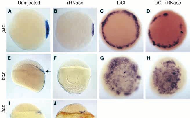
YSL and germ layer patterning 4683

Fig. 1. Injected RNase is limited to the YSL.
(A) 30% epiboly (4.7 hours) embryo injected
into the yolk cell at the 1K cell stage (3 hours)
with biotinylated dextran. Labeling is restricted
to the YSL (arrowhead, in brown) except for
some marginal cells and their descendants
(arrow). (B,C) spt expression in sphere stage
(4 hours) embryos injected with (B) dextran or
(C) dextran and RNase. No change in spt
expression is apparent in the RNase injected embryo compared to the dextran-injected embryo. (D) Uninjected embryo, fixed 10 minutes after
siblings were injected with RNase at oblong stage (3.7 hours), expresses boz in both the YSL and dorsal marginal blastomeres. (E) Embryo
fixed 10 minutes after injection with RNase expresses boz only in the dorsal marginal blastomeres. Arrows in D and E point to the YSL.
Embryos in panels A-C are animal pole views, D and E are side views, dorsal is to the right.

we could specifically target RNAs in the YSL and ask whether
or not the YSL is required for mesendoderm induction.

Injected RNase is limited to the YSL
Until the YSL is completely formed at the 1K cell stage (3
hours), marginal blastomeres retain cytoplasmic bridges with
the yolk cell (Kimmel and Law, 1985). We wanted to ensure
that material injected into the YSL at this stage would not be
inherited by the marginal blastomeres, which are also a source
of inducing signals, such as sqt and cyc (Erter et al., 1998;
Feldman et al., 1998; Gritsman et al., 2000; Rebagliati et al.,
1998). To determine the extent to which an injection of RNase
into the yolk cell at the 1K cell stage (3 hours) would populate
marginal blastomeres, we injected a similarly sized
biotinylated dextran molecule, which was visualized by biotin-
avidin peroxidase staining. We observed that the injected
dextran was mostly limited to the YSL, although a few
marginal cells and their descendents were labeled by 30%
epiboly (4.7 hours; Fig. 1A). Since the labeled blastomeres
represented a very small percentage of the marginal
blastomeres, we concluded that even if the RNase were to enter
a few marginal blastomeres, there were a large number of
marginal cells remaining that could induce mesendodermal
genes, if they had the capacity to do so.                               Fig. 2. RNase quickly and specifically eliminates RNA in the YSL.
   Although the dextran injections revealed that injected RNase         (A-C) boz expression in uninjected embryos fixed 0 min (A), 10 min
was likely to be restricted to the YSL, it was also important to        (B) and 20 min (C) after siblings were injected with RNase into the
establish that the expression of RNAs in the blastoderm that            yolk cell at at sphere stage (4 hours). (D-F) boz expression in
are not dependent on signals from the YSL are unaffected by             embryos fixed 0 min (D), 10 min (E) and 20 min (F) after injection
the injection. As spadetail (spt; Griffin et al., 1998) is              with RNase. Expression of boz is greatly reduced 10 min after
                                                                        injection (E; arrow) and completely eliminated 20 min after injection
ubiquitously expressed at sphere stage (4 hours), we used it            (F). Embryos in panels A-F are side views with dorsal to the right,
as a marker for YSL-independent gene expression. The                    when distinguishable. (G) Embryos injected with GFP protein.
expression of spt was indistinguishable in embryos that had             (H) Embryos injected with GFP and RNase. Injection of RNase did
RNase injected into their yolk cells when compared with                 not change the level of GFP fluorescence compared with embryos
embryos injected only with dextran (Fig. 1B,C). As a second             injected with GFP alone
control, we examined the expression of boz (Koos and Ho,
1998; Yamanaka et al., 1998) in embryos injected with RNase
at oblong stage (3.7 hours), when boz is expressed in both the          selectively eliminate RNAs from the YSL without degrading
YSL and dorsal blastomeres. boz is normally expressed only              transcripts in the blastomeres.
in the dorsal blastomeres at the 1K cell stage (3 hours), then
expands to include both the dorsal blastomeres and YSL until            Effects of RNase injection on epiboly
sphere stage (4 hours), after which it is restricted to the dorsal      Previous studies have suggested a role for the YSL in driving
YSL (Koos and Ho, 1998; Yamanaka et al., 1998). Whereas                 epibolic movements (Solnica-Krezel and Driever, 1994;
boz is observed in both the YSL and blastomeres of uninjected           Strahle and Jesuthasan, 1993). As was observed with the
embryos (Fig. 1D), RNase injection into the YSL abolished the           microtubule inhibitor nocodazole, RNase injections in the YSL
expression of boz in the YSL without affecting its expression           resulted in slow epibolic movements and the blastoderm lifting
in the dorsal blastomeres (Fig. 1E). These results, in addition         off the yolk cell (data not shown). These results suggested that
to those examining spt expression, demonstrated that we could           one or more transcripts in the YSL are necessary for the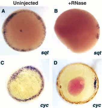
4684 S.-R. Chen and D. Kimelman
microtubule-driven process of epiboly.                                             Fig. 3. Ventrolateral mesendoderm induction
In order to ensure that the effects on                                             requires YSL RNA. (A-F) 30% epiboly (4.7 hours).
epiboly did not affect our analysis of                                             (A) ntl expression in an uninjected embryo. (B) ntl
mesendoderm induction, the studies                                                 expression in an embryo injected with RNase.
on mesendodermal markers described                                                 Ventrolateral ntl expression is eliminated by RNase
below were conducted prior to or at                                                injection. (C,D) Embryos stained with ntl alone
                                                                                   (pink), uninjected (C) or RNase injected (D).
50% epiboly (5.3 hours), before severe                                             (E,F) Embryos stained with ntl (pink) and gsc
morphogenetic effects of RNase                                                     (purple). (E) Uninjected embryo shows ntl and gsc
injections were observed.                                                          co-expressed on the dorsal side (arrow). (F) RNase
                                                                                   injected embryo has no ventrolateral ntl expression,
RNase rapidly and specifically                                                     and the remaining patch of ntl expression co-
eliminates RNAs in the YSL                                                         localizes with gsc expression. (G,H) 50% epiboly
To establish that RNase injection                                                  (5.3 hours) embryos stained for gta5 expression.
eliminates YSL RNAs and to                                                         (G) Uninjected embryo shows gta5 throughout the
determine how quickly this process                                                 endoderm. (H) RNase injected embryo shows only
occurs, we injected RNase and                                                      dorsal gta5 expression. (I-K) 30% epiboly (4.7
                                                                                   hours). (I) Uninjected embryo shows ntl expression
examined the expression of boz at a                                                throughout the margin. (J) Embryo injected with
time when it is restricted to the YSL.                                             RNase that has been inactivated by overnight
Injections were made into the yolk cell                                            incubation at 37°C with DEPC also shows normal
of sphere stage (4 hours) embryos and                                              ntl expression throughout the margin. (K) Embryo
then the embryos were fixed varying                                                injected with RNase incubated at 37°C overnight
times after injection. We found that                                               shows no ventrolateral ntl expression. (L) In vitro
boz expression was significantly                                                   analysis of the RNase mixtures injected in I-K.
reduced within 10 minutes of                                                       (lane 1) 0.5 µg of RNA substrate incubated at room
injection, and completely eliminated                                               temperature for two hours. (lane 2) 0.5µg RNA
20 minutes after injection (Fig. 2A-F).                                            substrate incubated for two hours with DEPC-
                                                                                   inactivated RNase is not degraded. (lane 3) 0.5µg
This demonstrated that RNAs in the                                                 RNA substrate treated with RNase that was
YSL are degraded within 20 minutes                                                 incubated overnight at 37°C alone is completely
of RNase injection.                                                                                                   degraded.
   To examine whether or not RNase                                                                                    (A-F, I-K) Animal
injections might have a general effect                                                                                pole views.
on protein stability, we injected                                                                                     (G,H) Side view
recombinant GFP protein with or                                                                                       with dorsal to the
without RNase into the yolk, and                                                                                      right, when
examined the embryos from oblong                                                                                      distinguishable.
stage (3.7 hours) to 30% epiboly
(4.7 hours) for the presence of
green fluorescent protein (GFP) by
fluorescence microscopy. There was no apparent change in the      confirmed that ventrolateral ntl expression was completely
level of GFP fluorescence over this time period in control        abolished, leaving only a dorsal region where gsc and ntl were
injected embryos, and there was no difference in GFP intensity    co-expressed (Fig. 3C-F). Dorsal expression of ntl could not
between embryos that were injected with or without RNase          be eliminated, even upon injection of ten times as much RNase
(Fig. 2G,H). Since we could not detect injections of five times   (data not shown). Examination of the endodermal marker, gta5,
less GFP, it seemed likely that there was not a significant       in RNase injected embryos at 50% epiboly (5.3 hours)
amount of degradation occurring in the embryos. These results     demonstrated that ventrolateral endoderm also requires signals
suggest that existing proteins in the YSL are not affected by     from the YSL (Fig. 3G,H). These results demonstrate that
the injection of RNase, and that any effects of RNase injection   induction of ventrolateral mesendoderm requires signals from
on mesoderm induction are due to eliminating existing             the YSL.
transcripts in the YSL and/or preventing new transcription.          In order to establish that the elimination of ventro-lateral ntl
                                                                  expression was a specific effect of RNase activity, we repeated
Ventrolateral mesendoderm induction requires a                    the injections with inactivated RNase. Since we were
YSL RNA between 1K (3 hours) and sphere stages                    concerned that any RNase inhibitor we would add to the RNase
(4 hours)                                                         could have its own effects when injected into embryos, we
We examined the effects of RNase injection at the 1K cell stage   inactivated the RNase using a volatile protein modifier, diethyl
(3 hours) on the expression of the pan-mesodermal marker no       pyrocarbonate (DEPC). Overnight treatment of RNase with
tail (ntl; Schulte-Merker et al., 1992) and the endodermal        DEPC at 37°C resulted in the inactivation of RNase, measured
marker gata5 (gta5; Rodaway et al., 1999) and found that ntl      in vitro on an RNA substrate (Fig. 3L). Treatment of the RNA
expression at 30% epiboly (4.7 hours) was almost completely       substrate with RNase that had been incubated at 37°C
absent, except for a patch of cells on the dorsal side (Fig.      overnight without DEPC resulted in its complete degradation
3A,B). Double-label in situ hybridization with the dorsal         (Fig. 3L, lane 3). Moreover, while fresh DEPC results in the
mesodermal marker goosecoid (gsc; Stachel et al., 1993)           degradation of the RNA substrate (data not shown) the DEPC
YSL and germ layer patterning 4685
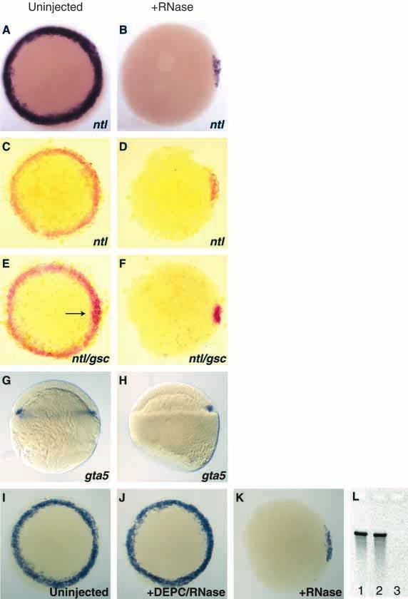
Fig. 4. Ventrolateral mesoderm induction
requires YSL RNA between the 1K cell
(3 hours) and sphere stages (4 hours).
(A-E) Expression of ntl at 50% epiboly
(5.3 hours) in (E) uninjected and (A-D)
RNase injected embryos. (A) Embryo
injected with RNase at the 1K cell stage
(3 hours) shows no ventrolateral ntl
expression. (B,C) Embryos injected with
RNase at oblong stage (3.7 hours)
include embryos that have (B) no ventrolateral ntl expression and those in which (C) some ventro-lateral ntl expression is retained. (D) Embryo
injected with RNase at sphere stage (4 hours) looks the same as (E) an uninjected embryo. (B,D) Pink color in the center of the embryo is a
portion of rhodamine-labeled dextran that has not diffused from the site of injection. All views are from the animal pole.
incubated overnight with RNase had no effect on the RNA
substrate (Fig. 3L, lane 2). We found that DEPC-inactivated
RNase had no effect on ntl expression when injected into
embryos (Fig. 3J). These results demonstrated that the
elimination of ventrolateral ntl expression is a result of RNase
activity.
Using RNase injections, we have shown that ventrolateral
mesendoderm induction requires a signal from the YSL. To
determine when this RNA is present in the YSL, we injected
embryos with RNase at different times and analyzed the effects
on ntl expression. Embryos were injected with RNase at the
1K cell (3 hours), oblong (3.7 hours) and sphere (4 hours)
stages, and examined at 50% epiboly (5.3 hours) for ntl
expression (Fig. 4A-E). Embryos injected at the oblong stage
(3.7 hours) showed a reduced effect compared with those
injected at 1K (Fig. 4C). Embryos injected at sphere stage
(4 hours) showed normal ntl expression (Fig. 4D,E), Fig. 5. Ventrolateral expression of the nodal-related genes requires a
demonstrating that an RNA in the YSL is required between 1K YSL signal. Expression of (A,B) sqt and (C,D) cyc at 30% epiboly
cell (3 hours) and sphere stage (4 hours). (4.7 hr). (A) sqt expression in an uninjected embryo. (B) RNase
injected embryo displays lack of ventrolateral sqt expression. (C) cyc
Ventrolateral expression of nodals requires YSL expression in an uninjected embryos. (D) RNase injected embryos
signal show no ventrolateral cyc expression. Brown color in the YSL is
Since RNase injections into the YSL eliminated ventrolateral biotin-avidin peroxidase staining of co-injected rhodamine-labeled
biotinylated dextran. All views are from the animal pole.
expression of the endodermal marker gta5, we wondered if this
would be accompanied by a reduction in the expression of the
nodal-related factors in the ventrolateral region. We observed 6I,J). As described earlier (Fig. 1E), boz expression was not
that expression of sqt (Erter et al., 1998; Rebagliati et al., 1998) observed in the YSL of RNase injected embryos. These results
and cyc (Rebagliati et al., 1998; Sampath et al., 1998) was suggest that induction of boz in the dorsal blastomeres is
eliminated by RNase injection at the 1K cell stage (3 hours) in independent of YSL signals, although the normal temporal
the ventrolateral marginal regions, just as ntl was (Fig. 5A-D). regulation of boz requires YSL signals.
Dorsal expression of both sqt and cyc remained in RNase In Xenopus, stabilization of β-catenin has been shown to be
injected embryos, as was observed for ntl, gsc and gta5 in required for the induction of dorsal mesoderm (reviewed in
RNase injected embryos (Fig. 3). These results demonstrate Moon and Kimelman, 1998). In zebrafish embryos, β-catenin
that the ventrolateral expression of the nodal-related factors is accumulates in the dorsal YSL and dorsal blastomeres
also dependent upon RNAs in the YSL. (Schneider et al., 1996). We hypothesized that the presence of
β-catenin in the dorsal blastomeres would allow these cells to
Stabilized β-catenin can induce dorsal mesoderm activate dorsal mesoderm independently of the dorsal YSL. In
independently of the YSL order to test this, we treated embryos with LiCl, which has been
The previous experiments established a role for the YSL in shown to result in ectopic stabilization of β-catenin throughout
induction of ventrolateral mesoderm, but clearly demonstrated the embryo (Schneider et al., 1996) by inhibiting GSK-3β
that dorsal mesoderm is regulated differently. Since boz has (Hedgepeth et al., 1997; Klein and Melton, 1996; Stambolic et
been shown to be required for dorsal mesoderm induction al., 1996). We expected that if ectopically stabilized β-catenin
(Fekany et al., 1999), we examined its expression in RNase did not require the YSL to induce dorsal mesoderm, embryos
injected embryos. We observed that boz was expressed in the treated with LiCl and injected with RNase would still express
dorsal blastomeres of RNase injected embryos, and that this ectopic dorsal mesodermal markers. As shown previously, LiCl-
expression persisted past the point at which boz expression in treated embryos ectopically express the dorsal mesodermal
uninjected siblings has been restricted to the dorsal YSL (Fig. marker gsc throughout the margin (Fig. 6C; Stachel et al., 1993).4686 S.-R. Chen and D. Kimelman
Fig. 6. Stabilized β-catenin in dorsal blastomeres can
induce dorsal mesoderm independently of YSL
signals. (A-H) Embryos shown are at 30% epiboly
(4.7 hours). (A) gsc expression in an untreated
embryo. (B) RNase injected embryo retains gsc
expression. (C) LiCl treated embryo shows ectopic
gsc expression at the margin. (D) LiCl treated and
RNase injected embryo shows ectopic gsc expression
at the margin. (E) Untreated embryo shows boz
expression in the dorsal YSL. (F) RNase injected
embryo has no boz in the YSL. (G) LiCl treated
embryo shows ectopic boz expression throughout the
blastoderm. (H) LiCl treated, RNase injected embryo
still shows ectopic boz expression throughout the
blastoderm. (I) boz expression in an embryo at sphere
stage (4 hours) is restricted to the YSL. (J) RNase
injected embryo retains boz in the dorsal blastomeres.
(A-D, G,H) Animal pole views, (E,F,I,J) Side view,
dorsal to the right, when distinguishable.
We observed that this ectopic expression is largely resistant to RNase injections
injection of RNase (Fig. 6D), just as dorsal expression of gsc is In order to address whether or not the YSL is required for germ
resistant in embryos not treated with LiCl (Fig. 6B). layer formation, we wished to eliminate the activity of the
We also examined the expression of boz in LiCl and RNase YSL. Dissection of the blastoderm away from the yolk cell in
injected embryos. As previously described, boz is restricted to order to separate it from yolk cell signals was not considered
the dorsal YSL at 30% epiboly (4.7 hours; Fig. 6E; Koos and a viable option. The dissection process typically results in loss
Ho, 1998; Yamanaka et al., 1998). In RNase injected embryos, or damage of the most marginal cells, which are the site of sqt
YSL expression of boz at 30% epiboly (4.7 hours) was and cyc expression (Erter et al., 1998; Feldman et al., 1998;
eliminated (Fig. 6F). At the same stage in LiCl-treated Gritsman et al., 2000; Rebagliati et al., 1998). This would
embryos, boz was ectopically expressed throughout the probably compromise the ability of the explanted blastoderm
blastoderm and not just restricted to the margin as was seen for to induce mesendodermal genes. Therefore, we wanted to
gsc (Fig. 6 compare G to C). LiCl-treated embryos, which were eliminate the capacity of the YSL to signal without removing
also injected with RNase, still show ectopic boz expression the blastoderm. As we hypothesized that the inducing signal
throughout the blastoderm at 30% epiboly (Fig. 6H), would be zygotic, based on studies in Xenopus, we originally
demonstrating that LiCl-dependent expression of boz does not sought to inhibit transcription in the YSL specifically. Since
require YSL signals. As boz is required for the formation of commonly available transcriptional inhibitors are membrane
the dorsal mesoderm downstream of β-catenin (Fekany et al., permeable, we chose to inject RNase. As proteins can not
1999), this agrees with our observations that induction of diffuse into the blastomeres after the YSL forms (Kimmel and
dorsal mesoderm can occur independently of YSL signals and Law, 1985), injection of RNase protein into the YSL would
we conclude that this is mediated by stabilized β-catenin. specifically degrade YSL RNAs. Although we have performed
injections with RNase A and RNase T1, most of our injections
were performed using a DNase-free RNase mix. We found that
DISCUSSION the RNase must be injected with a carrier to obtain consistent
results, and we used biotinylated dextrans as carriers, since this
While transplantation experiments have shown that the YSL is afforded us the option of visualizing the extent of RNase
sufficient to induce mesoderm and endoderm (Mizuno, 1996; diffusion using biotin-avidin peroxidase staining. We have
Ober and Schulte-Merker, 1999; Rodaway et al., 1999) demonstrated that RNase injections into the yolk cell at the 1K
whether or not the YSL was required for germ layer formation cell stage (3 hours) are limited to the YSL, do not affect
was not known. As genes encoding key regulatory factors ubiquitously expressed genes, and we present data that indicate
known to be expressed in the YSL are also expressed in the proteins in the YSL are unaffected. These experiments indicate
marginal blastomeres, mutants in those genes could not resolve that injection of RNase to the yolk cell after the YSL forms is
the issue of whether gene expression in the YSL is required, an effective technique for eliminating RNA transcripts in the
as mutants abolished their activities in both the YSL and YSL specifically.
marginal blastomeres. Therefore, it was necessary to devise a
technique to eliminate gene expression specifically in the YSL A role for YSL RNAs in epiboly
in order to demonstrate whether or not it was required. We We have presented evidence that suggests that injection of
have accomplished this by injecting RNase into the YSL, and RNase into the YSL eliminates RNAs without affecting the
have demonstrated that the YSL is required for ventrolateral stability of proteins. Consistent with studies that suggest a role
mesendoderm induction and the expression of nodal-related for the YSL in epiboly, we see the same phenotype in our
factors within the marginal blastomeres, while dorsal RNase-injected embryos as in embryos treated with UV or
mesoderm induction can occur independently of the YSL. nocodazole, which disrupt microtubules: epiboly is slowed,YSL and germ layer patterning 4687
Fig. 7. Model for mesendoderm induction.
Ventrolateral mesendoderm induction
requires a signal from the YSL (blue
arrow), which is present as at least one Endoderm (gta 5)
Ventrolateral mesendoderm
RNA between the 1K cell stage (3 hours)
inducing activity
and sphere stage (4 hours) to separately sqt cyc Mesoderm (ntl)
activate sqt and cyc, and ntl. Sqt and Cyc, Co-factor required for
Ventral
Dorsal
in turn, are required to induce ventro-lateral sqt expression YSL
endoderm. Dorsal mesendoderm inducing
factors are activated in the dorsal Stabilized β-catenin
blastomeres and YSL as a result of
stabilized β-catenin in both places (green
hatched region). β-catenin requires a co-
factor (yellow) that is restricted to the YSL
and marginal blastomeres in order to induce
sqt expression, but not boz (see text for
details).
and the blastoderm of RNase injected embryos constricts at the molecules. Overexpression of the TGFβ family member
margin and lifts off of the yolk cell when control embryos are antivin (Thisse and Thisse, 1999), which has been shown
past 50% epiboly (5.3 hours) (Strahle and Jesuthasan, 1993). to antagonize TGFβ signaling, abolishes mesendoderm
The manner in which the blastoderm lifts off of the yolk cell induction. Additionally, overexpression of the TGFβ signaling
was proposed to be due to both constrictive forces in the margin molecule activin, results in the induction of mesodermal genes
of the blastoderm, which normally facilitates blastopore (Schulte-Merker et al., 1992).
closure, and a weakening of the yolk cytoskeleton (Strahle and We do not consider activin a likely candidate for our
Jesuthasan, 1993). We observe that this process occurs more mesendoderm-inducing activity, since a study using an activin
rapidly in our RNase injected embryos, where the blastoderm cleavage mutant, which must interact with newly translated
lifts off the yolk cell by the time uninjected siblings reach activin ligand in order to function as a dominant negative, had
60-70% epiboly (6.7 hours). We suggest that this is because no effect on mesoderm induction in medaka (Wittbrodt and
cytoskeletal elements that are turned over after RNase injection Rosa, 1994). Since our RNase injection experiments indicated
cannot be replaced due to the presence of RNase. This results that RNA in the YSL is required for mesoderm induction, the
in a weaker cytoskeleton in RNase-injected embryos than in activin cleavage mutant described above should have also
embryos treated with UV or nocodazole, in which the eliminated ventrolateral mesoderm if activin transcripts were
cytoskeleton can recover to some degree after treatment the targets of the RNase.
(Strahle and Jesuthasan, 1993). The similarity between the We can also exclude nodal-related signals as candidates for
results with RNase injection, and UV and nocodazole the factor. Although sqt and cyc are required for endoderm
treatment suggests that the effects of RNase injection are induction (Rodaway et al., 1999), cyc is not expressed in the
limited to the YSL, and that the blastoderm itself can properly YSL, and sqt is only expressed in the dorsal YSL between the
undergo morphogenetic processes that are independent of the 1K cell stage (3 hours) and sphere stage (4 hours). Our data
YSL. In addition, our data indicate that there is one or more suggest that the RNase sensitive mesendoderm-inducing
RNA in the YSL that are essential for epiboly. activity is expressed as RNA throughout the YSL during these
stages. In addition, maternal and zygotic mutants in one-eyed
RNA in the YSL is required for ventrolateral pinhead, which is required for nodal signaling, still induce
mesoderm induction ventrolateral mesoderm (Gritsman et al., 1999), as do sqt;cyc
We have shown that expression of mesendodermal markers and double mutants (Feldman et al., 1998). This indicates that the
inducing genes requires at least one RNA in the YSL between nodal-related genes do not play a role in the initial induction
the 1K cell stage (3 hours) and sphere stage (4 hours). A of ventrolateral mesoderm (Feldman et al., 1998; Gritsman et
significant exception is dorsal mesendoderm, discussed below. al., 1999).
Moreover, the RNase-sensitive activity induces the expression In summary, our data demonstrate that there is an
of sqt and cyc, which are required for endoderm induction unidentified RNA required for mesendoderm induction that is
(Rodaway et al., 1999). Although sqt;cyc double mutants show expressed specifically in the YSL at the start of zygotic
defects in mesoderm formation during gastrulation, sqt and cyc transcription (Fig. 7).
are not required for ventrolateral mesoderm induction
(Feldman et al., 1998). Therefore, the activity must separately Dorsal mesoderm induction
induce the expression of ntl, either directly or through an Our data demonstrated that dorsal mesendoderm is induced
unknown pathway (Fig. 7). Whether or not the RNase sensitive through a different pathway from ventrolateral mesendoderm.
activity is zygotic or maternal is not known, as the injected We found that dorsal mesendoderm was not eliminated in
RNase would eliminate both. RNase injected embryos, and that dorsal mesoderm could be
Based on a number of investigations, likely candidates induced by LiCl treatment in the absence of a functional YSL.
for the RNase-sensitive inducing activity belong to the Although we can not rule out the possibility that the effects
transforming growth factor β (TGFβ) family of signaling of LiCl are due in part to changes in phosphoinositide4688 S.-R. Chen and D. Kimelman
metabolism (Ault et al., 1996), since LiCl treatment results in Evidence for a localized co-factor involved in dorsal
the ectopic stabilization of β-catenin in zebrafish embryos mesoderm induction
(Schneider et al., 1996a) and since stablized β-catenin has While stabilization of β-catenin is required in order to induce
been shown in Xenopus embryos to activate dorsal genes dorsal mesoderm, it is not sufficient to do so (Schneider et al.,
(reviewed in Moon and Kimelman, 1998), we conclude that 1996b). It has been previously shown in Xenopus that treatment
the stabilization of β-catenin in the dorsal blastomeres makes with LiCl results in ectopic stabilization of β-catenin
them independent of signaling from the YSL. Our results throughout the embryo, but that dorsal mesodermal markers are
correlate with a number of experiments that contribute to a restricted to the equatorial regions where the mesodermal layer
model in which dorsalizing factors, responsible for stabilizing is normally formed (Schneider et al., 1996b). Similarly, in
β-catenin, are localized to the vegetal pole of the oocyte and zebrafish we observed that LiCl-treated embryos show gsc
moved up to the future dorsal side via microtubule transport (Fig. 6C), ntl and sqt (data not shown) expression only at the
prior to the 16-cell stage (1.5 hours) (Jesuthasan and Stahle, margin even though boz expression is induced throughout the
1996; Ober and Schulte-Merker, 1999; Yamaha et al., 1998). blastoderm (Fig. 6G; Yamanaka et al., 1998). Both sqt and boz
We believe that these dorsalizing factors are delivered to a have been shown to be downstream of β-catenin (Shimizu et
region that includes both the dorsal YSL and dorsal al., 2000), which suggests that a co-factor required for sqt
blastomeres, thus accounting for the ability of the YSL to expression is responsible for restricting dorsal mesoderm
induce dorsal mesodermal genes in transplantation induction to the margin. We suggest that both the YSL and
experiments (Mizuno, 1996; Ober and Schulte-Merker, 1999; marginal blastomeres contain a co-factor required for
Rodaway et al., 1999), and of the blastomeres to induce dorsal expression of sqt, but not boz (Fig. 7).
mesodermal genes independently of the YSL (our results, Fig.
7). Consistent with this interpretation, ectopic gsc expression We thank Kevin Griffin and Carole Weaver for very helpful
was frequently less robust in LiCl-treated and RNase-injected comments on the manuscript and Kevin Griffin for many useful
embryos when compared with embryos treated with LiCl discussions. We also thank Will Talbot, Scott Dougan, and Alex
alone. This suggests that stabilization of β-catenin in the Schier for communicating unpublished results. This work was
dorsal YSL also plays a non-autonomous role in the induction supported by NSF grant IBN-9722949 to D. K. S. C. was supported
by an NSF pre-doctoral fellowship.
of dorsal mesoderm (Fig. 6, compare D with C)
One caveat to our interpretation is that there may be a signal
from the dorsal YSL that acts too early for our RNase
injections to interrupt. We do not believe this is the case, as we REFERENCES
inject RNase as soon as the YSL is formed and have shown
that injection of RNase eliminates most of the RNA in the YSL Ault, K. T., Durmowicz, G., Galione, A., Harger, P. L. and Busa, W. B.
(1996). Modulation of Xenopus embryo mesoderm-specific gene expression
within 10 minutes. In addition, nuclear localized β-catenin has and dorsoanterior patterning by receptors that activate the
been observed in marginal and non-marginal blastomeres at the phosphatidylinositol cycle signal transduction pathway. Development 122,
1K cell stage (3 hours) (S. Dougan, A. Schier and W. Talbot, 2033-2041.
personal communication). These observations suggest that the Erter, C. E., Solnica-Krezel, L. and Wright, C. V. (1998). Zebrafish nodal-
related 2 encodes an early mesendodermal inducer signaling from the
dorsal blastomeres are capable of activating dorsal mesodermal extraembryonic yolk syncytial layer. Dev. Biol. 204, 361-372.
genes independently of signals from the YSL via stabilized β- Fekany, K., Yamanaka, Y., Leung, T., Sirotkin, H. I., Topczewski, J.,
catenin. Gates, M. A., Hibi, M., Renucci, A., Stemple, D., Radbill, A. et al. (1999).
The expression of all mesendodermal markers examined, The zebrafish bozozok locus encodes Dharma, a homeodomain protein
sqt, cyc, ntl and gta5, was shown to be regulated differently on essential for induction of gastrula organizer and dorsoanterior embryonic
structures. Development 126, 1427-1438.
the dorsal side than in the ventrolateral margin. Our data Feldman, B., Gates, M. A., Egan, E. S., Dougan, S. T., Rennebeck, G.,
suggest that dorsal expression of these genes can be induced Sirotkin, H. I., Schier, A. F. and Talbot, W. S. (1998). Zebrafish organizer
downstream of stabilized β-catenin. This has been previously development and germ-layer formation require Nodal-related signals.
demonstrated for boz (Fekany et al., 1999; Yamanaka et al., Nature 395, 181-185.
Griffin, K., Patient, R. and Holder, N. (1995). Analysis of FGF function in
1998), sqt (Shimizu et al., 2000) and ntl (Kelly et al., 1995), normal and no tail zebrafish embryos reveals separate mechanisms for
and our data suggest it is also true for dorsal cyc and gta5 formation of the trunk and the tail. Development 121, 2983-2994.
expression. Griffin, K. J., Amacher, S. L., Kimmel, C. B. and Kimelman, D. (1998).
Molecular identification of spadetail: regulation of zebrafish trunk and tail
Regulation of dorsal mesodermal genes requires the mesoderm formation by T-box genes. Development 125, 3379-3388.
YSL Gritsman, K., Talbot, W. S. and Schier, A. F. (2000). Nodal signaling
patterns the organizer. Development 127, 921-932.
In addition to playing a cooperative role with the dorsal Gritsman, K., Zhang, J., Cheng, S., Heckscher, E., Talbot, W. S. and
blastomeres to induce dorsal mesoderm, our data suggest that Schier, A. F. (1999). The EGF-CFC protein One-Eyed Pinhead is essential
the YSL is required to regulate dorsal mesoderm formation. for Nodal signaling. Cell 97, 121-132.
Hedgepeth, C. M., Conrad, L. J., Zhang, J., Huang, H., Lee, V. M. Y. and
Although the dorsal blastomeres can induce the expression of Klein, P. (1997). Activation of the Wnt signaling pathway: A molecular
boz independently of the YSL, in the absence of YSL RNAs mechanism for lithium action. Dev. Biol. 185, 82-91.
the expression pattern of boz is altered such that it is still Jesuthasan, S. and Stahle, U. (1996). Dynamic microtubules and specification
expressed in the dorsal blastomeres when uninjected siblings of the zebrafish embryonic axis. Curr. Biol. 7, 31-42.
have restricted their expression of boz to the dorsal YSL (Fig. Kane, D. A. and Kimmel, C. B. (1993). The zebrafish midblastula transition.
Development 119, 447-456.
6I,J). This result suggests that dorsal YSL activity regulates boz Kelly, G. M., Erezyilmaz, D. F. and Moon, R. T. (1995). Induction of a
expression by inhibiting boz in the dorsal blastomeres during secondary embryonic axis in zebrafish following the overexpression of β-
epiboly. catenin. Mech. Dev. 53, 261-273.YSL and germ layer patterning 4689 Kimelman, D. and Griffin, K. J. (1998). Mesoderm induction: a postmodern catenin translocation into nuclei demarcates the dorsalizing centers in frog view. Cell 94, 419-421. and fish embryos. Mech. Dev. 57, 191-198. Kimmel, C. B. and Law, R. D. (1985). Cell lineage of zebrafish blastomeres. Schulte-Merker, S., Ho, R. K., Herrmann, B. G. and Nusslein-Volhard, C. II. Formation of the yolk syncytial layer. Dev. Biol. 108, 86-93. (1992). The protein product of the zebrafish homologue of the mouse T gene Klein, P. S. and Melton, D. A. (1996). A molecular mechanism for the is expressed in nuclei of the germ ring and the notochord of the early effect of lithium on development. Proc. Natl. Acad. Sci. USA 93, 8455- embryo. Development 116, 1021-1032. 8459. Shimizu, T., Yamanaka, Y., Ryu, S., Hashimoto, H., Yabe, T., Hirata, T., Koos, D. S. and Ho, R. K. (1998). The nieuwkoid gene characterizes and Bae, Y., Hibi, M. and Hirano, T. (2000). Cooperative roles of mediates a Nieuwkoop-center-like activity in the zebrafish. Curr. Biol. 8, Bozozok/Dharma and Nodal-related proteins in the formation of the dorsal 1199-206. organizer in zebrafish. Mech. Dev. 91, 293-303. Koos, D. S. and Ho, R. K. (1999). The nieuwkoid/dharma homeobox gene is Solnica-Krezel, L. and Driever, W. (1994). Microtubule arrays of the essential for bmp2b repression in the zebrafish pregastrula. Dev. Biol. 215, zebrafish yolk cell: organization and function during epiboly. Development 190-207. 120, 2443-2455. Melby, A. E., Kimelman, D. and Kimmel, C. B. (1997). Spatial regulation Stachel, S. E., Grunwald, D. J. and Myers, P. Z. (1993). Lithium of floating head expression in the developing notochord. Dev. Dyn. 209, perturbation and goosecoid expression identify a dorsal specification 2225-2237. pathway in the pregastrula zebrafish. Development 117, 1261-74. Mizuno, T., Yamaha, E., Wakahara, M., Kuroiwa, A. and Takeda, H. Stambolic, V., Ruel, L. and Woodgett, J. R. (1996). Lithium inhibits (1996). Mesoderm induction in zebrafish. Nature 383, 131-132. glycogen synthase kinase-3 activity and mimics wingless signalling in intact Moon, R. T. and Kimelman, D. (1998). From cortical rotation to organizer cells. Curr. Biol. 6, 1664-1668. gene expression: toward a molecular explanation of axis specification in Strahle, U. and Jesuthasan, S. (1993). Ultraviolet irradiation impairs epiboly Xenopus. BioEssays 20, 536-545. in zebrafish embryos: evidence for a microtubule-dependent mechanism of Ober, E. A. and Schulte-Merker, S. (1999). Signals from the yolk cell induce epiboly. Development 119, 909-919. mesoderm, neuroectoderm, the trunk organizer, and the notochord in Thisse, C. and Thisse, B. (1999). Antivin, a novel and divergent member of zebrafish. Dev. Biol. 215, 167-181. the TGF-β superfamily, negatively regulates mesoderm induction. Rebagliati, M. R., Toyama, R., Haffter, P. and Dawid, I. B. (1998). cyclops Development 126, 229-240. encodes a nodal-related factor involved in midline signaling. Proc. Natl. Westerfield, M. (1989). The Zebrafish Book. Eugene, OR: Institute of Acad. Sci. USA 95, 9932-9937. Neurosciences, University of Oregon. Rodaway, A., Takeda, H., Koshida, S., Broadbent, J., Price, B., Smith, J. Wittbrodt, J. and Rosa, F. M. (1994). Disruption of mesoderm and axis C., Patient, R. and Holder, N. (1999). Induction of the mesendoderm in formation in fish by ectopic expression of Activin variants: the role of the zebrafish germ ring by yolk cell-derived TGF-β family signals and maternal Activin. Genes Dev. 8, 1448-1462. discrimination of mesoderm and endoderm by FGF. Development 126, Yamaha, E., Mizuno, T., Hasebe, Y., Takeda, H. and Yamazaki, F. (1998). 3067-3078. Dorsal specification in blastoderm at the blastula stage in the goldfish, Sampath, K., Rubinstein, A. L., Cheng, A. M., Liang, J. O., Fekany, K., Carassius auratus. Dev. Growth Differ. 40, 267-275. Solnica-Krezel, L., Korzh, V., Halpern, M. E. and Wright, C. V. (1998). Yamanaka, Y., Mizuno, T., Sasai, Y., Kishi, M., Takeda, H., Kim, C. H., Induction of the zebrafish ventral brain and floorplate requires Hibi, M. and Hirano, T. (1998). A novel homeobox gene, dharma, can Cyclops/Nodal signalling. Nature 395, 185-189. induce the organizer in a non-cell-autonomous manner. Genes Dev. 12, Schneider, S., Steinbeisser, H., Warga, R. M. and Hausen, P. (1996). β- 2345-2353.
You can also read