Utilizing image and caption information for biomedical document classification
←
→
Page content transcription
If your browser does not render page correctly, please read the page content below
Bioinformatics, 37, 2021, i468–i476
                                                                                                         doi: 10.1093/bioinformatics/btab331
                                                                                                                            ISMB/ECCB 2021

Utilizing image and caption information for biomedical
document classification

                                                                                                                                                                            Downloaded from https://academic.oup.com/bioinformatics/article/37/Supplement_1/i468/6319676 by guest on 26 September 2021
Pengyuan Li1,*, Xiangying Jiang1,2, Gongbo Zhang1,3, Juan Trelles Trabucco4,
Daniela Raciti5, Cynthia Smith6, Martin Ringwald6, G. Elisabeta Marai4,
Cecilia Arighi1 and Hagit Shatkay1,*
1
 Department of Computer and Information Sciences, University of Delaware, Newark, DE 19716, USA, 2Amazon, Seattle, WA 98109,
USA, 3Google, Mountain View, CA 94043, USA, 4Department of Computer Science, The University of Illinois at Chicago, Chicago, IL
60612, USA, 5Division of Biology and Biological Engineering, California Institute of Technology, Pasadena, CA 91125, USA and 6The
Jackson Laboratory, Bar Harbor, ME 04609, USA
*To whom correspondence should be addressed.

Abstract
Motivation: Biomedical research findings are typically disseminated through publications. To simplify access to do-
main-specific knowledge while supporting the research community, several biomedical databases devote significant
effort to manual curation of the literature—a labor intensive process. The first step toward biocuration requires iden-
tifying articles relevant to the specific area on which the database focuses. Thus, automatically identifying publica-
tions relevant to a specific topic within a large volume of publications is an important task toward expediting the
biocuration process and, in turn, biomedical research. Current methods focus on textual contents, typically extracted
from the title-and-abstract. Notably, images and captions are often used in publications to convey pivotal evidence
about processes, experiments and results.
Results: We present a new document classification scheme, using both image and caption information, in addition
to titles-and-abstracts. To use the image information, we introduce a new image representation, namely Figure-
word, based on class labels of subfigures. We use word embeddings for representing captions and titles-and-
abstracts. To utilize all three types of information, we introduce two information integration methods. The first com-
bines Figure-words and textual features obtained from captions and titles-and-abstracts into a single larger vector
for document representation; the second employs a meta-classification scheme. Our experiments and results
demonstrate the usefulness of the newly proposed Figure-words for representing images. Moreover, the results
showcase the value of Figure-words, captions and titles-and-abstracts in providing complementary information for
document classification; these three sources of information when combined, lead to an overall improved classifica-
tion performance.
Availability and implementation: Source code and the list of PMIDs of the publications in our datasets are available
upon request.
Contact: pengyuan@udel.edu or shatkay@udel.edu

1 Introduction                                                                         (GXD) identify publications relevant to gene expression during
                                                                                       mouse development (Finger et al., 2017). Manually selecting bio-
Biomedical research findings are typically reported via publications.                  medical publications in such focus areas is often too labor-intensive
To simplify access to domain-specific knowledge, while supporting                      and slow for effectively detecting all and only the relevant articles
the research community, several biomedical databases [e.g. UniProt                     within a large volume of published literature. As such, automatically
(Bateman et al., 2021), BioGRID (Chatr-Aryamontri et al., 2017),                       identifying publications relevant to a specific topic is an important
Wormbase (Harris et al., 2020) and MGI (Blake et al., 2021)] invest                    task toward expediting biocuration and, in turn, biomedical
significant effort in expert curation of the literature. The first step in             research.
the biocuration process is to identify articles that are relevant to a                     The vast majority of current methods for categorization of bio-
specific area on which the biomedical databases focus. For example,                    medical documents focus on textual contents which are typically
biocurators at the Jackson Laboratory’s Gene Expression Database                       extracted from the title and the abstract of the publication. Several

C The Author(s) 2021. Published by Oxford University Press.
V                                                                                                                                                                  i468
This is an Open Access article distributed under the terms of the Creative Commons Attribution License (http://creativecommons.org/licenses/by/4.0/), which permits unre-
stricted reuse, distribution, and reproduction in any medium, provided the original work is properly cited.
Utilizing image information for biomedical document classification                                                                           i469

supervised learning methods, including Support Vector Machines             employ word embeddings (Moen and Ananiadou, 2013), for both
(SVMs) (Garcia et al., 2015), Decision Trees (Almeida et al., 2014)        caption-based and title-and-abstract-based document representa-
and Neural Networks (Burns et al., 2019; Fergadis et al., 2018),           tions. To utilize all three types of information sources (images, cap-
have been applied and studied to build document classifiers. Burns         tions and titles-and-abstracts), two information integration methods
et al. (2019) investigated the application of several word embedding       are introduced. The first combines Figure-words and features
methods using different neural network configurations for identify-        obtained from captions and title-and-abstract into a single larger
ing scientific literature containing information about molecular           vector for document representation; while the second employs a
interaction. Rule-based methods have also been proposed for docu-          meta- classification scheme.
ment classification (Hu et al., 2005; Karystianis et al., 2017). For in-       The rest of the paper presents the details of our method, and
stance, to identify epidemiological publications, Karystianis et al.       demonstrates its effectiveness through a series of experiments.
(2017) developed a set of rules based on syntactical patterns              Section 2 describes the complete framework of our method; Section
                                                                           3 presents experiments and results, assessing its performance;

                                                                                                                                                     Downloaded from https://academic.oup.com/bioinformatics/article/37/Supplement_1/i468/6319676 by guest on 26 September 2021
observed from the training documents. Notably, current methods
utilize only textual information, while important research processes       Section 4 discusses and analyzes the results, while Section 5 con-
and experimental results are often reported via images and their cap-      cludes and outlines directions for future work.
tions in publications.
     Figures and captions convey fundamental, essential information
in biomedical documents. As such, there is a growing interest in           2 Methods
storing, browsing and in utilizing images and their respective cap-
                                                                           Our goal is to identify biomedical documents that are relevant to a
tions as a source of knowledge. In particular, biomedical databases
                                                                           specific domain by utilizing images and captions along with titles-
are beginning to store and to display images as evidence for a variety
                                                                           and-abstracts. To do that, we first extract figures and their captions
of processes and for experimental results (Finger et al., 2017; Liechti
                                                                           from the PDF files of biomedical documents, by employing the pars-
et al., 2017). Notably, most current biomedical publications are
                                                                           ing tool that we have developed—and is now publicly available—
stored as Portable Document Format (PDF). An essential step to-
                                                                           PDFigCapX         (https://www.eecis.udel.edu/compbio/PDFigCapX)
ward making use of images is the extraction of figures and captions
                                                                           (Li et al., 2019). As many of the extracted figures are compound
from the PDF files of publications. Several systems have been devel-
                                                                           images comprising multiple panels, we also separate such figures
oped for identifying and extracting figures and captions from scien-
                                                                           into their constituent panels, using our previously developed FigSplit
tific documents (Clark et al., 2016; Li et al., 2019).
                                                                           (https://www.eecis.udel.edu/compbio/FigSplit) system (Li et al.,
     Another obstacle toward utilizing biomedical images is the
                                                                           2018) for compound image separation.
abundance of compound figures comprising multiple panels (see e.g.
                                                                               To represent images within biomedical documents, we first
Fig. 1), where each panel often conveys a distinct information type
                                                                           introduce an image taxonomy comprising 12 categories, which
obtained via one of several possible diverse modalities. For instance,     serves as framework for classifying biomedical figures. Next, we
both graphs and gel images may appear side-by-side as panels in a          train an image classifier to categorize the extracted panels. We intro-
single figure providing evidence for similar or for distinct findings.     duce a new image representation, namely, Figure-word, which enco-
In order to utilize the information from individual subfigures, it is      des the combination of different types of panels in a figure, and use
essential to segment compound images into their constituent panels.        it to generate an image-based representation of each document,
Identifying compound figures and their constituent panels is a topic       dIMG . Word embeddings, which convert a word to a numerical vec-
of much research (Chhatkuli et al., 2013; Santosh et al., 2015),           tor of a fixed number of dimensions have been prevalently used for
including our own (Li et al., 2018).                                       text representation (Mikolov et al., 2013). As such, we use word
     Image captions have been shown effective for document classifi-       embeddings pre-trained over a corpus of biomedical articles to gen-
cation in several studies (Burns et al., 2019; Jiang et al., 2017, 2020;   erate, for each document d, its caption-based representation, dCAP ,
Regev et al., 2002). For instance, Burns et al. (2019) compared clas-      as well as its title-and-abstract-based representation, dTA . We intro-
sification performance under different information sources, when           duce two information integration methods to utilize the information
identifying publications containing molecular interaction informa-         from images and captions, in addition to titles-and-abstracts. The
tion, relevant to the IntAct Molecular Interaction database (Kerrien       first method concatenates the representations dIMG ; dCAP and dTA
et al., 2012). Their experiments showed that a classifier utilizing fig-   into a single larger vector for representing each document, d. The se-
ure captions outperformed classifiers using information from either        cond is a meta-classification approach, combining the output of
the title-and-abstract, MeSH terms, body text or figure description        base classifiers that were trained separately over images, captions
from the body text. Our group is one of the first to use image con-        and titles-and-abstracts to train the final document classifier.
tent information for biomedical document classification (Ma et al.,
2015; Shatkay et al., 2006). Shatkay et al. (2006) first proposed to
use the class label of figures (such as: line chart, gel electrophoresis   2.1 Extracting figures, subfigures and captions from
and fluorescence microscopy) as image features to identify publica-        biomedical documents
tions that are relevant to the Gene Ontology annotation task per-          To utilize image information, we first extract images and their corre-
formed by the Mouse Genome Informatics at the Jackson                      sponding captions from the PDF file of biomedical publications.
Laboratory. A more recent work from our group integrates informa-          Extracting figures and captions is not a simple task due to the com-
tion extracted from figures using Optical Character Recognition            plex and diverse layout of biomedical publications and the varia-
with text information for identifying documents that are relevant to       tions in figure structure, texture and contents. To extract images
cis-regulatory modules (Ma et al., 2015). However, none of the cur-        and their captions from biomedical publications, which are primar-
rent methods use image captions and image contents together. Thus,         ily stored as PDF files, we use PDFigCapX (Li et al., 2019). Unlike
we aim to integrate information from both image contents and their         other methods that extract figures by handling raw encoded con-
respective captions, in addition to titles-and-abstracts, toward           tents of PDF documents, PDFigCapX begins by separating text from
improving biomedical document classification.                              graphical contents, utilizing layout information to detect and disam-
     Here we introduce a new scheme that utilizes information from         biguate figures and captions. Files containing the figures and their
images, captions and title-and-abstracts toward improved biomed-           associated captions are produced as output.
ical document classification. To do this, we first extract figures, sub-       The vast majority of the extracted figures are compound images
figures/panels and captions from the documents. In order to                consisting of multiple panels. In order to utilize image information
represent figures within biomedical documents, we propose a new            from each individual panel, we use our FigSplit tool (Li et al., 2018),
image representation, namely, Figure-word that encodes the com-            segmenting compound images into their constituent panels. Unlike
bination of different types of panels within a figure. An image tax-       other methods that segment images using gaps between panels,
onomy is also introduced and used to train a classifier for                FigSplit identifies panels based on Connected Component Analysis.
categorizing the extracted panels. For handling text-contents, we          It also overcomes the common issues of over- and under-
i470 P.Li et al.
Downloaded from https://academic.oup.com/bioinformatics/article/37/Supplement_1/i468/6319676 by guest on 26 September 2021
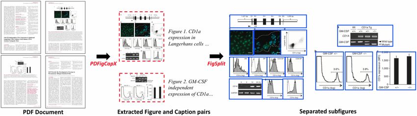
Fig. 1. An example of our pipeline for figures, subfigures and captions extraction. The original PDF document (Kobayashi et al., 2012) is shown on the left. Figures (dashed red
boxes) and captions are first extracted from the document using PDFigCapX. Figures then be further separated into subfigures (solid blue boxes) using FigSplit
segmentation by evaluating and self-correcting candidate segmenta-
tions that are likely to be inaccurate.
Both systems, PDFigCapX and FigSplit, were tested on existing
and on newly assembled datasets, demonstrating robustness and sig-
nificant improvement compared to other state-of-the-art methods
(Li et al., 2018, 2019). Figure 1 shows an example of our pipeline
for extracting figures, subfigures and captions from biomedical pub-
lications. The input to our pipeline is the original PDF document
shown on the left. By using PDFigCapX, figure and caption pairs are
extracted. The extracted images are shown in red dashed boxes. By
applying FigSplit, compound images are split into their constituent
panels—each shown within a solid blue box on the right.
2.2 Image-based document representation
Figures in biomedical publications are typically used to show the pro-
cess and results of experiments. Different types of images are used to
report certain types of experiments. For example, gel images are typic-
ally used in pull-down assays (Orchard et al., 2012). Class labels of
figures have been shown useful for document representation in bio-
medical document classification in our previous work (Shatkay et al.,
2006). As discussed in Section 2.1, the majority of figures within bio- Fig. 2. The image taxonomy used for panel classification
medical publications are compound images. Building upon our previ-
ous idea, we introduce here a new method to represent figures within Trained and tested via a 5-fold cross validation, the classifier dem-
documents based on class labels of their constituent panels here. onstrates 87.89% accuracy.
While several image taxonomies were proposed for classifying bio- Once the class label of each panel is obtained, we represent
medical images (De Herrera et al., 2016; Lopez et al., 2013; Shatkay each figure as an 11-dimensional binary vector < c1 ; c2 ; . . . ;
et al., 2006), as no standard exists, we extend the image taxonomy ci ; . . . ; c11 >, where ci is 1 if a panel from class i is present in the figure
previously proposed by our group, through collaboration with GXD and 0 otherwise. For instance, if the figure comprises only histograms
(Finger et al., 2017), Protein Information Resource (Wu et al., 2003) and fluorescence microscopy panels (panels of type1 and type6 re-
and WormBase (Harris et al., 2020), as shown in Figure 2. At the top spectively), its corresponding vector is: .
level, images are classified into Graphics, Molecular Structure, We refer to each such vector as a Figure-word. Figure 3 shows the pro-
Experimental and Other images. At the second level, Graphics are cess for converting figures into their corresponding Figure-words. As
classified into Histogram, Line Chart and Other Diagram. Molecular the number of classes in our image-taxonomy is 11, the total number
Structure images are classified into Macromolecule Sequence and 3D of possible Figure-words in our vocabulary is 211 (2048). A document
Structure images. Experimental images are further classified into d, in turn, is represented as a vector dIMG ¼ < I1 ; I2 ; . . . ; Ii ; . . . ; In >,
Fluorescence Microscopy, Light Microscopy, Whole Mount, Gel and where n ¼ 211 and Ii (1 i 2048) is 1 if the ith Figure-word
Plate images. We also note that figure legends or margins are some- appears in the document d, 0 otherwise.
times over-separated from their original images by the compound
image separation process, thus forming individual panels. We refer to
such panels formed by over-segmentation of compound images as sep- 2.3 Caption-based document representation
aration residuals. As these residuals do not belong to any of the in- Captions associated with figures provide another important source
formative taxonomy’s classes, we augment our taxonomy with a of information for biomedical document classification. In order to
separation residual class. make use of captions, we employ a standard preprocessing proced-
To automatically assign class label to individual panels, we build ure that includes named-entity recognition (NER), stemming and
an image classifier. A pre-trained Convolutional Neural Network, stop-words removal as we have done in our earlier work (Jiang
VGG16 (Andrearczyk and Müller, 2018; Simonyan and Zisserman, et al., 2017, 2020). For NER, we first identify all gene, disease,
2015), is used for image classification. To train the classifier, we use chemical, species, mutation and cell-line concepts using PubTator,
the annotated image dataset that was introduced by Lopez et al. which is widely used for annotations of biomedical concepts (Wei
(2013) based on the Molecular INTeraction database dataset et al., 2019). We then substitute each of the identified concepts by
(Licata et al., 2012). The image dataset consists of 34 876 pre- its respective generic terms ‘gene’, ‘disease’, ‘chemical’, ‘species’,
labeled panels; its statistics are shown in Table 1. In addition, a set ‘mutation’ or ‘cell-line’. We also stem words using the Porter
of 500 labeled whole mount images were provided by GXD. stemmer and remove standard stop words (Porter, 1980; Canese
Utilizing image information for biomedical document classification                                                                                                     i471

Table 1. Distribution of image types included in our experiments, based on the image dataset introduced by Lopez et al. (2013)

                   Histogram      Line       Other       Macromolecule            3D         Fluorescence      Gel/blot     Plate      Light          Other     Separation
                                  chart     diagram        sequence            structure      microscopy                             microscopy                  residual

No. of panels         4270        2664        3536              499              1424            5714           14865       508          1156          130          110

                                                                                                                                                                                Downloaded from https://academic.oup.com/bioinformatics/article/37/Supplement_1/i468/6319676 by guest on 26 September 2021
Fig. 3. The process for converting figures into their corresponding Figure-words. The set of figures extracted from the biomedical documents is shown on the left. The corre-
sponding Figure-words along with their vector representations indicating the types of comprising panels, are shown on the right. The images on the left are taken from (Li
et al., 2008, Fig. 4; Qiu and Dhe-Paganon, 2011, Fig. 6; Sugioka et al., 2014, Fig.3; Leung et al., 2012, Fig. 9; Heinonen et al., 2015, Fig. 4; Dai et al., 2012, Fig. 3)

and Weis, 2013). The concatenated captions are used as the caption-                            Under this scheme, to represent a document d, we simply concat-
based document representation.                                                             enate the vectors dIMG ; dCAP and dTA into a single vector dALL , thus
    Word embeddings map words to vectors of a fixed dimension so                           utilizing the information obtained from images, captions and titles-
that words appearing in similar contexts are mapped to similar vec-                        and-abstracts. Recall that the value of an entry in the dIMG vector is
tors. Such a vectorized representation has been widely used for text                       either 1 or 0 which indicates whether or not a Figure-word appears
classification (Moen and Ananiadou, 2013), and more recently for                           in a document, while the value of an entry in dCAP or dTA is obtained
biomedical named entity recognition and biomedical relation extrac-                        by calculating the mean of embedding vectors converted from words
tion (Lee et al., 2020). A word embedding model (Moen and                                  in a caption or a title-and-abstract. There is no specific limit on the
Ananiadou, 2013) has been pre-trained specifically on a biomedical                         range of embedding vectors. Therefore, the values of such entries are
corpus, which consists of PubMed titles-and-abstracts and PubMed                           at different scales from that of entries in dIMG . As such, we standard-
Central full text articles by employing the word2vec tool (Mikolov                         ize each feature within dALL by rescaling the features such that they
et al., 2013). We use such embeddings to represent the concatenated                        have a mean of 0 and a standard deviation of 1. For classifying the
captions. Each word, wi, within the concatenated captions is con-                          documents, we conducted experiments with several classification
verted to a word embedding vector w   ~i whose dimensionality is 200.                      schemes, including Random Forests, Naı̈ve Bayes (not shown here)
The document d is then represented as a 200-dimentional vector                             and SVMs. As SVMs have been commonly used for both image and
dCAP , calculated as dCAP ¼ 1n ðw
                                ~1 þ w~2 þ    þ w
                                                   ~n Þ, where n is the                    text classification, and have shown the best performance in this con-
total number of distinct words in the concatenated captions, and w   ~i                    text (Holzinger et al., 2014; Simpson et al., 2015), we use SVM as
is the embedding vector of the ith distinct word.                                          the model for classifying the final resulting document-vectors, and
                                                                                           denote this SVM classifier CombV.
                                                                                               (2) Integration via meta-classification
2.4 Title-and-abstract-based document representation                                           Another approach we propose toward integrating the multiple
The title and the abstract of articles are the text components most                        types of information is to employ a meta-classification scheme,
often used for biomedical document classification. To represent a                          which combines results obtained from multiple base classifiers into
document based on those, we first obtain the title-and-abstract of                         a single classification output. To do that, we first separately train
each publication. Similar to the steps described in Section 2.3, we                        three base classifiers CIMG ; CCAP and CTA using the representations
employ a standard preprocessing procedure that includes named-en-                          of images ðdIMG Þ, captions (dCAP ) and titles-and-abstracts (dTA ). By
tity recognition, stemming and stop-words removal to each title-                           applying a base classifier to a document d, we obtain the class label
and-abstract. The same word embeddings described in Section 2.3                            L and the probability P of document d to be assigned to the relevant
are employed to convert each word wi in the preprocessed text to a                         class. Each document d is then represented as a 6-dimensional vector
word embedding vector w ~i . The document d is then represented as a                       dCombC ¼ < LIMG ; PIMG ; LCAP ; PCAP ; LTA ; PTA >. This representation
200-dimensional vector, denoted as dTA , by calculating the mean of                        is then used for training another classifier, referred to as meta-classi-
embedding vectors that are associated with words in the prepro-                            fier, denoted as CombC, which assigns the final class label to each
cessed text.                                                                               document. Similar to the concatenation-based integration, we use
                                                                                           SVMs both as base classifiers and as the ultimate classifier in the
                                                                                           meta-classification.
2.5 Information integration for document classification
So far, we have introduced document representations based on
images (dIMG ), captions (dCAP ) and title-and-abstracts (dTA ). Next,
we present two schemes for integrating the information stemming
                                                                                           3 Experiments and results
from these three sources.                                                                  To evaluate our method we conduct two sets of experiments. The
   (1) Integration via concatenated vectors                                                first aims to compare the classification performance obtained when
i472 P.Li et al.
using only a single type of information to represent documents ver- associated with relevant publications and 41 015 panels obtained
sus the performance when employing a representation that combines from the 8414 figures associated with the irrelevant publications.
all three types of information. The classifiers using the representa- The second dataset used in our experiments, denoted DSP, was
tions of Figure-words, captions and titles-and-abstracts, are denoted introduced by Burns et al. (2019) for testing their system
as CIMG ; CCAP and CTA , respectively. CNNBiLSTM. It comprises 537 publications relevant to molecular
In the second set of experiments, we compare the performance of interactions and 451 irrelevant ones spanning the year range 1996-
our system when utilizing a representation that combines images, 2017. Only publications for which PDF files are available are used
captions and titles-and-abstracts to the performance attained by in our experiments. As such, out of the 537 relevant publications
three state-of-the-art systems. The first system to which we compare only 534 are used, while out of the 451 irrelevant ones only 448 are
is a random forest-based method (RFCAP) developed by Jiang et al. retained, as their PDF files were available for download online. We
(2017) for identifying publications that are relevant to GXD. This then apply PDFigCapX and FigSplit to identify and extract figures,
classifier uses features extracted from the title-and-abstract and
Downloaded from https://academic.oup.com/bioinformatics/article/37/Supplement_1/i468/6319676 by guest on 26 September 2021
captions and constituent panels of extracted figures. From the 534
from caption text. The second is a convolutional neural network tri- relevant publications, 3975 figures, 3912 captions and 21 421 pan-
age system (CNNBiLSTM ) presented by Burns et al. (2019) for identi- els are extracted, while 2928 figures, 2832 captions and 14 224 pan-
fying publications containing information about molecular els are extracted from the 448 irrelevant ones. Table 2 shows the
interactions. The CNNBiLSTM classifier uses captions only. The third
statistics for these two datasets.
is a hierarchical recurrent neural network classification system
The total time for PDFigCapX to process the GXD2000 dataset
(HRNN) developed by Fergadis et al. (2018) for identifying publica-
of 2000 publications is about 5.9 h (10.60 s per document, wall
tions containing information about protein-protein interactions
clock) where the average document contains 8.7 figures, 8.3 cap-
affected by genetic mutations; and uses title-and-abstract only. We
tions and is 7.2MB in size. It takes about 4.3 h (0.83 s per image,
compare all three systems using the code provided by their respective
wall-clock) for FigSplit to process all extracted figures where on
authors. For comparison, we run five complete rounds of 5-fold
cross validation with 5 different 5-way partitions of the dataset. All average 50.6 panels are extracted from each publication within the
experiments are conducted on a DELL machine that uses an Intel GXD2000 dataset. Over the DSP dataset, PDFigCapX takes about
Core i7-6700 processor, an Nvidia GTX 1070 GPU, 8 GB of RAM 2.4 h (8.69 s per document, wall-clock) to process all 982 publica-
and 256 GB of SSD. tions where the average document contains 7.0 figures, 6.9 captions
and the average file size is 2.8MB. FigSplit takes about 1.6 h (0.78 s
per image, wall-clock) to process all extracted figures where on aver-
3.1 Datasets and evaluation age 36.3 panels are extracted from each publication in the DSP
In our experiments, we use two datasets for which we have the dataset.
ground-truth class-labels. The first dataset, denoted GXD2000, is a To evaluate the document classification performance, we use
subset of the dataset used by Jiang et al. (2017), who is also the de- standard measures, Precision, Recall and F-score defined as:
veloper of RFCAP. The original dataset is a collection of 58 362 pub-
lications (provided as PDF), curated by the Jackson Lab’s GXD TruePositive
throughout the years 2012–2016. As a first test of our method, we Precision ¼ ;
TruePositive þ FalsePositive
selected at random 1000 relevant and 1000 irrelevant documents
from these publications, while retaining the same distribution of TruePositive
Recall ¼ ;
publication-years as in the larger GXD dataset. In order to use fig- TruePositive þ FalseNegative
ures and captions, we first apply PDFigCapX to the GXD2000 data-
set. 8939 figures and 8594 captions are extracted from the relevant 2 Precision Recall
F score ¼ :
publications, while 8414 figures and 8042 captions are extracted Precision þ Recall
from the irrelevant publications. We note that the number of figures
extracted exceeds that of the captions, as some pages display figures
(or parts of figures) without associated captions. FigSplit is then 3.2 Results
applied to separate compound figures into their constituent panels, Table 3 presents the classification performance attained when using
resulting in 60 194 individual panels extracted from the 8939 figures only a single type of information to represent documents, along with
Table 2. The number of figures, captions and panels identified and extracted from publications in the datasets used in our experiments
Datasets Classes No. of docs No. of figures No. of captions No. of panels
GXD2000 Relevant 1000 8939 8594 60 194
Irrelevant 1000 8414 8042 41 015
DSP Relevant 534 3975 3912 21 421
Irrelevant 448 2928 2832 14 224
Table 3. Classification performance attained by using information from images (CIMG ), captions (CCAP ), the title-and-abstract (CTA ), concaten-
ated vectors from all three types (CombV), and by using the meta-classifier (CombC). The highest values attained are shown in boldface.
Standard deviations are shown in parentheses.
GXD2000 DSP
Classifiers Precision Recall F-score Precision Recall F-score
CIMG 0.805 (.021) 0.770 (.026) 0.787 (.021) 0.679 (.018) 0.768 (.026) 0.731 (.026)
CCAP 0.886 (.027) 0.871 (.032) 0.878 (.021) 0.804 (.024) 0.809 (.034) 0.806 (.021)
CTA 0.875 (.021) 0.877 (.015) 0.876 (.013) 0.790 (.023) 0.807 (.023) 0.798 (.015)
CombC 0.887 (.019) 0.899 (.025) 0.893 (.008) 0.822 (.032) 0.826 (.044) 0.823 (.020)
CombV 0.894 (.019) 0.910 (.017) 0.902 (.008) 0.831 (.014) 0.834 (.031) 0.832 (.019)Utilizing image information for biomedical document classification i473
the performance when employing a representation that combines all 78.3%. The CNNBiLSTM system, whose author introduced the DSP
three types of information in our first set of experiments. The second dataset itself, reached 82.0% precision, 79.6% recall and 80.9% F-
to fourth columns in the table show results from experiments over score. Our classifier attained the highest performance of 83.1% pre-
the GXD2000 dataset. Our classifier CIMG , utilizing the Figure-words cision, 83.4% recall and 83.2% F-score over the DSP dataset.
alone, attained 80.5% precision, 77.0% recall and 78.7% F-score. Moreover, the differences between the results obtained by our classi-
By using information from the caption alone, our classifier CCAP fier and those attained by other state-of-the-art systems are statistic-
attained 88.6% in precision, 87.1% in recall and 87.8% in F-score. ally significant (P < 0.001, two-sample t-test).
A similar performance (87.5% precision, 87.7% recall and 87.6%
F-score) is attained by the classifier CTA when titles-and-abstracts
are used to represent documents. Notably, a significant improve- 4 Discussion
ment is attained by using the representation that combines informa-
Notably, Figure-words provide important information for document
Downloaded from https://academic.oup.com/bioinformatics/article/37/Supplement_1/i468/6319676 by guest on 26 September 2021
tion from images, captions and titles-and-abstracts. By employing
the meta-classification scheme, our classifier CombC attained classification. The image-based classifier, CIMG , which uses the
88.7% precision, 89.9% recall and 89.3% F-score. Our classifier newly proposed Figure-words alone for document representation
CombV attained the highest performance of 89.4% precision, attained 80.5% precision, 77.0% recall and 78.7% F-score over the
91.0% recall and 90.2% F-score when the concatenated vectors are GXD2000 dataset, while attaining 69.7% precision, 76.8% recall
used for document representation. The performance attained by and 73.1% F-score when applied to the DSP dataset (Table 3).
using the integrated information is statistically significantly higher Moreover, Figure-words provide information distinct from that cap-
than the performance attained based on the Figure-words, the cap- tured by captions or by titles-and-abstracts. Of the GXD2000 data-
tions or the titles-and-abstracts alone (P < 0.01, two-sample t-test set, 71 relevant publications (7.1% of the relevant publications)
for all measures). were correctly identified by CIMG , but incorrectly classified by CCAP ,
The three rightmost columns in Table 3 present the results while 71 publications were correctly identified by CIMG , but incor-
attained over the DSP dataset. When the Figure-words are used for rectly classified by CTA . Of the DSP dataset, 59 relevant publica-
document representation, our classifier CIMG attained 69.7% preci- tions (11.0% of the relevant data) were correctly identified by CIMG ,
sion, 76.8% recall and 73.1% F-score. The classifier CCAP attained but incorrectly classified by CCAP , while 56 publications (10.5% of
80.4% precision, 80.9% recall and 80.6% F-score. A similar per- the relevant data) were correctly identified by CIMG , but incorrectly
formance (79.0% precision, 80.7% recall and 79.8% F-score) is classified by CTA .
attained by CTA when titles-and-abstracts are used for document Another noteworthy point is that captions provide distinct infor-
representation. Again, a significant improvement is attained by mation from that provided by titles-and-abstracts for document clas-
using the integrated information. We attained 82.2% precision, sification. As indicated in Section 3.2, the performances attained
82.6% recall and 82.3% F-score when the meta-classification using a classifier based on captions or titles-and-abstracts alone
scheme is applied. The highest performance of 83.1% precision, (CCAP and CTA , respectively) are similar over both the GXD2000 and
83.4% recall and 83.2% F-score is attained when the concatenated the DSP datasets. However, the relevant publications identified by
vectors are used to represent documents. The performance attained classifiers CCAP and CTA are quite different. Of the GXD2000 data-
by classifiers that integrate information from images, captions and set, 60 relevant publications (6.0% of the relevant ones) were cor-
titles-and-abstracts is statistically significantly higher than the per- rectly identified by CCAP , but incorrectly classified by CTA , while 66
formance attained by classifiers that are based on single information distinct publications were correctly identified by CTA , but incorrect-
source (P < 0.01, two-sample t-test for all measures). Our results ly classified by CCAP . Of the DSP dataset, 38 relevant publications
demonstrate that our information integration schemes indeed im- (7.1% of the relevant data) were correctly identified by CCAP , but in-
prove biomedical document classification. correctly classified by CTA , while 37 distinct publications were cor-
Table 4 compares the performance of our classifier CombV to rectly identified by CTA , but incorrectly classified by CCAP . By their
that attained by the three other state-of-the-art systems, RFCAP, very nature, titles-and-abstracts form a high-level summary of an en-
CNNBiLSTM and HRNN. Over the GXD2000 dataset, RFCAP tire study, while captions present details of experimental processes
attained 82.9% recall, 87.8% F-score and the highest precision of and results. This difference is reflected in the vocabulary of titles-
93.4%. CNNBiLSTM achieved 87.6% precision, 85.0% recall and and-abstracts versus that of captions. For instance, words such as
86.2% F-score, while HRNN attained 85.5% in precision, 87.5% anterior, WT, dorsal, embryo, green, lateral and mount, are com-
in recall and 86.4% in F-score. While the precision (89.4%) attained monly found in captions of publications relevant to GXD when
by our classifier is slightly lower than that reached by RFCAP, our describing gene expression experiments in mouse embryos. As such,
classifier CombV attained the highest recall of 91.0% and the high- captions provide information that is distinct from that provided
est F-score of 90.2% over the GXD2000 dataset. Notably, recall is through titles-and-abstracts, thus supporting more effective
often viewed as more important than precision for biomedical docu- classification.
ment curation (Fang et al., 2012; Müller et al., 2004). Moreover, Figure 4 illustrates the respective classification results attained
the differences between the results obtained by our system and those by the classifiers CIMG , CCAP and CTA . Of the GXD2000 dataset, 18
attained by RFCAP, CNNBiLSTM and HRNN are statistically signifi- relevant publications are identified only by CCAP , while 24 relevant
cant (P < 0.001, two-sample t-test). The three rightmost columns in ones are identified only by CTA . Notably, 29 relevant publications
Table 4 presents the results attained over the DSP dataset. RFCAP can only be identified by CIMG using Figure-words for document
achieved 79.8% precision, 80.9% recall and 79.8% F-score, while representation. Of the DSP dataset, classifier CIMG identified 32
HRNN attained 75.4% precision, 81.8% recall and an F-score of relevant publications that are distinct from those identified by CCAP
Table 4. Classification performance Comparison with other state-of-the-art systems. The highest values attained are shown in boldface.
Standard deviations are shown in parentheses.
GXD2000 DSP
Classifiers Precision Recall F-score Precision Recall F-score
RFCAP 0.934 (.017) 0.829 (.037) 0.878 (.018) 0.798 (.068) 0.809 (.057) 0.798 (.030)
CNNBiLSTM 0.876 (.028) 0.850 (.031) 0.862 (.013) 0.820 (.023) 0.796 (.030) 0.809 (.018)
HRNN 0.856 (.044) 0.875 (.033) 0.864 (.010) 0.754 (.032) 0.818 (.054) 0.783 (.017)
CombV 0.894 (.019) 0.910 (.017) 0.902 (.008) 0.831 (.014) 0.834 (.031) 0.832 (.019)i474 P.Li et al.
and CTA . These findings strongly suggest that the three data sources, document classification over the DSP dataset (Fig. 5b). As such, our
namely, Figure-words, captions and titles-and-abstracts provide dis- newly proposed Figure-words compactly account for and convey
tinct and complementary information for document classification. certain types of biomedical experiments. Experimental evidence is
To better understand the contribution of Figure-words to important for identifying relevant biomedical documents (Burns
improved classification, we identify the most distinguishing Figure- et al., 2018; Han et al., 2006), thus our Figure-words can contribute
words by ranking them based on the Z-score Test (Myers et al., much informative evidence to the document classification task.
1993), as we have done before for identifying distinguishing text As discussed above, Figure-words, captions and titles-and-
terms (Jiang et al., 2017, 2020). Table 5a shows the top-5 scoring abstracts provide complementary information for document
Figure-words, along with their occurrence frequency in the relevant
and in the irrelevant publications of the GXD2000 dataset. There is a
significant difference between the Figure-word distribution in rele-
Downloaded from https://academic.oup.com/bioinformatics/article/37/Supplement_1/i468/6319676 by guest on 26 September 2021
vant publications and their distribution in irrelevant ones. For in-
stance, there are 1339 images consisting of fluorescence alone in
relevant publications of the GXD2000, while only 437 such images
in the irrelevant publications. Similarly, Table 5b shows that the dis-
tinguishing Figure-words identified with respect to the DSP dataset,
demonstrate a clear difference in Figure-word distribution between
relevant publications and irrelevant ones. Therefore, we believe that
our newly proposed Figure-words have much potential for improv-
ing biomedical document classification.
Figure 5 shows examples of Figure-words. Notably, the top scor-
ing Figure-words also correspond to images that are typically found
in distinct biomedical experiments. For instance, fluorescence mi-
croscopy images often appear in publications relevant to the GXD
as this is a common imaging technique for visualizing gene expres-
sion. As curators at the GXD focus on mouse embryo studies,
Figure-words containing embryonic whole-mount images are also
indicative of documents that are likely relevant to GXD. Similarly,
Co-inmmunoprecipitation, and Pull Down experiments are com-
monly used in studies relevant to molecular interactions, thus
Figure-words corresponding to the gel/blot images are important for
Fig. 5. Examples of images for top scoring Figure-words. The leftmost column
Fig. 4. Comparison of classification results between the classifiers CIMG ; CCAP and shows Figure-words along with their vector representations and their respective
CTA . The solid circle corresponds to the relevant publications that are correctly combination of panel types. The rest of the columns show examples of correspond-
identified by classifier CIMG . The relevant publications correctly classified by CCAP ing images. (a) Image examples from the GXD2000 dataset. Original images are
are indicated as a dashed circle, while those correctly identified by CTA are shown as taken from (Vogt et al., 2012, Fig. 1; Grimsley-Myers et al., 2012, Fig. 7;
a dotted circle. The region marked by horizontal stripes indicates the relevant publi- Yamaguchi et al., 2014, Fig. 5; Quattrocolo & Maccaferri, 2014, Fig. 1; Liu et al.,
cations identified by classifier CIMG only. The region marked by grid pattern corre- 2012, Fig. 6; Rooijen et al., 2012, Fig. S3). (b) Image examples from the DSP data-
sponds to the relevant publications classified by CCAP only, while the region shown set. Original images are taken from (Shinohara et al., 2005, Fig. 3; Cheng et al.,
in solid gray indicates those identified by CTA only. (a) The comparison over the 2016, Fig. 4; Mysling et al., 2016, Fig. 1; Yoshida et al., 2014, Fig. 3; Graef et al.,
GXD2000 dataset. (b) The comparison over the DSP dataset 2009, Fig. 6; Li et al., 2008, Fig. 4)
Table 5. Top scoring Figure-words that contribute to the document classification task. The left most column in each table shows the Figure-
words and their corresponding vectors indicating the types of comprising panels. The other two columns show the occurrence frequencies
of corresponding figures in the relevant and in the irrelevant dataset, respectively. (a) Top Figure-words identified over the GXD2000 data-
set. (b) Top Figure-words identified over the DSP dataset.Utilizing image information for biomedical document classification i475
classification. A significant improvement is obtained by using the document classification performance. It is noteworthy that the
representation comprising all three sources. In our experiments, we newly proposed Figure-words correspond to certain distinct types of
attained statistically significantly improved performance by employ- images used in reporting biomedical experiments. We will investi-
ing the meta-classification scheme CombC as well as by employing gate the potential usage of Figure-words for other tasks, such as bio-
the classifier, CombV, where concatenated vectors are used for medical image classification.
document representation, as compared to classification based on the
title-and-abstracts, the captions and the Figure-words alone.
In the second set of experiments when our system is compared Funding
against three state-of-the-art systems, over the GXD2000 dataset,
two classifiers (CNNBiLSTM, HRNN) utilizing only a type of infor- This work was partially supported by National Institutes of Health (NIH)/
mation source attained similar performance. The RFCAP classifier National Library of Medicine (NLM) awards [R56LM011354A and
which uses features extracted from combined title-and-abstract and R01LM012527]; NIH/National Institute of Child Health and Human
Downloaded from https://academic.oup.com/bioinformatics/article/37/Supplement_1/i468/6319676 by guest on 26 September 2021
caption text, indeed outperforms the systems that use just a single Development (NICHD) award [P41 HD062499 to M.R.].
type of information. Our method, which integrates the information
Conflict of Interest: none declared.
from Figure-words, captions and titles-and-abstracts attained the
highest recall and F-score over the GXD2000 dataset. When applied
to the DSP dataset, our method attained the highest score across all
measures. These results demonstrate that Figure-words provide in- References
formation distinct from that provided by titles-and-abstracts and by Almeida,H. et al. (2014) Machine learning for biomedical literature triage.
captions for supporting classification, and also prove the effective- PLoS One, 9, e115892.
ness of the integration methods that we introduced. Andrearczyk,V. and Müller,H. (2018) Deep multimodal classification of
While our method indeed improves classification performance, image types in biomedical journal figures. In: Proc. of CLEF, pp. 3–14.
there is still room for improvement, especially for the image tax- Avignon, France.
onomy. In the work reported here, we utilized the image taxonomy Bateman,A. et al. (2021) UniProt: the universal protein knowledgebase in
consisting of 11 categories, as there is no unique standard image tax- 2021. Nucleic Acids Res., 49, D480–D489.
onomy for categorizing biomedical research images yet. A more Blake,J.A. et al.; The Mouse Genome Database Group. (2021) Mouse
comprehensive taxonomy has the potential to support a more in- Genome Database (MGD): knowledgebase for mouse-human comparative
formative Figure-words vocabulary and as such improve the overall biology. Nucleic Acids Res., 49, D981–D987.
Burns,G. et al. (2018) Towards evidence extraction: analysis of scientific fig-
document classification results. In our future work, we plan to ex-
ures from studies of molecular interactions. In Proc. of ISWC, pp. 95–102.
pand and refine the image taxonomy we employ for categorizing
Monterey, CA, USA.
biomedical research images. We are already in the process of apply-
Burns,G.A. et al. (2019) Building deep learning models for evidence classifica-
ing our classification scheme to a larger dataset, namely the
tion from the open access biomedical literature. Database, 2019, baz034.
COVID-19 open research dataset comprising more than 50 000 Canese,K. and Weis,S. (2013) PubMed: the bibliographic database. In: The
articles (Chen et al., 2020; Wang et al., 2020), and plan to further NCBI Handbook [Internet]. 2nd edn. National Center for Biotechnology
apply it to the complete GXD dataset of more than 58 000 publica- Information. Bethesda, MD, USA.
tions used by Jiang et al. (2017). Chatr-Aryamontri,A. et al. (2017) The BioGRID interaction database: 2017
As our newly proposed Figure-words correspond to distinct update. Nucleic Acids Res., 45, D369–D379.
images used in certain types of biomedical experiments, our image Chen,Q. et al. (2020) LitCovid: an open database of COVID-19 literature.
representation method can help biocurators identify images accord- Nucleic Acids Res., 49, D1534–D1540.
ing to their experiment types. For example, a biocurator may want Cheng,L.F. et al. (2016) Incorporation of GM-CSF or CD40L enhances the
to identify images used in yeast two-hybrid experiments based on immunogenicity of Hantaan virus-like particles. Front. Cell. Infect.
the images used to describe such experiments. We will also investi- Microbiol., 6, 185.
gate the usage of our image representation for other tasks, such as Chhatkuli,A. et al. (2013) Separating compound figures in journal articles to
biomedical image classification. allow for subfigure classification. Proc. SPIE Med. Imaging, 8674, 86740J.
Clark,C. et al. (2016) Pdffigures 2.0: mining figures from research papers. In
Proc. of IEEE/ACM JCDL, pp. 143–152. Newark, NJ, USA.
5 Conclusion Dai,B. et al. (2012) A comparative transcriptome analysis identifying FGF23
regulated genes in the kidney of a mouse CKD model. PLoS One, 7,
We presented a new scheme for identifying biomedical documents e44161.
that are relevant to a certain domain, by using information derived De Herrera,A.G.S. et al. (2016) Overview of the ImageCLEF 2016 medical
from both images and captions, as well as from titles-and-abstracts. tasks. In: Working Notes of ImageCLEF, pp. 219–232. Evora, Portugal.
To do so, we first employed a pipeline for processing biomedical Fang,R. et al. (2012) Automatic categorization of diverse experimental infor-
documents, comprising two parts: PDFigCapX that extracts figures mation in the bioscience literature. BMC Bioinformatics, 13, 16.
with their captions from documents, and FigSplit for splitting the Fergadis,A. et al. (2018) Hierarchical bi-directional attention-based RNNs for
extracted compound figures into constituent panels, to biomedical supporting document classification on protein–protein interactions affected
documents. by genetic mutations. Database, 2018, bay076.
A new image representation, Figure-word that encodes the com- Finger,J.H. et al. (2017) The mouse gene expression database (GXD): 2017
bination of different types of panels is proposed for representing fig- update. Nucleic Acids Res., 45, D730–D736.
Garcia,M.A.M. et al. (2015) Biomedical literature classification using encyclo-
ures within documents. For captions and titles-and-abstracts, word
pedic knowledge: a Wikipedia-based bag-of-concepts approach. PeerJ, 3,
embeddings are employed to represent documents as vectors. To
e1279.
utilize both image and caption information, in addition to titles-
Graef,T. et al. (2009) KIR2DS4 is a product of gene conversion with
and-abstracts, we introduced two information integration methods.
KIR3DL2 that introduced specificity for HLA-A 11 while diminishing avid-
The first concatenates Figure-words and features obtained from cap- ity for HLA-C. J. Exp. Med., 206, 2557–2572.
tions and titles-and-abstracts into a single larger vector for docu- Grimsley-Myers,C.M. et al. (2012) Redundant functions of RAC GTPases in
ment representation; the second employs a meta-classification inner ear morphogenesis. Dev. Biol., 362, 172–186.
scheme. Our experiments demonstrate the effectiveness of the newly Harris,T.W. et al. (2020) WormBase: a modern model organism information
proposed Figure-words for representing images. Moreover, classifi- resource. Nucleic Acids Res., 48, D762–D767.
cation performance is improved through the integration of informa- Han,B. et al. (2006) Substring selection for biomedical document classifica-
tion from all three sources, namely, images, captions and titles-and- tion. Bioinformatics, 22, 2136–2142.
abstracts. Heinonen,H. et al. (2015) Identification of several potential chromatin binding
As part of future work, we plan to build a more comprehensive sites of HOXB7 and its downstream target genes in breast cancer. Int. J.
taxonomy for refining image classification and improving the Cancer, 137, 2374–2383.i476 P.Li et al.
Hu,Z.Z. et al. (2005) Literature mining and database annotation of protein Müller,H.M. et al. (2004) Textpresso: an ontology-based information re-
phosphorylation using a rule-based system. Bioinformatics, 21, 2759–2765. trieval and extraction system for biological literature. PLoS Biol., 2, e309.
Holzinger,A. et al. (2014) Biomedical text mining: state-of-the-art, open prob- Myers,S.L. et al. (1993) Probability and Statistics for Engineers and Scientists.
lems and future challenges. In Interactive Knowledge Discovery & Data Macmillan, New York.
Mining in Biomedical Informatics, Ch. 16. Springer, Berlin, Heidelberg, Mysling,S. et al. (2016) The acidic domain of the endothelial membrane pro-
271–300. tein GPIHBP1 stabilizes lipoprotein lipase activity by preventing unfolding
Jiang,X. et al. (2017) Effective biomedical document classification for identify- of its catalytic domain. Elife, 5, e12095.
ing publications relevant to the mouse Gene Expression Database. Orchard,S. et al. (2012) Protein interaction data curation: the International
Database, 2017, bax017. Molecular Exchange (IMEx) consortium. Nat. Methods, 9, 345–350.
Jiang,X. et al. (2020) Integrating image caption information into biomedical Porter,M.F. (1980) An algorithm for suffix stripping. Program, 14, 130–137.
document classification in support of biocuration. Database, 2020, Qiu,L. and Dhe-Paganon,S. (2011) Oligomeric structure of the MALT1 tan-
baaa024. dem Ig-like domains. PLoS One, 6, e23220.
Downloaded from https://academic.oup.com/bioinformatics/article/37/Supplement_1/i468/6319676 by guest on 26 September 2021
Karystianis,G. et al. (2017) Evaluation of a rule-based method for epidemio- Quattrocolo,G. and Maccaferri,G. (2014) Optogenetic activation of
logical document classification towards the automation of systematic Cajal-Retzius cells reveals their glutamatergic output and a novel feed-
reviews. J. Biomed. Inf., 70, 27–34. forward circuit in the developing mouse hippocampus. J. Neurosci., 34,
Kerrien,S. et al. (2012) The IntAct molecular interaction database in 2012. 13018–13032.
Nucleic Acids Res., 40, D841–D846. Regev,Y. et al. (2002) Rule-based extraction of experimental evidence in the
Kobayashi,C. et al. (2012) GMCSF-independent CD1a expression in epider- biomedical domain: the KDD Cup 2002 (task 1). ACM SIGKDD
mal Langerhans cells: evidence from human CD1A genome-transgenic mice. Explorations Newslett., 4, 90–92.
Rooijen,C. et al. (2012) Evolutionarily conserved requirement of Cdx for
J. Invest. Dermatol., 132(1), 241–244.
post-occipital tissue emergence. Development, 139, 2576–2583.
Lee,J. et al. (2020) BioBERT: a pre-trained biomedical language representa-
Santosh,K.C. et al. (2015) Stitched multipanel biomedical figure separation. In
tion model for biomedical text mining. Bioinformatics, 36, 1234–1240.
Proc. of IEEE CBMS, pp. 54–59. Sao Carlos, Brazil.
Leung,L. et al. (2012) Apolipoprotein E4 causes age-and sex-dependent
Shatkay,H. et al. (2006) Integrating image data into biomedical text categor-
impairments of hilar GABAergic interneurons and learning and memory
ization. Bioinformatics, 22, e446–e453.
deficits in mice. PLoS One, 7, e53569.
Shinohara,H. et al. (2005) PKCb regulates BCR-mediated IKK activation by
Li,P. et al. (2018) Compound image segmentation of published biomedical fig-
facilitating the interaction between TAK1 and CARMA1. J. Exp. Med.,
ures. Bioinformatics, 34, 1192–2299.
202, 1423–1431.
Li,P. et al. (2019) Figure and caption extraction from biomedical documents.
Simonyan,K. and Zisserman,A. (2015) Very deep convolutional networks for
Bioinformatics, 35, 4381–4388.
large-scale image recognition. In Proc. of ICLR, pp. 1409–1556. San Diego,
Li,Y. et al. (2008) Crystal structure of human XLF/Cernunnos reveals unex-
CA, USA.
pected differences from XRCC4 with implications for NHEJ. EMBO J., 27,
Simpson,M.S. et al. (2015) Literature-based biomedical image classification
290–300. and retrieval. Comput. Med. Imag. Graph., 39, 3–13.
Licata,L. et al. (2012) MINT, the molecular interaction database: 2012 up- Sugioka,K. et al. (2014) The roles of urokinase-type plasminogen activator in
date. Nucleic Acids Res., 40, D857–D861. leukocyte infiltration and inflammatory responses in mice corneas treated
Liechti,R. et al. (2017) SourceData: a semantic platform for curating and with lipopolysaccharide. Investig. Ophthalmol. Vis. Sci., 55, 5338–5350.
searching figures. Nat. Methods, 14, 1021–1022. Vogt,E.J. et al. (2012) Importance of the pluripotency factor LIN28 in the
Liu,Y. et al. (2012) Sexually dimorphic BDNF signaling directs sensory innerv- mammalian nucleolus during early embryonic development. Development,
ation of the mammary gland. Science, 338, 1357–1360. 139, 4514–4523.
Lopez,L.D. et al. (2013) An image-text approach for extracting experimental Wang,L. et al. (2020) CORD-19: the Covid-19 open research dataset. In Proc.
evidence of protein-protein interactions in the biomedical literature. In of NLP COVID-19 workshop at ACL, pp. 1–12. Seattle, WA, USA.
Proc. of ACM BCB, pp. 412–418. Washington, DC, USA. Wei,C.H. et al. (2019) PubTator central: automated concept annotation for
Ma,K. et al. (2015) Utilizing image-based features in biomedical document biomedical full text articles. Nucleic Acids Res., 47, W587–W593.
classification. In Proc. of IEEE ICIP, pp. 4451–4455. Quebec city, Quebec, Wu,C.H. et al. (2003) The protein information resource. Nucleic Acids Res.,
Canada. 31, 345–347.
Mikolov,T. et al. (2013) Distributed representations of words and phrases and Yamaguchi,T. et al. (2014) Skn-1a/Pou2f3 is required for the generation of
their compositionality. In Proc. of NIPS, pp. 3111–3119. Lake Tahoe, NV, Trpm5-expressing microvillous cells in the mouse main olfactory epithe-
USA. lium. BMC Neurosci., 15, 1–10.
Moen,S.P.F.G.H. and Ananiadou,T.S.S. (2013) Distributional semantics Yoshida,S. et al. (2014) Molecular cloning of rat and porcine retina-derived
resources for biomedical text processing. In Proc. of LBM, pp. 39–44. POU domain factor 1 (POU6F2) from a pituitary cDNA library. J. Reprod.
Tokyo, Japan. Dev., 60, 288–294.You can also read