1H, 13C and 15N backbone and side-chain resonance assignments of Drosophila melanogaster Ssu72
←
→
Page content transcription
If your browser does not render page correctly, please read the page content below
Biomol NMR Assign (2012) 6:57–61
DOI 10.1007/s12104-011-9325-2

 ARTICLE

1
 H, 13C and 15N backbone and side-chain resonance assignments
of Drosophila melanogaster Ssu72
Jon W. Werner-Allen • Pei Zhou

Received: 5 April 2011 / Accepted: 28 June 2011 / Published online: 6 July 2011
Ó Springer Science+Business Media B.V. 2011

Abstract Ssu72 helps regulate transcription and co-                     directed specifically at the pS5 position. Ssu72 activity is
transcriptional mRNA processing by dephosphorylating                    essential in yeast and affects the initiation, elongation and
serine residues at the 5th position in the heptad repeats of            termination stages of transcription as well as co-transcrip-
the C-terminal domain of RNA polymerase II. Here we use                 tional 30 -end processing of nascent mRNA. Recently, we
multidimensional, multinuclear NMR experiments to                       and others reported crystal structures of Ssu72 in complex
assign the backbone and side-chain resonances of the                    with pS5 CTD (Werner-Allen et al. 2011; Xiang et al.
23 kDa Ssu72 from Drosophila melanogaster in the                        2010), which show that Ssu72 contains the scaffold of the
phosphate-bound state, and use NMR titrations to examine                low molecular weight protein tyrosine phosphatases (LMW
the phosphate-binding properties of three active site                   PTPs) with unique additions, giving structural support for a
mutants.                                                                common catalytic mechanism of dephosphorylation. The
                                                                        complexes also reveal a remarkable substrate conformation
Keywords Ssu72  RNA Polymerase II  CTD                                with the pS5-P6 motif of the CTD in the cis isomer, which
phosphatase                                                             exists at only *10% in solution, providing the first
                                                                        example of a cis proline-specific enzymatic activity and a
                                                                        surprising explanation for the in vivo connection between
Biological context                                                      the activities of Ssu72 and the proline isomerase Ess1 (Pin1
                                                                        in humans).
The C-terminal domain (CTD) of RNA polymerase II                            Despite these recent findings, many aspects of Ssu72
consists of multiple copies of a heptad repeat with the                 function have yet to be elucidated at the molecular level.
consensus sequence Y1-S2-P3-T4-S5-P6-S7 and acts as an                  For example, Ssu72 binds to symplekin (Pta1 in yeast), a
unstructured platform for association with proteins that                scaffolding component of the large complex that catalyzes
regulate transcription and co-transcriptional processes.                30 -end cleavage and polyadenylation of mRNA transcripts,
Phosphorylation of the CTD ties the recruitment of specific             and this interaction significantly stimulates in vitro pS5
factors to the correct stage of transcription, with phos-               CTD dephosphorylation by Ssu72 (Xiang et al. 2010).
phorylation at the S5 position (pS5) predominating during               However, a Ssu72-symplekin complex structure shows that
initiation and S2 phosphorylation (pS2) increasing during               the symplekin binding site is far from the active site of
elongation. Ssu72 is a CTD phosphatase with activity                    Ssu72 (*25 Å) and that complex formation does not cause
                                                                        any significant changes in the Ssu72 structure (Xiang et al.
                                                                        2010), suggesting that dynamics may play a dominant role
Electronic supplementary material The online version of this
article (doi:10.1007/s12104-011-9325-2) contains supplementary          in the allosteric mechanism. Ssu72 has also been reported
material, which is available to authorized users.                       to physically interact with several other proteins, including
                                                                        the general transcription factor TFIIB. The association of
J. W. Werner-Allen  P. Zhou (&)
                                                                        Ssu72 with TFIIB may contribute to the interaction of
Department of Biochemistry, Duke University Medical Center,
270 Sands Building, Research Drive, Durham, NC 27710, USA               initiation factors with the 30 -end processing machinery
e-mail: peizhou@biochem.duke.edu                                        during gene looping, a proposed mechanism for

                                                                                                                          123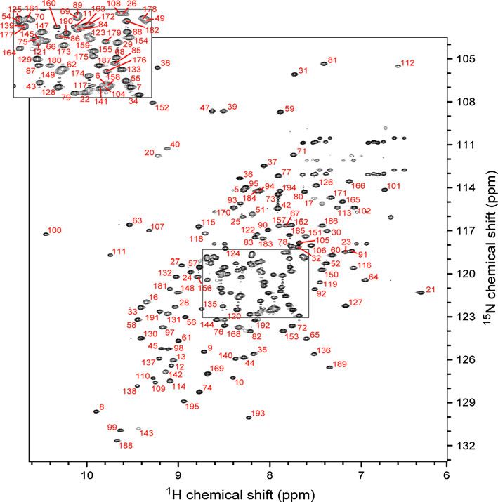
58                                                                                                 J. W. Werner-Allen, P. Zhou

transcription reinitiation that tethers the promoter and ter-   Ssu72 construct contains an N-terminal His tag followed by
minator regions of a gene. NMR should provide a crucial         a thrombin cleavage site. Proteolytic removal of the His tag
tool for mapping the protein–protein interaction sites of       leaves three extra residues N-terminal to the natural protein
Ssu72 and for understanding how these binding events            sequence, which are disordered and do not contribute peaks
affect the structure and dynamics of the enzyme in order to     to the 1H/15N-HSQC-TROSY spectrum. In the figures and
modulate its role in transcription.                             discussion below, residue numbering corresponds to the
                                                                natural protein sequence. NMR samples for resonance
                                                                assignment were prepared by extensive buffer exchange
Methods and experiments                                         into 25 mM sodium phosphate pH 8.0, 25 mM KCl, 2 mM
                                                                dithiothreitol, with either 5 or 100% D2O, and brought to a
Drosophila melanogaster Ssu72 protein was expressed and         final protein concentration of *1 mM. NMR samples for
purified as previously described (Werner-Allen et al. 2011).    titrations with inorganic phosphate were prepared in 25 mM
Isotopically enriched samples were prepared from cells          Tris–HCl, 25 mM KCl, 2 mM dithiothreitol and 5% D2O.
grown in M9 minimal media with 15N-NH4Cl and 13C-                   NMR data were collected on 600 and 800 MHz Varian
glucose as the sole nitrogen and carbon sources. Perdeu-        Inova spectrometers equipped with triple-resonance, cryo-
terated Ssu72 was expressed in D2O M9 minimal media             genically-cooled probes at 30°C. FIDs were processed with
with 15N-NH4Cl and 2H/13C-glucose, with the addition of         NMRPIPE (Delaglio et al. 1995) and datasets were ana-
85 mg [3-2H] 13C-a-ketoisovalerate and 50 mg [3,3-2H2]          lyzed with CARA (Keller 2004).
13
   C-a-ketobutyrate *1 h prior to induction for selective
protonation of ILV methyl groups (Goto et al. 1999). A
sample prepared from 10% 13C-glucose M9 minimal media           Assignments and data deposition
was used to stereospecifically assign valine and leucine
methyl groups (Neri et al. 1989). All isotopes were pur-        Backbone amide resonances of phosphate-bound D.
chased from Cambridge Isotope Laboratories, Inc. The            melanogaster Ssu72 were assigned with a 2H/13C/

Fig. 1 Assigned 1H/15N-
HSQC-TROSY spectrum of
phosphate-bound
D. melanogaster Ssu72
collected at 800 MHz and 30°C

123
1 13 15
H, C and N backbone and side-chain resonance 59
15
N-labeled protein sample using a suite of TROSY- were confirmed through analysis of a 4-D NH–NH
based, 3-D triple-resonance experiments—HNCO, the diagonal-suppressed TROSY-NOESY-TROSY experi-
‘just-in-time’ HN(CA)CO (Werner-Allen et al. 2006), ment (Werner-Allen et al. 2010), and through resonance
HNCA, HN(CO)CA, HN(CA)CB, and HN(COCA)CB— connectivity in TROSY-based HN(CA)HA and HN(CO-
and the PACES algorithm to identify resonance con- CA)HA experiments collected with a 13C/15N-labeled
nectivity (Coggins and Zhou 2003). Amide assignments sample. Overall, every identifiable backbone amide peak
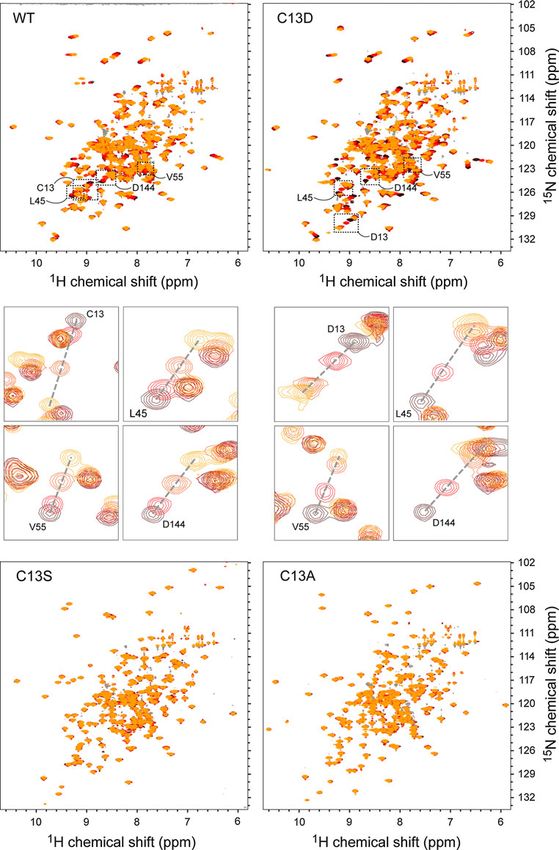
Fig. 2 The effect of active site
mutations on phosphate binding.
The phosphate titration of WT
dSsu72 is shown alongside
identical titrations of C13D,
C13S and C13A mutants.
Brown to yellow coloring
denotes 1H/15N-HSQC-TROSY
spectra for apo enzyme to a 25:1
molar ratio of phosphate to
enzyme, respectively. Close-up
views of peak perturbations for
four residues in the WT and
C13D titrations are shown
below the full spectra
12360 J. W. Werner-Allen, P. Zhou
in the 1H/15N-HSQC-TROSY spectrum was assigned interaction with phosphate. Furthermore, the set of per-
(Fig. 1), representing 93% (177 of 190) of the non-pro- turbed residues is very similar to the set of residues per-
line residues in Ssu72. Residues without amide signals turbed by titration of a catalytically-dead C13D/D144N
are found in flexible regions, including the N-terminus Ssu72 mutant with synthetic pCTD peptide (Werner-Allen
(residues 1–3) and the active site loop (residues 14–15 et al. 2011), suggesting that phosphate binding alone drives
and 18–19). Secondary structure predictions based on 1H an analogous conformational change.
and 13C backbone chemical shifts agree well with the Next, we tested C13A, C13S and C13D Ssu72 mutants
secondary structures observed in the crystal structure for phosphate binding. While C13D Ssu72 exhibited
(Supplemental Figure 1). The perdeuterated NMR sample phosphate-induced chemical shift perturbations similar to
also contained protonated valine c methyls and leucine the WT enzyme, neither C13A nor C13S showed any
and isoleucine d methyls, which were assigned with a perturbations (Fig. 2 and Supplemental Figure 2B). Addi-
4-D HC(CCO)NH experiment. tionally, no perturbations were observed in a titration of
Side-chain resonances were assigned using a 3-D HCCH C13S dSsu72 with a synthetic pS5 CTD peptide (data not
TOCSY experiment, which worked well for shorter side- shown). Notably, a catalytic cysteine to serine mutation in
chains with efficient magnetization transfer. Longer side- protein tyrosine phosphatase 1B (C215S PTP1B) has been
chain assignments were confirmed and extended using intra- reported to dramatically distort the apo conformation of the
residue NOEs in a 1H/13C-NOESY dataset. Aromatic ring 1H active site loop, causing it to flip open into the substrate-
and 13C resonances were assigned using a structure-guided binding space (Scapin et al. 2001). It is possible that the
analysis of NOEs in both aromatic and aliphatic 3-D 1H/13C- loss of a negatively charged catalytic side-chain in the
NOESY datasets; however, several of these ring resonances C13A and C13S mutants severely alters the conformation
were broadened or missing. Methyl groups of valine and and dynamics of the active site loop of Ssu72, rendering it
leucine residues were stereospecifically assigned using unable to bind substrate effectively. Interestingly, a recent
fractional 13C-labeling (Neri et al. 1989). As a result of structure of the catalytic cysteine to serine mutant of
backbone and side-chain resonance assignment, *82% of human Ssu72 in a ternary complex with pCTD substrate
the protons in Ssu72 were assigned, including 95% of the and the scaffolding protein symplekin (Xiang et al. 2010)
backbone protons and 77% of side-chain protons, along with shows that the active site loop properly recognizes the
80% of the 13C and 15N atoms. These chemical shifts have substrate phosphate group. Therefore, the requirement of a
been deposited in the BioMagResBank database (http:// negatively charged catalytic side-chain may be a species-
www.bmrb.wisc.edu) under BMRB accession number specific characteristic of D. melanogaster Ssu72. Alterna-
17510. tively, the active site loop of the human mutant may be
We also used NMR titrations to explore the phosphate- stabilized by the allosteric association with its symplekin
binding properties of wild-type Ssu72 and mutant enzymes binding partner in the ternary crystal complex.
with alanine, serine, and aspartate replacements of the
catalytic cysteine residue (C13). The catalytic cysteine is Acknowledgments This research was supported by NIH grant
GM079376 to P. Zhou. JWW is the recipient of the Kamin
part of a conserved CX5R motif also found in protein Fellowship.
tyrosine phosphatases. The C13 side-chain is held as a
negatively charged thiolate group and initiates dephos-
phorylation through nucleophilic attack of the substrate
References
phosphorous atom, while the five following residues form
the phosphate-binding active site loop. Together with the Coggins BE, Zhou P (2003) PACES: protein sequential assignment by
side-chain of the conserved arginine (R19), the backbone computer-assisted exhaustive search. J Biomol NMR 26(2):
amides of the active site loop hold the substrate phosphate 93–111
Delaglio F, Grzesiek S, Vuister GW, Zhu G, Pfeifer J, Bax A (1995)
group in place with strong electrostatic interactions. NMRPipe: a multidimensional spectral processing system based
Catalysis by Ssu72 also requires an aspartate residue on UNIX pipes. J Biomol NMR 6(3):277–293
(D144) on a flexible loop positioned near the active site Goto NK, Gardner KH, Mueller GA, Willis RC, Kay LE (1999) A
(‘the aspartate loop’), which protonates the leaving phos- robust and cost-effective method for the production of Val, Leu,
Ile (delta 1) methyl-protonated 15 N-, 13C-, 2H-labeled proteins.
phate to regenerate the enzyme. Surprisingly, we observed
J Biomol NMR 13(4):369–374
a large number of significant perturbations upon titration of Keller R (2004) The computer aided resonance assignment tutorial.
wild-type Ssu72 with inorganic phosphate (Supplemental CANTINA Verlag, Goldau
Figure 2A). Given the small size of the phosphate group Neri D, Szyperski T, Otting G, Senn H, Wuthrich K (1989)
Stereospecific nuclear magnetic resonance assignments of the
and the large number of residues affected, it is likely that
methyl groups of valine and leucine in the DNA-binding domain
these perturbations are caused by conformational changes of the 434 repressor by biosynthetically directed fractional 13C
associated with phosphate binding, rather than direct labeling. Biochemistry 28(19):7510–7516
1231 13 15
H, C and N backbone and side-chain resonance 61
Scapin G, Patel S, Patel V, Kennedy B, Asante-Appiah E (2001) The Werner-Allen JW, Lee CJ, Liu P, Nicely NI, Wang S, Greenleaf AL,
structure of apo protein-tyrosine phosphatase 1B C215S mutant: Zhou P (2011) cis-Proline-mediated Ser(P)5 dephosphorylation
more than just an S ? O change. Protein Sci 10(8):1596–1605 by the RNA polymerase II C-terminal domain phosphatase
Werner-Allen JW, Jiang L, Zhou P (2006) A ‘just-in-time’ Ssu72. J Biol Chem 286(7):5717–5726
HN(CA)CO experiment for the backbone assignment of large Xiang K, Nagaike T, Xiang S, Kilic T, Beh MM, Manley JL, Tong L
proteins with high sensitivity. J Magn Reson 181(1):177–180 (2010) Crystal structure of the human symplekin-Ssu72-CTD
Werner-Allen JW, Coggins BE, Zhou P (2010) Fast acquisition of phosphopeptide complex. Nature 467(7316):729–733
high resolution 4-D amide–amide NOESY with diagonal
suppression, sparse sampling and FFT-CLEAN. J Magn Reson
204(1):173–178
123You can also read