Brain Structure Morphology After Being Fixated With Ethanol on Electron Microscope - SciELO
←
→
Page content transcription
If your browser does not render page correctly, please read the page content below
Int. J. Morphol.,
38(2):305-308, 2020.

                Brain Structure Morphology After Being Fixated
                     With Ethanol on Electron Microscope

                                     Morfología de la Estructura Cerebral Después de la
                                        Fijación con Etanol en Microscopio Electrónico

                                      Arni Kusuma Dewi1; Chairul Anwar2 & Yoshihiro Komohara3

DEWI, A. K.; ANWAR, C. & KOMOHARA, Y. Brain structure morphology after being fixated with ethanol on electron microscope.
Int. J. Morphol., 38(2):305-308, 2020.

         SUMMARY: Fixation is one of the processes in preparing histology and pathology. The common material for fixation is buffered
formalin including paraformaldehyde. However, the effect of the damaged cells, which is fixed for a long time, causes the research for
other fixation materials to become necessary. In addition, paraformaldehyde is also harmful to human body and natural environment.
Ethanol is one of the alternative fixation materials, which has been used for two hundred years. It has been used for many purposes, both
in routine staining and immunohistochemistry. Nonetheless, no research confirms its effect on the electron microscope. The authors studied
the effect of 50 % of ethanol on the cell membrane, organelles, and nucleus of Purkinje cells (Neuron purkinjense) observed on a light
microscope and Transmitted Electron Microscope (TEM). Then it was compared to buffered formalin. In the light microscope, it shows that
both of fixations have no different effects of the morphology of the cell membrane, cytoplasm, the nucleus of Purkinje cells and the
neutrophils. We assume that our 50 % of ethanol concentration is almost the same as BF 10 % in the ability of hardening tissue and color
absorption based on the previous study. In TEM, the structure of the cell membrane, organelles, and cytoplasm of Purkinje cell look broken
in the cerebellum of 50 % of ethanol except for the nucleus. There was no significant difference diameter of the nucleus. It happened in
general because of the shrinkage effect of ethanol. However, the authors recommend using 50 % of ethanol for routine staining.

        KEY WORDS: Fixation, Brain, Organelle, Electron Microscope

INTRODUCTION

       Fixation is one of the processes in preparing histology                        The alternative fixation material that can be used to store
and pathology. The process in fixation includes the selection                specimens for a long period is alcohol. Alcohol has been used as
of the materials, the concentration used, the method, and                    a fixation material since 1922 by Freudenthal. His research was
the length of time required. It is an area of research that                  re-summarized in 1947 (Freudenthal). In general, he
continues to grow because it determines the results as                       recommended ethanol as a fixation material for all tissues and
expected.                                                                    cells except for fat staining. The research continues to find the
                                                                             effect of alcohol on each tissue since 1957 (Jarrett & Hardy, 1957).
       The use of buffered formalin (BF) as fixation mate-                   Then, in 1986, ethanol began to be investigated as fixation ma-
rial was found by F. Blum in 1893 (Freudenthal, 1947) and                    terial for histochemical staining (Battifora & Kopinski, 1986).
has been used in common practice until now. BF is widely
used because of its capability to hardening tissue without                           Ethanol is an alcohol group that is more widely used.
considering the size, the solubility in salt, and the capability            Based on several studies, ethanol provides excellent protection
to be used for various research purposes (Fish, 1896).                      for alkaline phosphatase enzymes, coir collagen, DNA, RNA,
However, the use of this fixation material for an extended                  and lipases (Stafford & Atkinson, 1948; Jarrett & Hardy; Su
period will cause tissue damage and is not suitable for                     et al., 2004). Based on the research, some modifications of
anatomy museum where which is stored in cold temperatures                   the technique to approve the continuous growth until now.
(Fish; Burke, 1933).                                                        However, none uses an electron microscope.

1
  Department of Health, Faculty of Vocational Studies, Universitas Airlangga Campus B, Surabaya, East Java, Indonesia.
2
  Department of Histology, Faculty of Veterinary Medicine, Universitas Airlangga (Campus C), Mulyorejo street, Surabaya, East Java, Indonesia.
3
  Department of Cell Pathology, Faculty of Live Science, Kumamoto University 1-chome-1-1 Honjo, Chuo-ku, Kumamoto, 860-0811, Japan.

                                                                                                                                                 305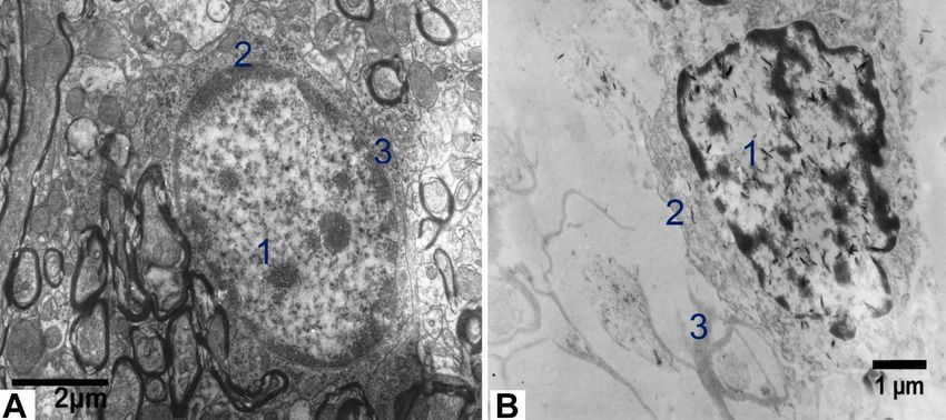
DEWI, A. K.; ANWAR, C. & KOMOHARA, Y. Brain structure morphology after being fixated with ethanol on electron microscope. Int. J. Morphol., 38(2):305-308, 2020.
MATERIAL AND METHOD Karnovsky solution was manufactured as a kit by Electron
Microscope Science (EMS) in Philadelphia. The authors
observed the structure of cell membranes, organelles, and
This study utilized healthy male balb/c mice (Rattus nucleus of the neuron cells. The authors would like to prove if
norwegicus) as the experimental animals, weighing 200-300 the denaturation effect of ethanol occurred in the nucleus by
g. The maintenance and treatment of the experimental analyzing the diameter by image raster3 tool and comparing
animals and the research process have received an ethical it with BF.
conduct certificate from the Ethics Commission of the
Faculty of Medicine, Universitas Airlangga. Most of the
research were conducted in the Department of Health, RESULTS
Faculty of Vocational Studies, Universitas Airlangga.
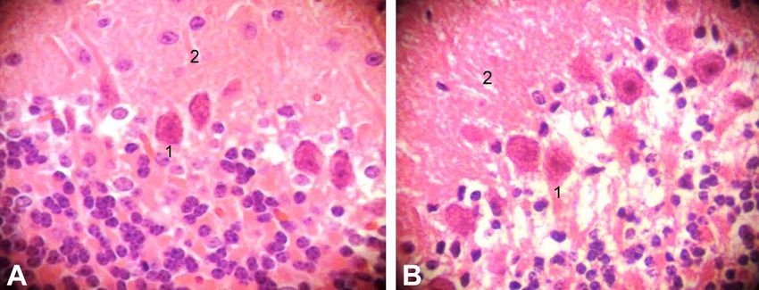
The two brain specimens were fixated by immersion There is a different result seen by a light microscope
for 24 hours at room temperature, each with 50 % of ethanol and TEM. In the light microscope. It shows that both of
and 10 % BF. Then, one of the hemispheres was processed fixation has no different effect of the morphology of the cell
with routine coloring for the light microscope and the other membrane, cytoplasm, the nucleus of Purkinje cells (Neuron
for Transmitted Electron Microscope (TEM). purkinjense), and no neutrophils are seen, as seen in Figure
1. The cell membrane and cytoplasm stained red and nucleus
The method by light microscope. The cerebellum of the blue. The neutrophils in both objects were sharp. In TEM,
hemisphere fixated with 50 % of ethanol was cut into 1 cm3 the structure of the cell membrane, organelles, and cytoplasm
to get embedded in paraffin, cleared dan stained using of Purkinje cells look broken in the cerebellum fixated with
Hematoxylin-Eosin (HE). The same process for the 50 % of ethanol, except for the nucleus (Fig. 2).
cerebellum of the hemisphere fixated in 10 % BF.
The authors analyzed the diameter of the nucleus of
Method and Analysis by TEM. Both cerebellum of in 50 % Purkinje cells in both fixations (Table I). The data were tested
of ethanol and 10 % BF were cut into one mm3 to get fixation for normal distribution with the Shapiro-Wilk test. Because
to Karnovsky 1⁄2 for 720 hours at 4 °C. This process took 720 the data were not normal, the authors continued with the
hours because the laboratory of Electron Microscope in Mann-Whitney test with IBM Statistic 20. It showed that
Eijkman Institute Universitas Indonesia-Cipto Mangunkusumo there was no significant difference in the diameter between
Hospitals for later steps must be removed at that time. The 50 % of ethanol and 10 % BF treatments (p>0.05).
Fig. 1. Cerebellum observed in a light microscope using 400 magnification, HE staining. A. 10 % BF; B. 50 % of ethanol (1. Purkinje
cell, considering to cell membrane and nucleus, 2. Neutrophil).
Table I. Description of the Effects of Fixation in TEM.
Variable Mean ± SD (µm) Mann-Whitney test
Nucleus diameter on BF fixation 10086.50±116.67 (n=2) Asymt Sig = 0.121
Nucleus diameter on ethanol fixation 14808.52±176.27 (n=2)
306DEWI, A. K.; ANWAR, C. & KOMOHARA, Y. Brain structure morphology after being fixated with ethanol on electron microscope. Int. J. Morphol., 38(2):305-308, 2020.
Fig. 2. Cerebellum observed in TEM. A. 10 % BF; B. 50 % of ethanol (1. Nucleus of Purkinje Cells, 2. Cytoplasm (consider to
organelles), and 3. cell membrane)
DISCUSSION
The results of the cerebellum preparation with HE the morphology would not happen if the ethanol was used
staining between 50 % of ethanol and 10 % BF were not as a wash after fixation (Llewellyn-Smith & Minson,
different. Recent research found that 50 % of ethanol had 1992).
the capability to hardening tissue to increase contrast in
micro-CT (Patzelt et al., 2019). Based on these findings, The morphology and diameter of the nucleus did
the authors assume that 50 % of ethanol concentration is not seem to differ between 50 % of ethanol and BF. The
almost the same as BF 10 % in terms of their capability of authors tried measuring the diameter of the nucleus Purkinje
hardening tissue and color absorption. Some studies prefer cells and found no difference in the nucleus diameter based
using ethanol with a concentration of 70 % for routine or on the statistical results. However, preliminary research
immunohistochemical staining (Freudenthal; Jarrett & proved that the RNA yield from brain tissue fixated with
Hardy; Schutte et al., 1987; Su et al.). However, there are BF could only be protected by 5 % on average, compared
also those who claim that ethanol shows better results to 70 % ethanol (Su et al.). BF makes a cross-linking
(Battifora & Kopinski). Therefore, until now, research is reaction between nucleotides and proteins so that less
still developing to determine the best protocol for ethanol perfect separation and high temperatures can reduce the
concentration and duration of fixation. detected RNA yield (Su et al.).
Different results were indicated in the observation
with TEM. The cell membrane and organelles in the ACKNOWLEDGMENT
Purkinje cell cytoplasm at 50 % of ethanol appeared to be
degraded even though the storage applied Karnovsky 1⁄2
at 4 °C for 720 hours. Research showed that storing The authors would like to extend their utmost
specimens in Karnovsky 1⁄2 more than six months did not gratitude to the Univesitas Airlangga and Eijkman Institute
cause any damage to the tissue and cell structures (Mount Universitas Indonesia-Cipto Mangunkusumo Hospitals to
et al., 1997). According to Patzelt et al., the effect of 50 % support the facilities. The authors also would like to
of ethanol shrinkage is very low, compared to other ethanol acknowledge to Department of Cell Pathology and the
fixation concentration. This notion had never been Department of Histology, the Faculty of Live Science,
conformed with an electron microscope. However, the Kumamoto University for supporting them with the
previous study showed that the effect of ethanol to damage knowledge.
307DEWI, A. K.; ANWAR, C. & KOMOHARA, Y. Brain structure morphology after being fixated with ethanol on electron microscope. Int. J. Morphol., 38(2):305-308, 2020.
DEWI, A. K.; ANWAR, C. & KOMOHARA, Y. Morfología Patzelt, M.; Mrzilkova, J.; Dudak, J.; Krejci, F.; Zemlicka, J.; Karch, J.;
Musil, V.; Rosina, J.; Sykora, V.; Horehledova, B.; et al. Ethanol fixation
de la estructura cerebral después de la fijación con etanol en method for heart and lung imaging in micro-CT. Jpn. J. Radiol.,
microscopio electrónico. Int. J. Morphol., 38(2):305-308, 2020. 37(6):500-19, 2019.
Schutte, B.; Reynders, M. M.; Bosman, F. T. & Blijham, G. H. Effect of
RESUMEN: La fijación es uno de los procesos en la tissue fixation on anti-bromodeoxyuridine immunohistochemistry. J.
preparación de muestras para histología y patología. El mate- Histochem. Cytochem., 35(11):1343-5, 1987.
rial más común para la fijación es la formalina tamponada. Sin Stafford, R. O. & Atkinson, W. B. Effect of acetone and alcohol fixation
and paraffin embedding on activity of acid and alkaline phosphatases
embargo, el daño a las células que se mantienen en formalina
in rat tissues. Science, 107(2776):279-81, 1948.
durante mucho tiempo, hace necesario buscar otros materiales Su, J. M. F.; Perlaky, L.; Li, X. N.; Leung, H. C. E.; Antalffy, B.; Armstrong,
de fijación. Además, el paraformaldehido también es perjudi- D. & Lau, C. C. Comparison of ethanol versus formalin fixation on
cial para el cuerpo humano y el medio ambiente natural. El preservation of histology and RNA in laser capture microdissected brain
etanol es uno de los materiales de fijación alternativos que se tissues. Brain Pathol., 14(2):175-82, 2004.
ha utilizado durante muchos años, con diversos objetivos, tan-
to en la tinción de rutina como en la inmunohistoquímica. Sin
embargo no se ha confirmdo su efecto con microscopio elec-
trónico. Los autores estudiaron el efecto del 50 % de etanol Corresponding author:
sobre la membrana celular, los orgánulos y el núcleo de las Arni Kusuma Dewi
células de Purkinje observados en un microscopio óptico y un Department of Health
microscopio de transmisión electrónico (TEM). Luego se com- Faculty of Vocational Studies
paró con la formalina tamponada. En el microscopio óptico se Universitas Airlangga (Campus B)
observó que ambas fijaciones no tienen efectos diferentes a la Jalan Dharmawangsa Dalam No. 28-30
morfología de la membrana celular, el citoplasma, el núcleo Surabaya
de las células de Purkinje y los neutrófilos. Suponemos que East Java
nuestra concentración de 50 % de etanol es casi la misma que INDONESIA
BF 10 % en la capacidad de endurecer el tejido y la absorción
de color según el estudio anterior. En TEM, la estructura de la
membrana celular, los orgánulos y el citoplasma de la célula de Email: arni-k-d@vokasi.unair.ac.id
Purkinje presentaban daño en el cerebelo con un 50 % de etanol,
a excepción del núcleo. No hubo diferencia significativa en el
diámetro del núcleo. En general lo anterior se debió al efecto Received: 21-05-2018
de contracción del etanol. En conclusión los autores recomien- Accepted: 24-07-2019
dan usar 50% de etanol para la tinción de rutina.
PALABRAS CLAVE: Fijación; Cerebro; Organelo;
Microscopio electrónico.
REFERENCES
Battifora, H. & Kopinski, M. The influence of protease digestion and
duration of fixation on the immunostaining of keratins. A comparison
of formalin and ethanol fixation. J. Histochem. Cytochem., 34(8):1095-
100, 1986.
Burke, F. V. The pH of formalin - A factor in fixation: adjustment and
stabilization of the hydrogen ion concentration of formalin solutions.
Am. J. Pathol., 9(6):915-20, 1933.
Fish, P. A. The use of formalin in neurology. Trans. Am. Microsc. Soc.,
17:319-30, 1896.
Freudenthal, W. Recent Advances in Clinical Pathology. London, Churchill,
1947. pp.388.
Jarrett, A. & Hardy, J. A. The value of alcohol for fixation of skin. Stain
Technol., 32(5):225-33, 1957.
Llewellyn-Smith, I. J. & Minson, J. B. Complete penetration of antibodies
into vibratome sections after glutaraldehyde fixation and ethanol
treatment: light and electron microscopy for neuropeptides. J.
Histochem. Cytochem., 40(11):1741-9, 1992.
Mount, S. L.; Schwarz, J. E. & Taatjes, D. J. Prolonged storage of fixative
for electron microscopy: effects on tissue preservation for diagnostic
specimens. Ultrastruct. Pathol., 21(2):195-200, 1997.
308You can also read