EFSUMB History of Ultrasound
←
→
Page content transcription
If your browser does not render page correctly, please read the page content below
Ultrasound of the liver ….              04.01.2021 10:23                                 1

                  EFSUMB History of Ultrasound
                                 Editor: Christoph F. Dietrich

                                    Ukraine (UA)

                 Oleh Dynnyk1, Eugen Barannik 2, Vladimir Medvediev 3

1Director,   Institute of Elastography, PhD, MD, Kyiv (obdynnyk@gmail.com).
2Professor,   Karazin Kharkiv National University, UA (evgenij.a.barannik@karazin.ua).
3Professor,   Department of Radiology, Shupyk National Medical Academy of Postgraduate
Education, Kyiv (vmedvediev@gmail.com).

Corresponding author:
1Anatoliy    Marusenko, chief designer, Ultrasign Ltd., Kharkiv
2
Yuriy Chirkov, MD, PhD, Oberig Ltd., Kyiv
3
Valeriy Kalashnikov, Assistant professor, Department of Ultrasound Diagnoctic, Kharkiv
National Medical Academy of Postgraduate Education, Kharkiv
Acknowledgment:
Authors acknowledge colleagues who have provided comments and corrections in draft:
Irina Bekalo, MD, sekretar of the UAUDS, Intosana Ltd., Odessa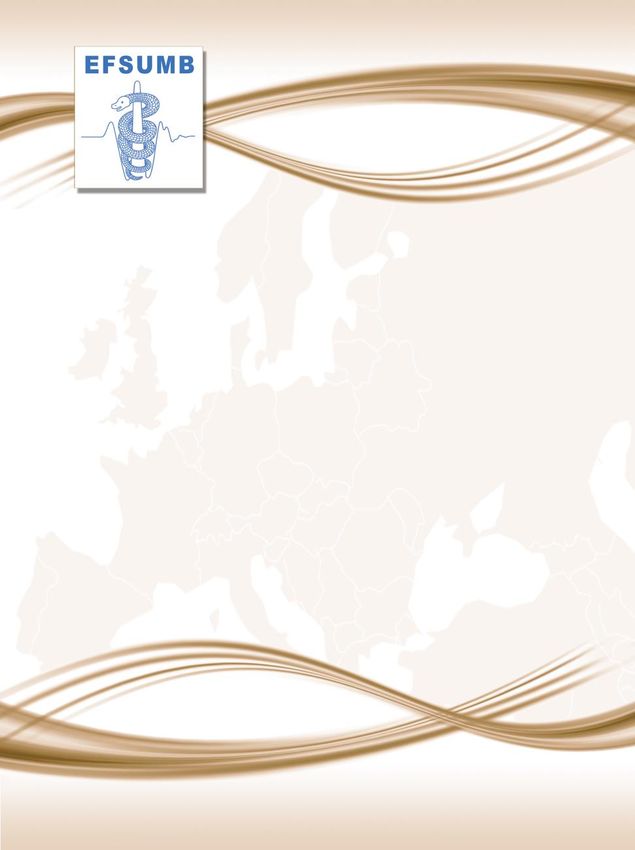
EFSUMB History of Ultrasound Ukraine     04.01.2021 10:23                                       2

Introduction

The development of ultrasound diagnostics (USD) in Ukraine began in the 70s of the 20th
century. It is worth considering 2 fields: 1 - the clinical application of ultrasound (US) in
medicine by doctors and 2 - technical, engineering solutions, which eventually led to the
creation of modern domestic US devices with excellent B-mode, sensitive Doppler, all types
of elastography and steatometry.

Clinical implementation of ultrasound

In the USSR in the clinic, the beginning of the use of US was laid in neurology in the 70s on the
basis of the application of the A-method for visualizing the width of the 3rd ventricle of the
brain and other intracranial structures - echoencephaloscopy. The A-method is based on
visualizing the amplitude modulation curve of ultrasonic wave reflections on the device
screen. These reflections of various amplitudes occurred at the boundaries of tissues with
contrasting acoustic impedance (skull bones, dura mater, brain matter and cerebrospinal fluid
in the cerebral ventricles). US insonation was performed by transtemporal approach. The
signal of the highest amplitude M-echo was obtained from reflections of the midline
structures of the brain, located perpendicular to the US beam in the sagittal plane (third
ventricle, pineal gland, crescent of the dura mater, pallid membrane, septum). In the case of
an increase in intracranial pressure, the cavity of the 3rd ventricle expanded. In case of mass
pathology, the displacement of the median structures of the brain relative to the signals from
the skull and M-echo was assessed (a symptom of dislocation of the median structures). A-
method of visualization with the Echoencephaloscope ЭЭС-01 device (USSR) was also used for
abdominal visualization. It was first mastered in 1983 by Dr. Dynnyk O.B. at the Department
of Internal Diseases No. 1 of the Bogomolets Kyiv Medical Institute and later, since 1985, used
the B-mode on the Aloka SSD-256, Japan (Department of Clinical Pathophysiology, Kavetsky
Institute of Experimental Oncology, National Academy of Sciences of Ukraine).
In 1981, the B-method began to be applied by the pioneers of US in Ukraine from the group
of Professor Medvediev V.E. (fellow workers of the Laboratory of Functional Methods of
Studying the Digestive Organs of the Kyiv Research Institute of Clinical and Experimental
Surgery of the Academy of Medical Sciences of Ukraine (Director - Academician O.O.
Shalimov). They used US devices with mechanical sector scanning - Aloka SSD-120 (Japan) and
EFSUMB History of Ultrasound Ukraine 04.01.2021 10:23 3
linear electronic scanning - Aloka SSD- 630 (Japan). At the same institute, a group of doctors:
Grigorash G.A. (head) and Hooch A.О. began to use Doppler for vascular examination (1987 -
Pocket Dopp, 1991 - Ultramark 9 (ATL, USA) and Multi-Dop (Compumedics DWL, Germany).
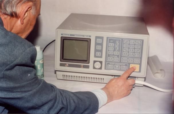
Intervention
Prof. Medvedev’ team for the first time in the USSR since 1983 applied methods of
interventional ultrasound in solving problems, first of all, in abdominal surgery: Medvedev V.E.
(leader), Tarasyuk B.A., Novikova M.М., Terzova T.B., Buchneva L.V (Figure 1).
Figure 1 Pioneers of ultrasound in Ukraine.
Professor V.E. Tarasyuk B.A. Novikova M.М. Terzova T.B.
Medvedev
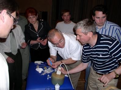
Since 1996 in Odessa doctor Boyko A.V. applied interventional methods of treatment of
parasitic (echinococcus) and nonparasitic liver cysts under ultrasound control. Since 1993, a
group of surgeons using interventional ultrasound began to work in the regional Donetsk
diagnostic center - DDC (director BD Kolesnikov): Zubov О.D. (leader), Chirkov Yu.E.,
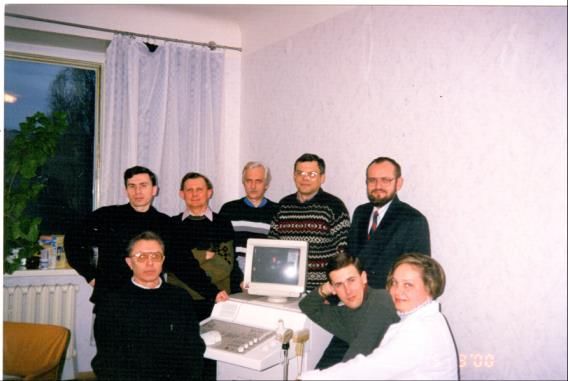
Belonenko G.A., Uspensky D.A., Cherednichenko S.I., Gubanov D.M (Figure 2).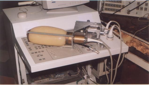
EFSUMB History of Ultrasound Ukraine       04.01.2021 10:23                              4

Figure 2      A group of surgeons using interventional US at the DDC.

Professor Konjkova M.B. founded her school of interventional ultrasound in Donetsk.
(Department of Oncology, Radiation Methods of Diagnostics and Treatment of Donetsk
National Medical University) (figure 3).

Figure 3      Professor Konjkova M.B.

In Zaporizhzhya, the methods of intervention under the US control were actively introduced
by Associate Professor Fedusenko O.A. (Department of Radiation Diagnostics of the
Zaporizhzhya Academy of Postgraduate Education).
EFSUMB History of Ultrasound Ukraine     04.01.2021 10:23                                       5

Endocrinology

In Kyiv, a group of doctors led by Professor Epshtein E.V. was engaged in the issues of US
imaging of the thyroid gland and fine-needle puncture aspiration biopsy at the Research
Institute of Endocrinology and Metabolism of the Academy of Medical Sciences of Ukraine.
This direction developed especially intensively after the accident at the Chernobyl nuclear
power plant in 1986. This event contributed to the intensive introduction of US, especially in
the diagnosis of the state of the thyroid gland after exposure to nuclides of millions of
Ukrainians. Then, soon, the intervention of the thyroid gland under the control of US was
introduced in many cities of Ukraine (devices from Toshiba and Aloka (Japan), Siemens
(Germany), GE (USA), B / K (Denmark), Kretz Technic (Austria)).

Pediatrics

Neurosonography began its development with the delivery of 20 devices to newborn
departments in the cities of Ukraine - Microimager-1000 with sector sensors in 1987. The
pioneers of neurosonography were the doctor Makarova О.О. (“OKHMATDET” hospital and
then the Ukrainian Medical Center for the Rehabilitation of Children with Organic Damage of
the Nervous System of the Ministry of Health of Ukraine) and Associate Professor Krygin Yu.A.
(Department of Radiation Diagnostics, Shupik National Academy of Postgraduate Education).
US of the thymus gland in pediatrics was developed thanks to the initiative of the professor of
the Department of Radiology of Shupik NMAPE O.A. Gonchar and the chief radiologist in Kyiv
Urina L.K. Rapid introduction was received by US of the hip joint in newborns and young
children by the method of Graff (1985) modified by Research Institute of orthopedics and
traumatology of the Academy of Medical Sciences of Ukraine (Vovchenko A.Ya., 1995).
Fundamental achievements of pediatrics US were made by the team of the Research Institute
of Pediatrics, Obstetrics and Gynecology of the Academy of Medical Sciences of Ukraine
(Professor Lukyanova I.S.).

Musculoskeletal US

In Ukraine the development of the musculoskeletal US has led to the formation of several
schools with leaders: in Kyiv - Vovchenko A.Ya., Urina L.K., Golovko T.S.; in Kharkiv - Abdullaev
EFSUMB History of Ultrasound Ukraine     04.01.2021 10:23                                      6

R.Ya.; in Sumy - Gapchenko V.V., in Lviv - Kucher A.R. A special direction was US of the spine
according to the method of Kindzersky A.Yu.

Oncology

The introduction of oncology US took place through the efforts of specialists from the Kyiv
Research Institute of Clinical and Experimental Surgery of the Academy of Medical Sciences of
Ukraine (Kyiv), the National Cancer Institute of the Ministry of Health of Ukraine (Kyiv), the
Institute of Medical Radiology of the Academy of Medical Sciences of Ukraine (Kharkiv),
Donetsk Medical University (Donetsk), etc.

Doppler

A team of doctors from the Romodanov Neurosurgery Institute, National Academy of Medical
Sciences of Ukraine made a significant contribution to the development of US imaging
methods in neurology and neurosurgery. Doppler research methods have been actively
introduced since 1993 on the basis of the Functional Diagnostics Department of the Institute.
First devices: Transcranial Doppler - "TC 2-64 EME" (Germany), "Sonomed 300" (Russia) and
since 1995 - "Multigon 500M" (USA) with conventional Doppler, Transcranial Doppler and
bilateral Transcranial Monitoring modes (doctors Globa M.V., Mikhalj A.V. and Vaschenko
V.V.). Since the 2000s, the department has been equipped with duplex US diagnostic systems
ode "Sonoline G-50" (Siemens, Germany, 2003), "Toshiba Aplio MX" (Toshiba, Japan, 2015),
"Toshiba Aplio 400" (Toshiba, Japan, 2018). Since 2015, intraoperative Doppler studies have
been introduced for transcranial surgeries to turn off arterial aneurysms of the brain - contact
microvascular Doppler with 16 MHz transducers (Angiodin PC, Bioss, Russia). In 1998, the
Medical Scientific and Practical Association "Medbud" introduced conventional methods of
duplex scanning of the vessels, and especially: kidneys, splanchnic vessels (diagnosis of renal
and portal hypertension), HABR by HDI-5000 (ATL, USA). It introduced transcranial Doppler
monitoring of cerebral blood flow and embolodetection by Angiodin ПК (Bioss, Russia). As well
as TEE and stress echocardiography with dobutamine on the HDI-5000 apparatus (ATL, USA)
(doctors Dynnyk O.B. (head), Korichensky О.М., Senatorova L.М. and Mostovoy S.E.
EFSUMB History of Ultrasound Ukraine     04.01.2021 10:23                                    7

Cardiology

Professor Stadnyuk L.A. began to perform Echocardiography since 1979 using US machines of
Smith Kline Instruments at the Strazhesko Research Institute Cardiology of the Academy of
Medical Sciences of Ukraine (Kyiv). After then he worked on the US machines of ATL, Toyota,
Kontron (Figure 4).

Figure 4      The pioneer of echocardiography in Ukraine - Professor Stadnyuk L.A.

Transesophageal echocardiography (TEE) in Ukraine was first introduced by doctors Ivaniv
Yu.A. (Lviv), Beshlyaga V.M. (Amosov Institute of Cardiovascular Surgery of the National
Academy of Medical Sciences of Ukraine, Kyiv). Professor Ivaniv Yu.A. heads the Department
of Radiation Diagnostics, Faculty of Postgraduate Education, Danylo Galitsky Lviv National
Medical University.
The school of the improvement of Doppler echocardiography and functional topographic
anatomy of the heart according to the author's methods "ECHO 3P + 4C" and "3D TRIPLAN"
was created by a native from the Cherkassy Cardiological Center, cardiologist Miroshnik M.
(employee of the Hôpital Européen Georges Pompidou PARIS - France). He introduced original
pedagogical techniques, including: virtual 3D modeling of the anatomy of the heart, navigation
on the echosimulator.
Dobutamine stress echocardiography has been actively implemented into routine practice
since 2004 at Medical center "Medbud" (cardiologist Mostovoy S.E.) and in 2007 at the Heart
Institute of the Ministry of Health of Ukraine (Kyiv). The pioneer of the method of myocardial
EFSUMB History of Ultrasound Ukraine 04.01.2021 10:23 8
deformation analysis (Strain Rate) is associate professor Molodan O.V. of the Zaporizhzhya
Academy of Postgraduate Education.
Obstetrics and gynecology
The introduction of US in obstetrics and gynecology practice began in 1993 by Corresponding
Member of the Academy of Medical Sciences of Ukraine, Professor Grechanina E.Yu., being
the head of the Department of Clinical Genetics and Ultrasound Diagnostics at Kharkiv Medical
University.
In the Interregional Center of Medical Genetics and Prenatal Diagnostics (Kryvyi Rig,
Dnepropetrovsk region), for the first time in Ukraine in 1980, genetic amniocentesis was
performed and prenatal diagnostics of chromosomal abnormalities in the fetus was started by
the pioneer of US in prenatal diagnostics Veropotvelyan M.P.
At the base of the Kyiv Regional Hospital, in early 1983, he performed amniocentesis in the
third trimester of pregnancy to determine lung maturity in the fetus using Aloka SSD-202
(Japan) in order to select a safe puncture approach. After 1.5 months training on US in
obstetrics and gynecology in the laboratory of Professor Demidov V.N. at the All-Union Center
for Maternal and Child Health (Moscow) in 1984. Veropotvelyan Mykola began to widely
practice echography at the City Maternity Hospital No. 1 and the Interregional Medical
Genetic Center in Kryvyi Rig. The first US scanners were Toshiba SAL-35 and SAL-77 (Japan),
and for invasive prenatal procedures portable devices Medata ADR 2002 (USA) Toshiba SAL-
32В Dr. Veropotvelyan M. has mastered and implemented the entire arsenal of invasive fetal
procedures performed under ultrasound control - amniocentesis, transcervical and
transabdominal chorionic villus sampling, cordocentesis, cardiocentesis, aspiration of fetal
cysts and intrauterine blood transfusions. Today Veropotvelyan M.P. is the General Director
of the Interregional Center for Medical Genetics and Prenatal Diagnostics in Kryvyi Rig and a
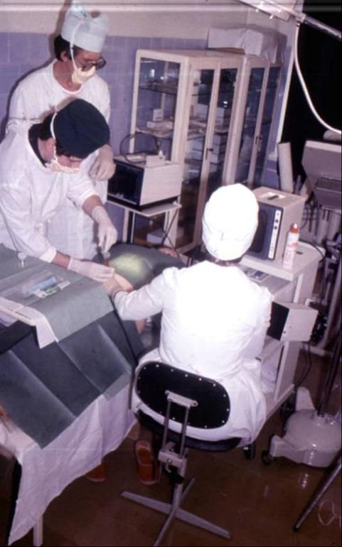
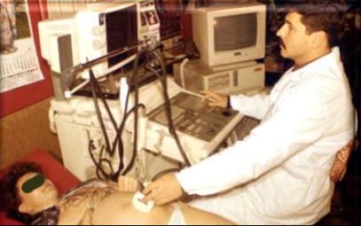
teacher for many young professionals (Figure 5).
Figure 5 Prenatal screening is carried out by Dr. Veropotvelyan M. (a). Amniocentesis
under ultrasound guidance in Interregional Center for Medical Genetics and
Prenatal Diagnostics in Kryvyi Rig (b).
a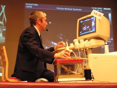
EFSUMB History of Ultrasound Ukraine    04.01.2021 10:23                                      9

b

Ian Donald's school was held in Ukraine twice, the last time in February 2020.

Urology and lithotripsy

On 09.07.1990, a lithotripsy room was opened in the Donetsk Diagnostic Center (DDC, director
B.D. Kolesnikov). Lithotriptor MPL-9000 manufactured by Dornier Medizintechnik GmbH was
used by a team of medical and engineering personnel trained and certified in clinics Stuttgart,
Munich, Ulm (Germany). Doctors urologists Reznikov D.B., Roshchin Yu.V., Fedorishin R.P.,
Reznikov G.D. mastered the basics of US navigation in lithotripsy and biopsy. Since 2007, shock
EFSUMB History of Ultrasound Ukraine      04.01.2021 10:23                                   10

wave lithotripsy has been performed on the “Modulith SLK” lithotripter by “Storz Medical”
(Switzerland). The ultrasound scanner Aloka SSD-1000 (Japan) and the X-ray apparatus "BV
Libra" (Philips, Holland) are used for navigation.
Transperineal prostate biopsy in DDC was performed from 1992 to 1994 on the Aloka SSD-630
device (linear sensor). Transrectal prostate biopsy has been performed since 1994 on devices:
Dornier AI 2200 and Aloka SSD-3500, equipped with a rectal probe. Kidney biopsy under US
control in adults and children is widely introduced by the doctor Chirkov Yu.E. in hospitals in
Donetsk and Kyiv. Today, prostate biopsy is performed with Fusion technology in ten hospitals
in Ukraine (mentor Solodovnik O.V.).

Elastography

Significant development of methods of US elastometry and steatometry became possible in
Ukraine due to the development and production of Ukrainian US devices with these innovative
technologies (firms "Radmir" and "Ultrasign", Kharkiv) (Figure 6).

Figure 6      The team of developers of elastography and steatometry from “Radmir” and
              “Ultrasign”, headed by chief designer Marusenko A.I. and doctor Linska G.V.

Experimental elastography studies on rats and pigs were carried out by different groups. We
used Ukrainian ultrasound devices (Radmir, Ultima SM and Ultrasign, Soneus P7) for shear
wave elastography (SWE) in modeling cirrhosis, fatty liver disease and Guerin's tumor [1-6].
There were collectives of the Department of Clinical Pathophysiology of the Bogomolets
Institute of physiology of the Ukrainian National Academy of Sciences, the Division of
EFSUMB History of Ultrasound Ukraine 04.01.2021 10:23 11 Endocrinology, Bogomolets National Medical University; the National Cancer Institute of the Ministry of Health of Ukraine (Kyiv), Shevchenko National University (Kyiv). Education Physicians have been trained in the basics of US diagnostics 1981 in the Laboratory of Functional Methods for the Study of the Digestive System of the Research Institute of Clinical and Experimental Surgery of the Academy of Medical Sciences of Ukraine (Chief - Professor Medvediev V.E., employees Tarasyuk B.A., Novikova M.M., Terzova T.B., Buchneva L.V.). The systematic teaching of US diagnostics was laid in Kyiv at the Department of Radiology of the Shupik National Academy of Postgraduate Education (head of the department - Professor Koval G.Yu., the course was conducted by Prof. Gonchar A.O., associate professors Yatsyk V.I. and Krygin Yu.A.) since 1989. Now it is the Department of Radiology (head professor Babkina T.M., head of the US course professor Medvediev V.E., teachers - professor Kazarenko T.M., associate professors Volyk N.K., Gladka L.Yu., etc.). The history of education in the field of US diagnostics in Kharkiv began in 2000, when Professor Abdullaev R.Ya. began teaching the fundamentals of US diagnostics for interns of the Department of Radiation Diagnostics of the Kharkiv Academy of Postgraduate Education (KhMAPO). In 2008, Professor Abdullaev R.Ya. created the first and so far the only Department of US Diagnostics in Ukraine at KhMAPO (Figure 7). Figure 7 Professor Abdullaev R.Ya.
EFSUMB History of Ultrasound Ukraine     04.01.2021 10:23                                     12

The teachers of the US Department are associate professors Kalashnikov V.I., Sysun L.A.,
Ponomarenko S.A. and others. Recently, the staff of the department has been working a lot in
the direction of improving Doppler methods in the diagnosis of pathology of various organs
and systems. In particular, the latest research diagnostic methods for vertebral arteries and
cerebral veins, a comprehensive assessment of the state of vascular regulation in various
neurological and cardiovascular diseases have been introduced into clinical practice. A special
stimulus to the development of multimodal thinking of US doctors in Ukraine was played by
the help of the m “Friends of Radiologists of Ukraine” group from the Ukrainian diaspora of
the USA and Canada. They are members of RSNA (O. Baltorovich, L. Volyansky, M. Poznyak, B.
Pollard, etc.). At the suggestion of O. Baltorovich, in the 90s, several ultrasound doctors were
trained at the Education and Research US Institute (director Professor Barry Goldberg) of the
Jefferson Philadelphia University (Pennsylvania, USA). Doctors of this group in Ukraine have
become leaders in the areas of US diagnostics (Ivaniv Yu.A. - TEE, Volyk N.K. - gynecology,
Serdyuk V.O. - endocrinology).
With the aim of teaching the basics of ultrasound in Ukraine, phantom simulators of an original
design were created: for ultrasound intervention (Zubov O.D. and colleagues), for Doppler
(Dynnyk O.B.), for elastography (strain and shear wave elastography) (Dynnik O.B.,
Zhaivoronok M.M.) and a multiparametric/multimodal phantom for US elastography,
steatometry and evaluation of tissue viscosity of the liver and MR steatometry (PDFF) (Dynnyk
O.B., Solodovnik O.V, Omelchenko O.M.). Studies were carried out on devices Canon Aplio
i800, Japan (2D SWE, Propagation, Share Wave Dispersion (SWD), Attenuation Coefficient
(AC)) and Ultrasign Soneus P7, Ukraine (2D SWE, 2D Attenuation Coefficient Measurement
(ACM)) (Figure 8).
EFSUMB History of Ultrasound Ukraine        04.01.2021 10:23                                13

Figure 8      Different phantoms for interventions, Doppler and multiparametric imaging.

 Intervention US Phantom (a)           Doppler Phantom (b)        3D Angio of the Doppler
                                                                  Phantom (c)

 Multiparametric liver phantom: 2D SWE, Propagation, SWD,         ACM by Ultrasign Soneus
 AC by Canon Aplio i800 Liver Package (d)                         P7, Ukraine (e)

The Ukrainian Association of Ultrasound Diagnostics Specialists (UAUDS)

The Ukrainian Association of Ultrasound Diagnostics Specialists (UAUDS) was registered on
May 29, 1998 (No.1032) as an all-Ukrainian public organization. The initiative group of US
doctors of various directions included Medvediev V.E., Sopko S.O., Gordienko E.Yu., Klimenko
O.F., Tarasyuk B.A., Dynnyk O.B., Novikova M.M., Golovko T.S., Lukyanova I.S., Dugan I.V.,
Volyk N.K., Pogodaeva G.A., Globenko T.A., Slobodyanyuk I.M. The first president of UAUDS is
Professor Medvediev V.E., and from 2004 until today – Dynnyk O.B. In April 2006, on the basis
of an agreement, UAUDS became an Associate member of the Association of Radiologists of
the Ukrainian (ARU), and UAUDS members thus joined the European Society of Radiologists
(ESR). Ukrainian US specialists became active participants in radiological meetings and
congresses in Ukraine and the European Congress of Radiology (ECR). On May 29, 2019 UAUDS
became a member of EFSUMB (President - Dynnyk O.B., Secretary - Bekalo I.S., Treasurer -
Zhayvoronok M.M.).
EFSUMB History of Ultrasound Ukraine 04.01.2021 10:23 14
With the aim of widespread introduction of the US Doppler among doctors in Ukraine, an
initiative group (Dynnyk O.B., Hooch A.O., Korichensky O.M., Kalashnikov V.I., Linska G.V.,
Slobodyanyuk I.M., Globa M.V., Dugan I.V.) in 2000 the Ukrainian Doppler Club (UDC) was
established. Members of the Russian Doppler Club provided great support in the
implementation of Doppler technologies. The first president of the UDC is Dynnyk O.B., and
from 2004 until today - Hooch A.O.
Development of US medical technologies and US instrumentation in Ukraine
The development of US technologies and the creation of equipment for medicine in Ukraine
occurred thanks to the work of 3 groups of Ukrainian engineers and scientists from Kyiv
("Eskulap-UST” Ltd, "Exim Ltd”) and Kharkiv (firm "Radmir", Department of Nuclear and
Medical Physics of the Physics-Technical faculty of Karazin Kharkiv National University,
"Ultrasign Ltd").
A team of US developers and engineers from Kharkov
Since 1980, in Ukraine, research in the field of application of US technologies in medicine has
been carried out at the School of Physics and Technology of Kharkiv State University named
O.M.Gorkiy (now V. N. Karazin Kharkiv National University, KhNU). Despite the insufficient
state funding in the USSR of developments on the creation of modern US medical equipment,
this line of research was very promising and relevant. The reason is that in the medical clinics
of the USSR, US medical equipment was practically absent, while in Western Europe, the USA,
Japan and others it has already become widespread and has proven its high diagnostic
efficiency. Research at KhNU concerned acoustic bio effects [11], ultrasound transducer [12],
as well as the possibility of using harmonics of ultrasonic radiation and nonlinear waves of
combination frequencies for medical diagnostics [13, 14]. Currently, research on medical US
is being carried out at Department of Medical Physics and Biomedical Nanotechnologies.
One of the leading enterprises for the development and production of space technology in the
USSR was the Research Development Institute of Radio Engineering Measurements (RDIREM),
Kharkiv. In the early 90s, after the collapse of the USSR and the deterioration of funding for
the space industry, RDIREM was forced to look for ways to preserve and develop its scientificEFSUMB History of Ultrasound Ukraine 04.01.2021 10:23 15 potential, highly qualified personnel and science-intensive technologies. An area related to the development and serial production of complex electronic medical equipment was also chosen as an alternative promising direction. At the same time, cooperation in the field of medical applications of US between RDIREM and KhNU began, which laid the foundation for further equal partnership between engineers, physical scientists and clinicians. In 2005, a group of engineers from RDIREM headed by Marusenko A.I., who were directly involved in the development of US diagnostic equipment, became a division of “RADMIR” Сompany, a subsidiary of RDIREM, specializing in the development, manufacture and sale of medical equipment. RADMIR has prepared and launched into serial production a range of US medical devices with electronic scanning from portable to expert class devices. In 2014, the same group of engineers, with the participation of the Makena Medical AG Company (Switzerland), founded a new company Ultrasign Ltd (Kharkiv, Ukraine), which is engaged in the development and implementation of US diagnostic technology in clinical practice. Diagnostic devices 1992 was the year of release of the first Ukrainian ultrasound diagnostic machine with mechanical transducers, developed in RDIREM, and in 1996 a prototype of a digital system with electronic scanning and a linear ultrasonic transducer was manufactured. After that, diagnostic scanners were mass-produced: ТИ-628А, ТИ-628М, Radmir М, ULTIMA Pro-10, ULTIMA Pro-30. As well as the diagnostic scanner ULTIMA PA Expert, which has all modern diagnostic and imaging modes and has a wide range of clinical applications (multibeam scanning, second tissue and inverse harmonics, 3D and panoramic scanning, elastography family, Doppler technologies (CDF, PDF, directed PDF, CFM of pulsating flow, Dynamic Flow, tissue Doppler (CFM, M-mode, spectrum-mode)). Subsequently, Ultrasign Ltd developed and launched into serial production the US diagnostic scanner Soneus P7 of low weight (13 kg), in which, along with all conventional diagnostic modes, technologies of 2D shear wave elastography and real-time elastometry, Strain elastography (Active and Natural Strain) and technology for calculating the Attenuation Coefficient Measurement (ACM) in soft tissues (in particular for quantitative staging of hepatic steatosis) (Figure 9).
EFSUMB History of Ultrasound Ukraine 04.01.2021 10:23 16
Figure 9 The first Ukrainian ultrasound diagnostic scanner ТИ-628 (a), diagnostic scanner
ULTIMA PA Expert (b) and Soneus P7 (c).
a b c
Shear Wave Elastography / Doppler
At the first stage of cooperation between KhNU and RDIREM, the goal was to theoretically and
experimentally study the characteristics of the ultrasonic Doppler response of biological
objects for further development and implementation of Doppler diagnostic modes in
Ukrainian US diagnostic scanners. The results of these studies and references to earlier works
concerning US Doppler since 1992 are given in [15]. The studies carried out made it possible
to optimize the parameters of pulsed and continuous-wave Doppler diagnostic modes in
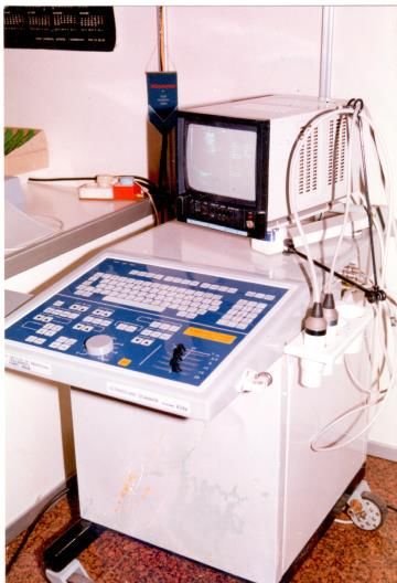
Ukrainian diagnostic devices (Figure 10).EFSUMB History of Ultrasound Ukraine 04.01.2021 10:23 17
Figure 10 Temporal behavior of the shear displacement at various locations within the
focal plane (a) recorded with a usage of modified scanner ТИ-628А and soft
tissue ultrasound phantom (b)
a b
At the same time, physicists from the School of Physics and Technology, headed by Barannik
E.O., were also looking for ways to determine the viscoelasticity properties of biological
objects, which could be very effective for diagnosing a large number of soft tissue pathologies.
In 1998, the first joint research project between KhNU and ArtannLabs Inc (NJ, USA, director
Sarvazyan A.P.) began, in the course of which the possibility of ultrasonic Doppler recording
of soft tissue movement during the passage of a shear wave, as well as the ability to determine
using SWE is the value of Young's modulus based on the shear wave propagation velocity [16,
17]. These studies were carried out in cooperation with RDIREM, which prepared a modified
version of the commercial US scanner TI-628A, with the help of which all experimental studies
related to SWE were carried out at KhNU. Unlike the American group of physicists headed by
K. Nightingale, who also developed the method of US elastography within the framework ofEFSUMB History of Ultrasound Ukraine 04.01.2021 10:23 18 the ARFI (Acoustic Radiation Force Impulse) technology [18] using speckle-tracking, the Ukrainian scanner TI-628A used the original phase tracking method (phased tracking) for precise determination of the magnitude of tissue movement and blood flow rate. During the next joint research with ArtannLabs Inc, experiments were carried out with soft tissues in vitro to determine the effect of viscosity on the magnitude of tissue displacement and the speed of shear waves [16, 19]. At the final stage of joint work with ArtannLabs Inc, a prototype of a portable ultrasonic Doppler motion detector with A-line mode was manufactured, which was successfully tested at ArtannLabs Inc. The results of the research were intensively discussed during several visits by Barannik E.A. to Sarvazyan A.P., Chief Scientist of ArtannLabs Inc, and were also presented at conferences NOISE-CON 2000, USA, “Ultrasonics International 2001”, Netherlands, and 5th World Congress on Ultrasonics WCU 2003, France. In experiments in vitro and especially in vivo, carried out to create a version of SWE, suitable for clinical implementation, an essential contribution was made by Linska G.V., who worked as a researcher at the Institute of Neurology, Psychiatry and Narcology of the National Academy of Medical Sciences of Ukraine. As a result, in 2011, just a year later than the French company SuperSonic Imagine, firm RADMIR released the first Ukrainian commercial scanners with 2D SWE technologies and colour mapping of soft tissue elasticity distribution. Ultrasound Attenuation Coefficient Measurement (ACM) During the 2010s, several research groups around the world have been involved in improving methods for measuring ultrasound attenuation in soft tissue. This interest is due to the fact that staging of hepatic steatosis is extremely important. Non-alcoholic fatty liver disease (NAFLD) has become a pandemic, affecting up to 30% of the population. NAFLD is an independent predictor of the development of cardiovascular and cerebrovascular accidents. In accordance with this, Ultrasign Ltd engineers in collaboration with physicists from KhNU have developed an original method for measuring the attenuation coefficient [20] and colour mapping of the attenuation coefficient distribution, which are successfully used now in clinical practice not only in Ukraine.
EFSUMB History of Ultrasound Ukraine    04.01.2021 10:23                                     19

"Eskulap-39 Ltd" / "Eskulap-UST Ltd"

"Eskulap-39 Ltd" and later "Eskulap-UST Ltd" was born in the bowels of the Kyiv Research
Institute "GidroPribor", which was created in Soviet times for the development of hydro
acoustic equipment for military purposes. In 1996, under the leadership of the director V.V.
Yaroshenko (now director Bykova O.T.) the team of "Esculap-39" Ltd has created an US device
with four line-scanning probes of its own production for studies of abdominal organs,
obstetrics and gynecology, urology, as well as superficial organs - "USDS-05" (Figure 11).

Figure 11 US apparatus "USDS-05" (a). The first director and founder of "Esculap-39" Ltd,
              Yaroshenko V.V. (b).

a

b
EFSUMB History of Ultrasound Ukraine 04.01.2021 10:23 20 The creation of original domestic ultrasonic probes was the result of the R&D "Flora" in 1991. This was especially important in connection with the growth of tumors of the thyroid and mammary glands after the Chernobyl accident. "Eskulap-UST" Ltd (designers Bykova O.T., Boyko V.F. and others) produces special phantoms for metrological verification of Doppler modules and the resolution of the B-mode of US devices: "Measure of blood flow velocities - MCK-03" and "Measurer of acoustic lengths - MAД-05". By order of the President of the UDC Dynnyk O.B., at “Eskulap-UST” Ltd, he created a training simulator - a Doppler phantom with three-dimensional reconstruction of vessels and presentation of the main Doppler phenomena in angiology: stenosis, aneurysm, deviation, turbulence, aliasing, redistribution, collateral blood flow. "Eskulap-UST Ltd" since 1993 and up to now has been producing domestic "Gel contact" for US. "Exim Ltd" The third group of developers and creators of equipment for US diagnostics was the team of "Exim Ltd" (since February 2002). In collaboration with developers from Vilnius (Lithuania), a series of portable (hand-held US device) US devices was created - SonoFly-3000, SonoFly- 3000M, incl. and for general practitioners (family doctors) and for telemedicine in 2003. These US devices were built on the basis of WINDOWS (first 98, and later XP) and used standard operating system tools for remote control and/or information transfer. The interface of the US devices was controlled remotely with a mouse or trackball. The RemoteAdmin program completely transferred the remote screen, mouse, keyboard to the doctor-consultant's computer. This program is the predecessor of today's TeamViewer. The SonoFly-3000 device has passed clinical trials in the Regional Clinical Hospital and Central District Hospital (Kolomyya), in the Rivne Regional Clinical Diagnostic and Treatment Center, and in the Zhytomyr Regional Consulting and Diagnostic Center. As a result, the SonoFly-3000 device was recognized as ready for implementation for conducting US telemedicine consultations.
EFSUMB History of Ultrasound Ukraine      04.01.2021 10:23                                    21

Conclusion

The development of US diagnostics in Ukraine is directly related to the development of world
US technologies. All the most modern US technologies and equipment are now available to
doctors in Ukraine. An important and crucial point is the mastering of knowledge and skills by
doctors for the most effective use of this equipment. An important fact is that UAUDS became
a member of EFSUMB. This event led to an intensive exchange of information and opened up
opportunities for doctors in Ukraine to honorably respond to the challenges of the time. In
particular, this illustrates the instant implementation of the protocols for US of the lungs and
the issues of safe conduct of US by WFUMB / EFSUMB in the conditions of COVID-19 by
Ukrainian doctors.

References

1. Age-related changes of liver stiffness in normal rats according to the method of ultrasonic
shear wave elastography / N. Kobyliak, O. Dynnyk, V. Berezovsky [et al.] // XVIII Annual Russian
Congress "Hepatology today", March 25-27, 2013: thesis. – Moscow. – Russian Journal of
Gastroenterology, Hepatology, Coloproctology. – 2013. – Vol. 23, N1 (ap. 40). – p. 168.
2. Diagnosis of experimental steatohepatosis using ultrasound shear wave elastography / P.M.
Bodnar, O.B. Dynnik, G.P. Mykhalchyshyn [et al.] // Current Issues in Pharmacy and Medical
Sciences. – 2013. – Vol. 26, N 1. – p. 97–101.
3. Performance of new ultrasound method for assessing liver stiffness - Share Wave
Elastography Imaging in rats with experimental obesity / Kobyliak N., Bodnar P., Dynnyk O.,
Mykhalchyshyn G., Kondro M. // 48th annual meeting of the European Association for the
Study of the Liver «International Liver CongressTM», April 24 - 28, 2013: thesis. – Amsterdam.
– Journal of Hepatology. – Vol. 58, Supl. 1. – p. 512.
4. Orel V., Mitrelias T., Tselepi M., Golovko T., Dynnyk O., Nikolov N., Romanov A., Rykhalskiy
A., Barnes C.C et all. Imaging of Guerin Carcinoma During Magnetic Nanotherapy //J
Nanopharmaceutics and Drug Delivery. – 2014; 2.- Р.1–11.
5. Dynnyk O., Kobyliak N., Berezovsky V., Yanko R. Echoelasto-morphological parallels: impact
of age-related hepatic lobule cytoarchitectonics changes on the liver stiffness measurement
in rats. // XXI Annual Russian Congress "Hepatology today", March 18-20, 2016: thesis. –
EFSUMB History of Ultrasound Ukraine     04.01.2021 10:23                                   22

Moscow. – Russian Journal of Gastroenterology, Hepatology, Coloproctology. – 2016. – Vol.
26, N1 (ap. 47). – p. 111.
6. Dynnyk O., Kobyliak N., Berezovsky V. Non-Invasive markers of liver fibrosis. The influence
of injection on liver stiffness measurment according to shear wave elastography in pigs // XXI
Annual Russian Congress "Hepatology today", March 18-20, 2016: thesis. – Moscow. – Russian
Journal of Gastroenterology, Hepatology, Coloproctology. – 2016. – Vol. 26, N1 (ap. 47). – p.
168.
7. Dynnyk O., Kobyliak N., Fedusenko A. Attenuation coefficient measurement (ACM) as a
newest mode for ultrasound quantitative hepatic steatosis assessment. Poster No.: B-1245.
Congress: ECR 2017. DOI: 10.1594/ecr2017/B-1245.
8. Dynnyk O., Fedusenko A., Kobyliak N. Multiparametric ultrasound (mp-US) in the chronic
diffuse liver disease diagnosis / Poster No.: C-2865. Congress: ECR 2017.
DOI: 10.1594/ecr2017/C-2865.
9. Dynnyk O., Marunchyn N. Diagnostic value of mp-US of the non-alcoholic fatty liver disease
in the patients with type 2 diabetes mellitus / Poster No.: C-0736. Congress: ECR 2019. DOI:
10.26044/ecr2019/C-0736
10. Dynnyk O., Omelchenko O., Solodovnik O., Marunchyn N. Multimodal multi-parametric
phantom of the fatty liver disease; Kiev/UA.// European Congress of Radiology 2020. Book of
abstracts. Scientific Programme. Physics in Medical Imaging. RPS 1013. Advances in MRI. RPS
1013 (11).- B 377.
11. Barannik EA, Girnyk SA, Tovstiak VV. Ultrasonic stimulation of gramicidin building into
bilayer. Biofizika 1988;33(2):364-366. (in Russian)
12. Barannik EA, Gromov AA, Kadnikov OG, Papakitsa VV, Stervoedov NG, Tovstiak VV,
Yakovlev AV. USSR Patent 1520682. Ultrasound transduser. In: Bul 7; 1990. (in Russian)
13. Barannik EA, Zaliubovskiy II, Kadnikov OG, Kobizskoy VI. USSR Patent 1300379. Method for
ultrasound influence on the object in opaque medium. In: Bul 12; 1987. (in Russian)
14. Barannik EA, Kadnikov OG, Papakitsa VV. On the scattering of sound by sound at
compositing focused wavebeams. Akusticheskij Zhurnal 1986;32(4):513-517. (in Russian)
15. Barannik EA. Pulsed Doppler flow-line spectrum for focused transducers with apodized
apertures. Ultrasonics 2001;39(2):311–317.
EFSUMB History of Ultrasound Ukraine         04.01.2021 10:23                               23

16. Barannik EA, Girnyk SA, Tovstiak VV, Marusenko AI, Emelianov SY, Sarvazyan AP. Doppler
ultrasound detection of shear waves remotely induced in tissue phantoms and tissue in vitro.
Ultrasonics 2002;40:849-852.
17. Barannik EA, Girnyk SA, Tovstiak VV, Marusenko AI, Volokhov VA, Sarvazyan AP, Emelianov
SY. The influence of viscosity on the shear strain remotely induced by focused ultrasound in
viscoelastic media. J Acoust Soc Am 2004;115(5):2358-2364.
18. K. Nightingale, M. Palmery, R. Nightingale, and G. Trahey. On the feasibility of remote
palpation using acoustic radiation force. J Acoust Soc Am 2001;110:625-634.
19. Girnyk SA, Barannik AE, Tovstiak VV, Barannik EA, Marusenko AI. The estimation of
Elasticity and Viscosity of Soft Tissues in vitro Using the Data of Remote Acoustic Pulpation.
Ultrasound Med Biol. 2006;32(2):211-219.
20. Barannik EA, Marusenko AI, Linska G.V. Pupchenko VI, Dynnyk OB, Kobyliak NM. Patent of
Ukraine 111234. Method and apparatus for detecting ultrasound attenuation parameter in
real time. In: Bul 7; 2016. (in Ukrainian)
You can also read