February 2021 Hanta Virus Infection - Indian Immunologicals Ltd
←
→
Page content transcription
If your browser does not render page correctly, please read the page content below

Volume X   Issue 2

                                                   February 2021

                     Hanta Virus Infection

                FelineTriaditis :                Formulation and
       Cholangio-hepatitis, Pancreatitis and         Delivery
   Inflammatory Bowel Disease - An Overview    of Vaccines in Wildlife
Reader’s Desk

                     RTR is covering a wider range of interesting             Inclusion of more clinical cases of different
                     articles including some topics related with              species will enrich this magazine.
                     surgery. This booklet having a very good
                     presentation.                                            Dr Pratyush Batabyal
                                                                              Malda, West Bengal
                     Dr A S Parihar
                     Indore, Madhya Pradesh.

                                                                              The magazine “Raksha Technical Review” is
                     It's amazing and excellent journal covering              dam informative. The articles provide vast
                     lots of knowledge with variety of disease                knowledge and strengthen the practical
                     information always helps in welfare of farmers           veterinary mind in field to farm animal level. I
                     and animal lovers. Finally, it's excellent journal       would greed to know the safety margins of
                                                                              Dewormers in various stages of animal life.
                     Dr Vijendra Singh Verma
                     Suroth- Karauli, Hindaun, Rajasthan                      Dr Anil Kumar Saini
                                                                              Bhilwara, Rajasthan

                     It is complete & comprehensive                           RTR is a compact publication for field
                     knowledgeable magazine in the field of Animal            veterinarian which keeps the field veterinarian
                     Husbandry. No suggestions.                               update.

                     Dr Pankaj Mohan Tripathi                                 Dr Subhash Fagera
                     Jaleli, Jodhpur, Rajasthan                               Bhilwara, Rajasthan

                     Need more content regarding Bovine & Small               This publication is of immense knowledge for
                     Animal. We need more information about                   veterinarians. I suggest to add more articles
                     clinical equipment.                                      on canines.

                     Dr Hanuman Ram Siyag                                     Dr Guranshpreet Singh Sethi
                     Jodhpur, Rajasthan                                       Jalandhar, Punjab.

                     It is advised to cover the topics related to field       Every reference in article is to be cited in
                     oriented diagnostic and their procedure so that          reference list. Yours field products are also
                     it is easy to apply & proceed further.                   valuable to the veterinarian for prescription to
                                                                              sick animals. Excellent magazine for field vets.
                     Dr B R Choudahry
                     Jodhpur, Rajasthan                                       Dr S K Nagyan
                                                                              Jaipur, Rajasthan

Vol.X | Issue 2 | Feb. 2021                                               2
Editorial Board
Contents
Reader’s Desk 02
Rajendra Lingala
Vice President - R & D From the Editor’s desk 04
Karnati Srinivas Managing Director's Message 05
General Manager - AH QC
Large Animal Section
Hanta Virus Infection 06
Honorary Members Subacute Ruminal Acidosis in Cattle 12
Ultrasonographic Diagnosis and Treatment of Follicular
Cyst in Kankrej Cattle 17
Abdul Samad
Former Director & Dean Pervaginal Delivery of Arthrogryposis and Hydrocephalus
MAFSU, Nagpur Foetus in a Primiparous Heifer 20
Cryptosporidiosis: An Emerging Zoonotic Infectious Threat 22
S Prathapan
Former Dean, TANUVAS
CVS, Tirunelveli Grazers and Browsers
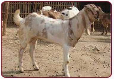
Pantja: Tarai Goat breed of Uttarakhand 31
K Sadasiva Rao
Former Associate Dean, Artificial Insemination in Goats: Techniques and Factors
NTR CVS, Gannavaram Affecting Fertility 32
H K Verma
Former Director of Extension Companion Animal Section
GADVASU, Ludhiana Management of Radio-Ulnar Ischemic Necrosis in a
Spayed Female German Shepherd Dog 36
Sub - Editorial Committee Successful V-Plasty Surgical Ablation of Lower Eyelid Margin
Tumor in Dog: A Case Report 38
Feline Triaditis: Cholangio-hepatitis, Pancreatitis and
Inflammatory Bowel Disease - An Overview 42
V Surya Prasad
Recent Approaches in Diagnosis and Management of
Sr Manager - Veterinary Services
Canine Atopic Dermatitis: A Review 49
Z Hasan
Manager - AH PMT General Articles
Formulation and Delivery of Vaccines in Wildlife: A Review 57
D Prabhakar
Intellectual Property Rights for Animal
Manager – CRM & CSR Genetic Resources in India 60
Microbiome Signature – A New Aid in Disease Therapy 64
Effect of Stress on Dairy Animals 66
Sourced & Published by
S Sobhan Babu Feedback 69
General Manager - Animal Health
Guidelines to Authors 70
From the Editor’s desk

Zoonotic transmission can occur either in contact with animals or consumption of animals, animal
products, or animal derivatives. Zoonotic studies are interesting because of the existence of
unrecognized diseases or due to virulence in population lacking immunity. Hanta is a globally emerging
group of virus which causes varying syndromes in Humans. The article Hanta Virus Infection has
highlighted the disease and its origin. Cryptosporidiosis: An Emerging Zoonotic Infectious Threat is
another article of zoonotic importance, caused by a protozoan parasite.

Rumen acidosis is a disorder of rumen in ruminants that cause many health problems in animals. The
article Subacute Ruminal Acidosis in Cattle has given elaborate information and concludes that its
detection and prevention is challenging. This article suggests dietary management to help in reducing
the problem.

Large animal section contains articles Ultrasonographic Diagnosis and Treatment of Follicular Cyst in
Kankrej Cattle and Pervaginal Delivery of Arthrogryposis and Hydrocephalus Foetus in a Primiparous
Heifer.

Pantja: Tarai Goat Breed of Uttarakhand and Artificial Insemination in Goats: Techniques and Factors
Affecting Fertility are two interesting articles of importance in Grazers and Browsers section.

Milford Veterinary Clinic (MVC), USA, has been a regular contributor of articles dealing with Canine and
feline veterinary practice. In this issue, MVC brought forward their experiences in handling various
issues. In case of canines, MVC presents the articles dealing with Radio-Ulnar Ischemic Necrosis
(RUIN) in a Spayed Female German Shepherd and V-Plasty Surgical Ablation of Lower Eyelid Margin
Tumor in Dog. MVC also reviews the Feline Triaditis namely Cholangiohepatitis, Pancreatitis and
Inflammatory Bowel Disease. The article highlights the management of these diseases. We hope these
examples will be beneficial for pet practitioners.

Atopic Dermatitis is an allergic skin disease in dogs. The article Recent Approaches in Diagnosis and
Management of Canine Atopic Dermatitis, discusses the end-to-end of Atopic Dermatitis.

General Articles Section brings forth seasonal stress factors affecting production and reproduction in
dairy animals in the article Effect of Stress on Dairy Animals. The articles Formulation and Delivery of
Vaccines in Wildlife: A review, signifies importance of vaccination of wildlife especially the endangered
species to save their lives from deadly infectious diseases and Intellectual Property Rights for Animal
Genetic Resources in India gives a brief idea on the rights in India and the world. The article Microbiome
Signature on Dairy Animals details about the recent approach to treat the disease condition.

Enjoy Reading! Stay Safe!! Stay Happy!!!
Managing Director’s Message

Dear Patrons,

Greetings!!

Coming out from a tough year 2020, year 2021 appears to be even tougher with the SARS-COV2
virus changing its nature. India and the World is facing the worst of times with COVID-19 at its
peak of 2nd Wave. In these hard times, community support and our experience of 2020 is handy
in dealing with the current scenario albeit with abundant caution.

Vaccine manufacturers and the Government of India are running against time to ensure
continuous production of COVID vaccine alongside looking for opportunities of enhancing the
manufacturing capabilities across the country. IIL, on its part, is actively pursuing the vaccine
manufacturing for COVID19 in two ways: 1) Augmentation of capacities for vaccines already
developed by existing manufacturers e.g., Bharat Biotech, 2) Technical Collaboration with Griffith
University, Australia for IIL’s own vaccine;

We are optimistic that IIL, India and the World will be back to normal by end of 2021.

Warm Regards

Dr K Anand Kumar
LARGE ANIMAL SECTION

                                                                       Hanta Virus Infection
                                     Anugraha Mercy Easaw and K Vijayakumar
   Dept. of Vet. Epidemiology and Preventive Medicine, College of Veterinary and Animal Sciences, Mannuthy, Kerala.

Introduction                                                   clinical syndromes are caused by different viruses
                                                               and those which cause HFRS rarely causes HPS
At present, mankind is facing the threat of
                                                               and vice versa. In Europe, a mild form of HFRS is
increasing outbreaks of infectious epidemics, out of
                                                               reported and is referred to as nephropathia-
which majority of the diseases are zoonotic.
                                                               epidemica (NE). Few important viruses which
Emerging viral epidemics are being reported from
                                                               cause HFRS include Hantaan, Seoul, Tula,
different parts of the globe. Emerging diseases are
                                                               Thailand, Thottapalayam, Bowe and Sangassou
those which were previously unknown, that caused
                                                               viruses Puumala, Amur-Soochong, Dobrava-
an outbreak or known infectious diseases whose
                                                               Belgrad and Gou viruses. The viruses that cause
incidence has increased or cause uncontrollable
                                                               HPS include Sin Nombre, Andes, Laguna Negra,
persistent infections. One such globally emerging
                                                               Black Creek Canal, Bayou, Cano Delgadito, Choclo
group of viruses includes Hanta viruses which
                                                               etc and variants of some of these viruses. Puumala
cause varying syndromes in humans. Hanta viruses
                                                               virus or the Saaremaa variant of Dobrava-Belgrade
are RNA viruses which belongs to the genus
                                                               virus is implicated to cause nephropathia-
OrthoHantavirus in the family Hantaviridae and
                                                               epidemica(3).
order Bunyavirales. They are referred to as newly
emerging viruses, and have been reported to have               Phylogenetic studies have revealed three main
caused illness during the American Civil war times.            lineages. Old World lineage is associated with those
The virus was first isolated during the 1970s from             viruses which cause HFRS whereas HPS causing
the tissues of field mice near the Hantaan River in            viruses has a common New World lineage.Viruses
the Republic of Korea. The disease is asymptomatic             belonging to New World lineage are linked to a
in rodents which harbor the virus, but can cause               single rodent subfamily Sigmodontinae (4). It was
clinical signs in humans. In humans two important              found from serological studies, genetic evaluation
syndromes have been reported, i.e., Haemorrhagic               and association between host and reservoir that few
Fever with Renal Syndrome (HFRS) and                           of these sigmodontine-derived viruses are
Hantavirus Pulmonary Syndrome (HPS). Severity                  independent species. Other species are still under
of the disease varies wherein it can cause mild                evaluation for its classification and assortment. The
illness with complete recovery or severe syndromes             viruses isolated from Europe and Asia are included
which can cause a case fatality rate up to 30 per              in the Old World Hanta viruses which include
cent or even higher.                                           Puumala virus (PUUV), Dobrava–Belgrade virus
                                                               (DOBV), Hantaan virus (HTNV) etc. The New World
Etiology
                                                               Hanta viruses are found mostly in America which
Hanta viruses are antigenically, genetically and               commonly comprises of Sin Nombre virus (SNV)
epidemiologically related to Bunyaviridae. Other               and Andes virus (ANDV), and Choclo virus (CHOV)
members of this family like Bunyavirus, Nairovirus,            etc. Human to human transmission has been
Tospovirus and Phlebovirus are arthropod borne                 reported only in the case of Andes Virus.
viruses except for Hanta virus. The virus particle is
                                                               Epidemiology
spherical with a diameter of 80-110 nm (1).The virus
is enveloped with negative-strand ribonucleic acid             Hanta viruses have worldwide distribution, in which
wherein the genome is three-segmented.The three                the known reservoirs include rodent, insectivores
segments are denoted as L, M, and S which are the              like shrews, moles etc, and bats. Generally each
large, medium, and small single-stranded RNA                   species of Hanta virus is associated with a single
genomes, respectively. The viral RNA polymerase,               reservoir host species. Therefore the distribution of
the glycoprotein and the nucleocapsid protein are              the virus in a particular geographical area is directly
encoded by S, M, and L segments respectively (2).              linked and restricted by the specific host species.
Even though many of the Hanta viruses are                      Long-termcoevolutionary relationship between host
unclassified, almost 40 Hanta viruses are identified           and viruses is supported by the high association
out of which 22 are found to be zoonotic. These 22             between host and viral phylogenies. From the
viruses are transmitted to humans wherein rodents              numerous reports it has been found that several
act as the reservoirs.                                         members of the same genus of host species can be
                                                               infected with the same virus. This is related to the
From the different outbreaks that has occurred in
                                                               possibility of high rodent density, spillover
different parts of the world it is seen that the two

Vol.X | Issue 2 | Feb. 2021                               6
RAKSHA TECHNICAL REVIEW                                                           LARGE ANIMAL SECTION

     Geographical representation of hantaviral disease incidence by country annually. Courtesy of
                              Douglas Goodin, Kansas State University.
transmission and increased chances of                      whereas HFRS peaks during agricultural activities
interspecies encounters (4).                               during spring and fall. Reports worldwide estimates,
                                                           150,000 to 200,000 humans to be hospitalized with
The viruses affecting the Western hemisphere
                                                           HFRS annually(3). Majority of these cases are from
usually cause HPS, with the exception of Seoul
                                                           Asia followed by Europe and Russia. HPS is more
virus (SEOV). Even though other viruses prevail in
                                                           frequently reported in South America as compared
these geographical areas, Sin Nombre virus causes
                                                           to North America and Canada with 11-50 cases
disease in North America and in South America
                                                           reported in the United States and 0-13 cases in
Andes viruses are seen to cause the disease. In the
                                                           Canada annually. Probably due to under reporting
Eastern Hemisphere, Hanta viruses are associated
                                                           and under diagnosis, clinical cases are rarely
with HRFS. These include Dobrava-Belgrade and
                                                           identified in Africa. Severity of cases varies with the
Puumala viruses in Europe, and Hantaan, Amur-
                                                           clinical manifestation, the viruses causing the
Shoochong, Gou, Thailand and Thottapalayam
                                                           disease and the health care facilities provided to the
viruses in Asia, Seoul and Tula virus in Europe and
                                                           affected individuals. Case fatality rates (CFR) have
Asia, and Sangassou, Bowe and Uluguru viruses in
                                                           reduced remarkably over the years due to early
Africa. Even though seropositive rodents have been
                                                           detection and supportive therapy. HPS is more life
identified in Australia, no evidence of disease has
                                                           threatening than HFRS with a CFR of 25% to 40%
been reported in Australia. Other animals, like non-
                                                           for most viruses.The CFR for HFRS varies with
human primates, dogs, cats, horses, cattle, etc can
                                                           viruses, for example Puumala virus causing
be incidental hosts and are found to have antibodies
                                                           nephropathiaepidemicahas a CFR < 0.5% to < 1%,
to Hantavirus. Hantaviral antigens have been
                                                           1-2% in case of Seoul virus and 5% by Hantaan
isolated from avian species including passerine
                                                           virus. Seroprevalance is seen in many countries
birds, owls, herons and doves. Experimental
                                                           wherein people are asymptomatic or have a mild
infection in pigs has also been reported in China.
                                                           form of the disease (5).
The occurrences of human cases are associated
                                                           Hantaviral infection in India
with rodent densities, seasonal fluctuations and
human activities with high risk of exposure to             The virus isolated from India is the Thottapalayam
rodents. Epidemic years are linked to favorable            virus which was isolated from the spleen of non
climate and high yield of seed crops leading to            rodent, house shrew in 1964. This was collected
increasing number of rodents. This is attributed to        during studies on Japanese Encephalitis in Vellore,
increased breeding and rate of survival of rodents.        South India. Researchers found prevalance in
HPS is reported during late spring or early summer         individuals wherein they found both healthy blood

Vol.X | Issue 2 | Feb. 2021                            7
RAKSHA TECHNICAL REVIEW                                                              LARGE ANIMAL SECTION

donors and individuals with febrile illness had                has been reported. Molecular studies from India
antibodies to Hanta virus (anti- HV IgM). Another              have aided in identifying isolated cases of Hanta
study has revealed high levels of Ig G titre in the risk       viral infection and reveal the ongoing scenario in the
group compared to healthy blood donor group (the               nation.
control group). Individuals with nephropathies in
                                                               Transmission
South India, like in Cochin and Chennai mimicking
leptospirosis were found to have 12% SEOV-                     In reservoir hosts, the transmission is by direct and
positive antibodies and 5% PUUV-positive                       aerosol route, especially when there is close
antibodies(6).This shows the possibility of                    contact during grooming, fighting and eating food.
asymptomatic and symptomatic illness in the south              Viruses are shed by rodents in saliva, urine and
Indian population. Other important reports of HV               faeces, and these rodents can be infected for a
cases include outbreak in Irula community, Vellore             lifetime. Arthropod borne transmission is not well
in 2008 affecting 28 individuals in the community              established. Mites were found to be potential
and in Mumbai 2016, a case of pulmonary                        vectors naturally and experimentally. Dependent
haemorhage was confirmed as a case of HV                       upon different factors like temperature, humidity,
infection. In the past two decades multiple cases              organic matter and exposure to sunlight, the virus
with ocular involvement, HV infection in conjunction           can persist in the environment for few days to weeks
with tuberculosis with renal involvement,                      together.
postpartum HPS from different parts of the country

Humans acquire the infection from direct contact               were isolated from urine and blood. Nosocomial
with infected reservoir hosts or by contact with their         transmission has not been reported in case of viral
excreta. Inhalation of aerosolized dust from rodent            Hanta infection(4).
excreta including urine, droppings or even nests
                                                               Pathogenesis
disturbed is one of the most common causes. Other
routes of transmission include exposure of virus via           The virus is capable of affecting multiple organs, but
broken skin or mucous membrane, rodent bite and                the virus has predilection for kidneys or lungs
even by ingestion. No reports of vertical                      endothelial cells and macrophages in both humans
transmission are by far reported, but possibility of           and animals. The clinical manifestations of viral
transmission of virus via breast milk was identified           Hanta infection are attributed to increased
in South America. Horizontal transmission in                   permeability of vascular endothelium and acute
humans has only been reported in case of Andes                 thrombocytopenia. The viral replication occurs in
virus even though in cases of HFRS Hanta viruses               the vascular endothelium even though there is no

Vol.X | Issue 2 | Feb. 2021                                8
RAKSHA TECHNICAL REVIEW                                                              LARGE ANIMAL SECTION

cytopathic effect. There is no acute viremia since            Clinical Signs
there is slow viral replication which takes an
                                                              In Animals
average of 5-10 days. The persistent survival of
viral particles allows the infiltration of inflammatory       The reservoir hosts do not have any overt disease
cells. The multi-organ injury is caused by infiltration       and show no particular clinical signs. In affected
of cells and response of the immune system to the             wild mice and bats, there is reduced weight gain and
viral infection.                                              survival rate and in experimentally affected infant
                                                              rodents, neurological signs are exhibited (3). In
Hanta viruses infect the endothelial cells through
                                                              certain species like, Syrian hamsters pulmonary
different integrin receptors. The complete
                                                              and renal involvement was observed. Most of the
dissemination of Hanta virus is not completely
                                                              adult rats and mice were asymptomatic. Other
understood, β-integrin receptors on the target cell
                                                              animals are said to be asymptomatic as well, except
membrane and viral proteins (Gn and Gc) have
                                                              non primates wherein at times naturally and
been found to interact with each other. These
                                                              experimentally renal and pulmonary signs are seen
receptors on the immature dentritic cells aid in the
                                                              which was reported in China.
spread of virus and acts as a vehicle to enter the
endothelial cells via the lymphatics and then into the        In humans, the clinical signs in case of viral Hanta
regional lymph nodes. The endothelial cells allow             infection can include asymptomatic, mild signs
viral replication and hence activate the immune               including febrile illness, HPS and HFRS (3,10).
system particularly via macrophages and CD8+ T                Humans usually show either one of the clinical
cells (7). There is also activation of delayed type I         signs, but rarely HPS and HFRS can occur in a
interferon response generated in response to                  single individual.
higher pathogenic viral load. This intensifies the
                                                              HFRS
response of the infected host cells by producing
inflammatory cytokine and chemokines which                    HFRS causes mild to severe diseases with the
worsens the condition(8).Hence it can be                      above mentioned five different phases in
concluded that the pathophysiology in case of viral           pathogenesis. There is an abrupt start of clinical
Hanta infections is multifactorial, with a combination        signs with fever, headache, chills, prostration etc.
of multiple complex factors including disruption in           Along with these signs, gastrointestinal signs like
the regulation and function of endothelial cell               vomiting and abdominal pain are also reported.
barrier, intense response from host immune system             Nonspecific signs like congested mucous
to the infection and platelet dysfunction.                    membrane, rashes over trunk and palate,
                                                              photophobia are exhibited, and rarely temporal
In case of HFRS the incubation period is seen to be
                                                              impairment of vision is observed. During the first
one to six weeks. Five phases of the illness in case
                                                              phase, i.e., the proteinuric phase there is
of HFRS include febrile, hypotensive, oliguria,
                                                              hypotension and in acute cases death is observed
diuretic and convalescent. In cases of NE, there is
                                                              during shock. The proteinuric phase is followed by
more of a renal impairment than haemorhaggic
                                                              oliguria, later by polyuria when the kidney function
syndrome. In HPS the incubation period is one to
                                                              improves. Death is common during the first two
seven weeks, and the clinical manifestation
                                                              stages. Lung involvement is rarely observed, but
involving the thoracic cavity with no haemorhaggic
                                                              not as severe as HPS, with mild symptoms of
episodes or renal damage. The pathogenesis and
                                                              pulmonary damage and pleural effusion.
clinical signs involved in case of Hantaviral infection
                                                              Haemorhaggic signs like petechiae melena and
is attributed to capillary leak. The virus is said to
                                                              haematuria are attributed to thrombocytopenia and
have affinity for the vascular endothelium but there
                                                              are very common. Sequele vary from having
is no cytopathic effect. The immune response to the
                                                              complete recovery with normal kidney function to
virus includes activation of antigen differentiation
                                                              hypotension, chronic kidney disease, and
cells and elevated level of cytokines which causes
                                                              permanent neurological condition. Recovery
the different symptoms. Post mortem findings of
                                                              usually takes few weeks to months.
fatal cases of HFRS show haemorhagges in
visceral organs including kidney, pancreas, skin,             HPS
meninges and pituitary gland. Retroperitoneal
                                                              In HPS, lung involvement predominates and can
oedema and even pulmonary oedema has been
                                                              cause high mortality. Initially, nonspecific signs like
reported. NE cases reveal signs similar to acute
                                                              that of HRFS are seen as similar to the prodromal
interstitial nephritis. In HPS, oedematous lung,
                                                              stage. Signs include cough, tachypnea followed by
pleural effusion and splenomegaly are consistent
                                                              pulmonary oedema and hypoxia. Rarely cardiac
findings(9).
                                                              signs are seen, and following this cardiopulmonary
                                                              phase, condition of the patient deteriorates. Signs
                                                              similar to HRFS related to thrombocytopenia, renal

Vol.X | Issue 2 | Feb. 2021                               9
RAKSHA TECHNICAL REVIEW                                                             LARGE ANIMAL SECTION

damage and neurological signs are reported.                  individuals require fluid therapy and inotropic
Recovered cases usually possess full lung                    agents to monitor blood pressure. Dialysis is
capacity.                                                    performed in severe renal damage. In HPS
                                                             patients, fluid therapy, administration of antibiotics
Some Hanta viruses cause mild illness with clinical
                                                             and vasopressors, and proper oxygenation is
signs similar to that of the prodromal stages of
                                                             followed. Antiviral drugs like Ribavirin is said to have
HFRS or HPS with no specific signs of the two
                                                             effect during the early stages of HFRS, but further
conditions. Fever of unknown origin is linked to
                                                             study is required. In HPS cases Ribavirin is said to
Hantaviral infection. Pulmonary involvement can
                                                             have no effect. Trials in South America have shown
occur in rare conditions with no abnormalities
                                                             good response to administration of antiserum to
observed in radiography.
                                                             affected individuals. Specific therapeutic agents
Diagnosis                                                    can be categorized as i) antiviral agents target the
                                                             host ii) drugs that the boost host immune system, iii)
Hantaviral infections are diagnosed by clinical and
                                                             antiviral agents that target the Hantavirus. Antiviral
epidemiological data and laboratory tests. The
                                                             agents which target the Hantavirus include,
clinical manifestations include high fever,
                                                             Ribavirin, Favipiravir, iron-binding glycoprotein
headache, malaise, myalgia, body pain and
                                                             lactoferrin, nucleoside analogues like ETAR and
increased hypotension. The major pathological
                                                             Vandetanib. Most of these drugs are found to have
findings include thrombocytopenia, leucocytosis,
                                                             good efficacy in in vitro studies and in laboratory
elevated serum creatinine, albuminuria and
                                                             conditions. There is a need for further studies to get
haematuria. Many at times the affected individuals
                                                             approval of FDA and use in humans.
might be asymptomatic or would only have mild
symptoms which make it difficult to solely rely on           Prevention and Control
clinical and pathological findings. Hence laboratory
                                                             Prime feature of prevention of Hantaviral infection
tests are employed for early and accurate detection.
                                                             include avoiding any form of contact with reservoir
At the beginning of infection the viral proteins of
                                                             hosts (3). Most of the clinical cases are associated
Hanta viruses (Gn, Gc and N) initiates production of
                                                             with being in enclosed spaces wherein rodents are
IgM antibodies, which are identified by laboratory
                                                             present and humans involving in activities where
tests. Specific IgM or rise in the IgG titer aids in
                                                             they can come in contact with their excreta like
diagnosis, since the antibody titers can be detected
                                                             agricultural activities, hiking, camping etc. It is
when the clinical signs have started. Serological
                                                             important to prevent entry of rodents in households.
tests such as ELISAs, immunofluorescent antibody
                                                             Rodenticides, traps can be used, at the same time it
tests and immunochromatographic tests are
                                                             is important to keep away all food materials which
commonly employed. ELISA tests are used to
                                                             can attract rodent. Guidelines are provided by CDC,
detect IgM antibodies in the acute phase of the
                                                             WHO, OIE for safe cleaning of rodent infested
disease which could persist for 3-6 months and IgG
                                                             areas. CDC recommendation suggests:seal up,
antibodies can be detected even for years.
                                                             trap up, and clean up.
Commercial kits are available for ELISA and
immunoblot assays for certain viruses (3).                   It is important not to aerolise the virus and to avoid
                                                             activities like sweeping and vacuum cleaning.
The above mentioned tests are only able to detect
                                                             Always wear gloves while cleaning and it is
whether an individual has been infected with Hanta
                                                             important to wet the contaminated area with
virus or not and cannot differentiate the different
                                                             disinfectants or even household bleach before
Hanta viruses. Specific viruses can be detected
                                                             wiping off the excreta (11). It is commended to use
using Focal reduction neutralization test (7).Viral
                                                             wet mopping or wet paper towels. The virus is
neutralization test has its own limitation since it
                                                             susceptible to disinfectants such as 1% sodium
needs live virus for performing the test, and is
                                                             hypochlorite, 70% ethanol and 1-5% peracetic acid.
expensive and time consuming. Virus isolation is
                                                             In heavily contaminated areas 10%sodium
not preferred due to the risk of handling such
                                                             hypochlorite is recommended. Individuals who are
viruses and inability to culture certain viruses. For
                                                             exposed to such situations in the work environment
experimental purposes the virus particles are
                                                             must wear proper protective gear including
cultivated in Vero E -6 cells. Immunohistochemistry
                                                             goggles, masks and gloves and must cover
can identify antigens in tissues and are employed
                                                             scratches or wounds if any on the body surface.
often. RT- PCR aids as a good means to detect the
antigen in affected individuals by identifying viral         Commercial inactivated vaccines are available in
RNA in blood, urine and saliva.                              South Korea and China by the National Extended
                                                             Immunization Program wherein two million doses of
Treatment
                                                             vaccines are administered regularly (12, 7).
The key to better recovery of affected individuals is        Inactivated vaccines grown in cell culture have
proper supportive therapy. HFRS affected                     been used for bivalent vaccination for two sub-types

Vol.X | Issue 2 | Feb. 2021                             10
RAKSHA TECHNICAL REVIEW LARGE ANIMAL SECTION
of Hantavirus. This includes SEOV & HTNV grown a successful vaccination strategy is being
in Vero cells. Recombinant vaccines are found to be developed. Trials using Virus-like particles (VLP) as
effective in rodents. Administration of naked DNA vaccines are ongoing and the efficacy as well as
has evoked good immune response in rodents and safety are yet to be analysed.
Inactivated Hantavirus Vaccines Used In China And Korea
Virus strains Generated in Vaccination Protection Country
Programme
HTNV MGKC+ 3 basic doses + 1booster >90% China
SEOV GHKCC++ 2 basic doses + 1booster >95% China
HTNV SMB+++ 3 basic doses + 1booster >90% China
HTN/SEO MGKC 2 basic doses + 1booster 100% China
HTNV SMB 2 basic doses + 1booster 75-100% Korea
+MGKC : Mongolian Gerbil Kidney Cell
++GHKC : Gold Hamster Kidney Cell
+++SMB : Suckling Mouse Brain
Conclusion 4. Pan American Health Organization.(1998).
Hantaviruses in the Americas: Guidelines for
Hantaviral infection is an emerging zoonotic
diagnosis, treatment, prevention and control.
disease and has great public health significance.
33p.
Even though the first case in India was reported in
1964, there have been over 40 reports of cases 5. Yanagihara, R., Gu, S. H., Arai, S., Kang, H. J.
from India as of 2020, which show that there is an and Song, J. W. (2014). Hantaviruses:
increasing occurrence which can be attributed to rediscovery and new beginnings. Virus
factors like human activities, climatic conditions, research. 187: 6.
rodent population etc. Limited and priced diagnostic
6. Hangaragi, P. S. (2020). Hantavirus: An
aids, lack of awareness and multiprofessional
emerging global threat. Asian Journal of Oral
collaborations increases the potential risk of an
Health and Allied Sciences. 10.
outbreak. Better epidemiological studies including
global monitoring and prediction is required to 7. Munir, N., Jahangeer, M., Hussain, S.,
tackle this disease which can easily become a Mahmood, Z., Ashiq, M., Ehsan, F., Akram, M.,
pandemic causing a huge mortality than that of the Ali Shah, S.M., Riaz, M. and Sana, A. (2020).
present Covid-19 pandemic. Medical professionals Hantavirus diseases pathophysiology, their
integrated with public health specialists must be diagnostic strategies and therapeutic
properly informed and be aware about the different approaches: A review. Clinical and
syndromes caused by hantavirus, its diagnosis, Experimental Pharmacology and Physiology.
treatment, prevention and control in endemic areas.
8. Mir, M. A. (2010). Hantaviruses. Clinics in
References laboratory medicine. 30: 67.
1. Schmaljohn, C. and Hjelle, B. (1997). 9. Rev. sci. tech. Off. int. Epiz. (2000). 19: 64.
Hantaviruses: a global disease problem.
10. Lee, J. A Review on OrthoHantavirus
Emerging infectious diseases. 3: 95.
(Hantavirus). Prevent. 6, p.7.
2. Lednicky, J. A. (2003). Hantaviruses: a short
11. C e n t e r s f o r D i s e a s e C o n t r o l a n d
review. Archives of pathology & laboratory
Prevention.(2013). Facts about Hantaviruses:
medicine. 127: 30.
What you need to know to prevent the disease
3. Centers for Disease Control and Prevention Hantavirus pulmonary syndrome (HPS).
Fact sheet: Spickler, Anna Rovid. (2018).
12. Bi, Z., Formenty, P. B. and Roth, C. E. (2008).
H a n t a v i r u s . Av a i l a b l e a t
Hantavirus infection: a review and global
http://www.cfsph.iastate.edu/DiseaseInfo/fact
update. The Journal of Infection in Developing
sheets.php.
Countries. 2: 003.
Vol.X | Issue 2 | Feb. 2021 11LARGE ANIMAL SECTION
Subacute Ruminal Acidosis in Cattle
Rupesh Chaurasiya, Pooja Dixit and Alok Kumar Dixit
Dept. of Veterinary Medicine, College of Veterinary Science & A.H., Kuthuliya, Rewa, Madhya Pradesh and Dept. of
Veterinary Parasitology, College of Veterinary and Animal Sciences, SVPUAT, Modipurum, Meerut, Uttar Pradesh.
Introduction protozoa present in rumen. These bacteria are of
two types Amylolytic and Cellulolytic. Cellulose and
Rumen acidosis is a disorder of rumen in ruminants
hemicellulose have β (1-4) glycosidic linkages,
in which increase in the amount of grain fed leads to
which are broken by cellulolytic bacteria i.e.
increase in milk production initially, but later
Rumenococcus spp, Bacteroids spp especially
becomes the cause of many health problems in
Butyrovibrio genera in a strict anaerobic condition,
animals. In our country in the dry period, when the
leading to formation of volatile fatty acid (VFA) i.e.
animal is not lactating we usually neglect the
acetic acid, propionic acid and butyric acid which
animal, especially its diet. When the lactation starts
are readily absorbed across the rumen epithelium
we abruptly increase the grain amount in order to
and used by the ruminants. Similarly, the starch
increase the milk yield, which leads to disturbance
present in the grains is also utilized by amylolytic
in the rumen physiology leading to increase weak
bacteria in a strict anaerobic condition leading to
acid absorption and acidemia. This condition is
formation of lactic acid along with VFA and it is ten
called ruminal acidosis (1, 2). It occurs in two forms;
times stronger than VFA. Protozoal populations
acute and subacute ruminal acidosis
also do not survive extended exposure to pH below
Due to sudden change in the diet at the start of 5.5 (8).
lactation and where we neglect feeding of the
When there is abrupt increase in starch or grain diet
animal and it approaches to easily fermentable diet,
to increase milk production. It will lead to increase
all these can lead to acute form of acidosis. This
lactic acid in rumen leading to low rumen pH. Saliva
condition is difficult to diagnose. Sometimes animal
production, Endogenous buffering and reduced
is found dead in the field, sudden death occurs in
starchy feed intake are the compensation
this condition which can be confused with poisoning
mechanisms of ruminants to overcome this problem
cases. In this condition rumen enlargement can
(9). But when all these mechanisms fail or the lactic
lead to pressure on diaphragm resulting in
acid production is more, it will not be absorbed from
respiratory arrest, which sometimes leads to
the rumen wall leading to rumenitis (10).
haemoptysis and oozing of blood from the mouth
that can be confused with Anthrax (3, 4). Increase lactic acid in rumen further decrease
proportion of cellulolytic bacteria and increase
Subacute Ruminal Acidosis (SARA) is usually not
concentration of G+ve bacteria especially
seen in individual animals and progresses slowly
Streptococcus bovis Which will ferment glucose to
usually in an on-off form. Many workers have
lactate and increase lactic acid concentration.
reported that pH below 5.6 for more than
Increase lactate concentration leads to increase in
180min/day is SARA (5). Dairy herds usually suffer
lactate utilizing bacteria Lactobacillus sp. which
this problem and a number of animals are affected
ferment lactate to other VFA (i.e.Valerate).
instead of one or two. It usually occurs in early
lactation to mid lactation. Most of the times, it As cellulolytic bacteria are G-ve bacteria and their
escapes the diagnosis because acidic episodes damage leads to production of lipopolysaccharide
occur only for 3-5 hrs/dayfollowed by normal which goes to liver via portal circulation and may
ruminal pH rest of the day. In a dairy herd, less milk cause endotoxicosis, pyelonephritis, arthritis and
production, less feed intake and lameness of chronic inflammatory disease.
varying degree are the signs, which indicate
Risk Factors
towards SARA, besides other problems due to
lipopolysaccharides (6, 7). Severe economic losses The cow at risk to develop SARA includes cows in
occur due to this condition mainly because of the early lactation, primiparous cows and cows
reduction in milk production and health grazing or fed with rapidly fermentable low fiber
compromises. grass (11). Cows in the early lactation have the
instability of the bacterial population. According to
Pathophysiology
Stone (12) cows might be at greatest risk for SARA
Before understanding the pathophysiology of immediately postpartum due to diminished size and
ruminal acidosis, we have to first see normal rumen absorptive capacity of rumen papillae following
physiology. Normal rumen pH is 6.8 to 7. Rumen feeding of lower energy density diets during the dry
fermentation of diet depends on bacteria and period. Enemark et al (13) reported that
Vol.X | Issue 2 | Feb. 2021 12RAKSHA TECHNICAL REVIEW LARGE ANIMAL SECTION
Primiparous cows were generally more prone to low 7) Milk Fat Depression
rumen pH, higher ruminal concentrations of volatile
Low ruminal pH causes milk fat depression by
fatty acids and possibly to metabolic acidosis, than
inhibiting the bacteria (24) which is responsible for
multiparous cows. Krause and Otzel (14) also
fatty acid biohydrogenation in the rumen.
indicated higher prevalence of SARA in primiparous
cows than in multiparous cows. Cows are 8) Rumenitis
apparently at higher risk for SARA in the summer
Rumen epithelial cells are stratified squamous
due to lack of ruminal buffering caused by heat
epithelium and not protected by mucous. So it is
stress, increased respiratory rate, respiratory
susceptible to acidity. Corrosive effects of
alkalosis, and low blood bicarbonate
increasing amount of acids in rumen are the most
concentrations.
likely feature of acidosis leading to rumenitis and
Adverse Health Effects of SARA on Animals later to erosion or ulceration. The adhesion
between rumen epithelial cells becomes weak
1) Low Rumen pH
leading to translocation of rumen microbes to liver
The main effect of SARA is low rumen pH because via portal circulation. In chronic rumenitis there is
organic acids accumulate in the rumen produced by thickening of rumen epitheliumi.e. Parakeratosis
rumen fermentation of carbohydrates(15). The low which decreases further acid absorption (25).
pH will lead to some adverse effects on rumen
9) Hindgut Acidosis
mucosa and will reach blood to cause transient
acidemia (16). Excessive intake of fermentable carbohydrates
may cause both rumen and hindgut acidosis.
2) Increased Ruminal Valerate
Several studies have reported reduced faecal pH
Low rumen pH increases the concentration of during induced SARA (26). Sometimes diarrhoea
lactate and thus lactate utilizing bacteria which work due to malabsorption and sloughing of epithelial
on lactate to produce other VFA i.e. Valerate. This cells of large intestine has been reported(27, 28).
further contributes to acidosis(17).
10) Lameness
3) Reduced Dry Matter Intake
The mechanism of lameness in cattle in SARA is not
Though sometimes SARA is a self limiting problem, well understood, but a direct effect of systemic
the adverse effects of SARA are many. It decreases lipopolysaccharides on capillaries in the hoof has
the Dry Matter (DM) intake. If DM intake is less it been reported (29). In horses, it has been reported
reduces acid production and allows rapid that activation of metalloproteinase enzyme
restoration of normal rumen pH. In this way animal produced by Streptococcus bovis is responsible for
tries to compensate the acidosis. It creates loss of laminitis in the lamellar structure of hoof (30).
production and the health of animal deteriorates as Exotoxins are responsible for activation of the
well. The frequency and amplitude of rumen metalloproteinase enzyme(31, 32).
contractions decrease in SARA(18).
11) Liver Abscess Formation
4) Cow Behaviour
Damage of gram negative bacterial cell wall
SARA causes some behavioural changes in cow lipopolysaccharide leads to endotoxin production
like decrease rumination activity, though standing which goes to liver via portal circulation and causes
time, lying time and feeding time were almost liver abscess(33). A slaughter survey of
normal during bouts of rumen acidosis(19). predominantly Holstein cows reported 32% liver
abscess.
5) Inflammation
12) Impaired Immune Function
When the rumen pH is low G-ve bacterial cell wall
damages and release of endotoxins (LPS) occur SARA causes decrease feed intake that leads to
which goes to different organs and cause damage immune suppression and increased susceptibility
like inhibition of fat synthesis in mammary glands, to other infectious diseases. Decreased dry matter
inflammation in the uterus (metritis) and intake will reduces the energy of the animal leading
hepatocellular damage(20, 21). to decrease immune function (34).
6) Reduced Feed Efficiency 13) Other Infectious Disease Secondary to
Rumenitis
Decreased rumen pH reduces the number of
cellulolytic bacteria which decreases fiber Ruminal bacterial fragments cross the liver and
digestibility and may be a cause of reduced milk reach different organs via systemic circulation.
production(22, 23). These may colonize lungs, heart valves, kidneys or
joints. In SARA, as the immunity of the animal is
Vol.X | Issue 2 | Feb. 2021 13RAKSHA TECHNICAL REVIEW LARGE ANIMAL SECTION
also reduced, endocarditis, pyelonephritis, arthritis from G-ve bacteria in feces (39). Li et al (26) found
and other problems are associated (35). that experimental induction of SARA increases the
LPS concentration in faeces.
Clinical Signs
5) Biochemical Profile
The feed intake is reduced as the acidosis develops
as a compensating mechanism to decrease Biochemical profile can also be done for diagnosis
acidosis. It is reflected in the form of clinical signs for of SARA.
example feed intake reduction. Lameness of
a) Milk Fatty Acid Profile
varying degree in some animals of the herd is also
indicative of this problem. Lowered milk production Low rumen pH alters the rumen environment such
is seen in SARA cases but all these signs only that biohydrogenation is altered to favour the
indicate the problem, not confirm the problem. formation of specific fatty acids. However, there are
Affected animals remain bright, alert and no commercially available tests for these specific
responsive but may have transient anorexia with milk fatty acids (40, 41).
signs of mild to moderate dehydration. Rumen
b) Acute Phase Proteins
motility is reduced, but diarrhoea and signs of mild
abdominal pain are inconsistent. This problem is Several studies have shown that grain induced
not easily seen in individual animal, mainly seen in SARA causes an increase in acute phase proteins
dairy herds. If the whole herd is reviewed keenly, it in blood such as Haptoglobin and serum amyloid A,
can be easily detected. which is an indicator of inflammation(42).
Diagnosis Prevention of SARA in dairy herd
Diagnosis of SARA is difficult as the low rumen pH is 1) Feeding a Total Mixed Ration
not a consistent sign, the pH often fluctuates. It can
All feed stuffs fed to the animal should be properly
be diagnosed on herd basis on the basis of clinical
weighed, blended and a complete ration is formed
signs like lameness, low production, variation in
before feeding to the animal. It will provide
appetite etc. Examination of the ruminal fluid pH can
adequate nourishment to dairy animals. All forages,
also be done, but at times it may not be low.
grains, protein supplements, minerals and vitamins
Collection of ruminal fluid and pH estimation can be
should thoroughly mixed before feeding.
done with the help of following procedures.
2) Maximizing Rumen Buffering
1) Ororuminal Probe
Endogenous buffers are produced by the cow and
The rumen fluid can be collected by ororuminal tube
secreted into the rumen with saliva flow during
which is inserted upto the depth of about 200 cm so
eating and ruminating. Endogenous buffering is
that the end of the sampling tube reaches the
maximized when cows are provided adequate long
central rumen (36) but this method is not perfect
forage particles in the diet due to more chewing.
because of chances of contamination of saliva (37).
Diet high in Na and K relative to Cl and Shave a
2) Rumenocentesis higher dietary cation anion difference (DCAD) and
promote higher ruminal pH and increase Dry matter
In rumenocentasis, the cow is restrained and a
(DM) intake and milk yield (43).
1.2mm diameter (16 guage) needle is inserted
approximately 100 mm long (4inch) into the rumen Buffers such as NaHCO3 and K2CO3 can be added
and 1ml of ruminal fluid is aspirated and then the pH in diet to increase DCAD. Optimal DCAD expressed
is checked. as [{Na+K}-{Cl+S}] in lactating diets is about 275 to
450 m Eq / kg of diet DM.
3) Indwelling pH Sensors
3) Allowing for adequate rumen adaptation to
Indwelling wireless pH sensors are used in
higher concentrate diets
research studies with SARA. The advantages of
wireless sensors are that they can be administered Rumen adaptation to diet, high in fermentable
to noncannulated cows, allowing them to eat and carbohydrate depends on two things - microbial
behave as they normally would, throughout the day adaptation(changes in microflora) and rumen
while their ruminal pH is recorded but it can be used papillae length. In dry period due to less feeding the
only in commercial herds (38). rumen papillae length is lower and after calving it
increases as a result of adaptation. In microbial
4) Faecal Lipopolysaccharide
adaptation, the lactate utilizing bacteria grow more
Feeding high-grain diets to induce subacute slowly than the lactate producing bacteria. Longer
ruminal acidosis in dairy cows has been associated papillae promote greater volatile fatty acid
with the increase in the concentration of absorption and thus maintain ruminal pH. If we
lipopolysaccharide (LPS) endotoxin originating abruptly increase the grain amount, instead of
Vol.X | Issue 2 | Feb. 2021 14RAKSHA TECHNICAL REVIEW LARGE ANIMAL SECTION
lactate utilizing bacteria, lactate producing bacteria References
develop quickly. So we should gradually increase 1. Owens, F.N., Secrist, D.S., Hill, W.J. and Gill, D.R. (1998).
the grain amount. Acidosis in cattle: a review. Journal of Animal Science. 76
(1): 275.
4) Changes in Feed
2. Radostits, O. M., Gay, C. C., Hinchcliff, K. W. and
a) Increasing Dietary Sugars Constable, P. D. (2006). Veterinary Medicine - A textbook
of the Diseases of Cattle, Horses, Sheep, Pigs and Goats.
Feeding sugar as a partial replacement for dietary Elsevier Health Sciences. pp. 314.
starch lowers the risk for SARA because sugars 3. Nocek, J. E. (1997). Bovine acidosis: implications on
ferment more rapidly to volatile fatty acid in the laminitis. Journal of Dairy Science. 80(5): 1005.
rumen than starch. 4. Nordlund, K. V., Garrett, E. F. and Oetzel, G. R. (1995).
Herd-based rumenocentesis-a clinical approach to the
b) Yeast Supplementation diagnosis of subacute rumen acidosis. American
Yeast selectively stimulates the growth of lactate Association of Bovine Practitioners. 17: S48.
utilizing bacteria i.e. Megaspheraelsdenii and 5. Plaizier, J. C., Li, S., Danscher, A. M., Derakshani, H.,
Selenomonas ruminantium in the rumen(44). Andersen, P. H. and Khafipour, E. (2017). Changes in
microbiota in rumen digesta and feces due to a grain-
These will help in maintaining rumen pH and this pH based subacute ruminal acidosis (SARA) challenge.
will not decrease. Yeast supplementation will Microbial Ecology. 74(2): 485.
reduce risk of SARA(45, 46). 6. Kleen, J. L., Hooijer, G. A., Rehage, J. and Noordhuizen, J.
c) Monensin P. T. (2003). Subacute ruminal acidosis (SARA): a review.
Journal of Veterinary Medicine. 50(8): 406.
Monensin an ionophore antibioticmay improve 7. Gozho, G. N., Plaizier, J. C., Krause, D. O., Kennedy, A. D.
rumen pH because it inhibit most lactate producing and Wittenberg, K. M. (2005). Subacute ruminal acidosis
rumen bacteria while leaving lactate consuming induces ruminal lipopolysaccharide endotoxin release
bacteria unaffected(47). Use of antibiotic is for and triggers an inflammatory response. Journal of Dairy
Science. 88(4): 1399.
controlling the lactate production, mainly S. bovis
and Lactobacillus spp. Amoxicillin or Tetracycline 8. Quinn, L. Y., Burroughs, W. and Christiansen, W. C.
(1962). Continuous culture of ruminal microorganisms in
like antibiotic can also be used in acute cases(48). chemically defined medium. II. Culture medium studies.
d) Dicarboxylic Acids Applied Microbiology. 10(6): 583.
9. Van Soest, P. J. (1994). Nutritional Ecology of the
Dicarboxylic acid such as aspartate, fumarate and Ruminant, 2nd Edn. Cornell University Press, Ithaca, New
malate may help to reduce the risk for SARA by York, 476p.
increasing rumen lactic acid uptake and favouring 10. Oetzel, G. R. (2007). Subacute ruminal acidosis in dairy
production of propionate. Mechanism of action is herds: physiology, pathophysiology, milk fat responses,
via enhanced lactate utilization by Selenomonas and nutritional management. In: Compendium AABP 40th
ruminantium (49). Annual Conference, Vancouver, BC, Canada, American
Association of Bovine Practitioners. pp 89.
e) Flavonoids and Essential Oils 11. Li, S., Danscher, A. M. and Plaizier, J. C. (2013). Subactue
These are plant derived compounds. Flavonoids ruminal acidosis (SARA) in dairy cattle: new
developments in diagnostic aspects and feeding
appear to improve fermentation and increase the management. Journal of Animal Science. 94(1): 353.
number of lactate consuming microbes in the
12. Stone, W. C. (2004). Nutritional approaches to minimize
rumen. subacute ruminal acidosis and laminitis in dairy cattle.
Conclusion Journal of Dairy Science. 87: E13.
13. Enemark, J. M. D., Jorgensen, R. J. and Kristensen, N. B.
Detection and prevention of SARA is challenging in (2004). An evaluation of parameters for the detection of
herds. Subacute ruminal acidosis is a challenging subclinical rumen acidosis in dairy herds. Veterinary
problem, as its recognition and prevention is Research Communications. 28(8): 687.
difficult. Causes of the disease may vary, but the 14. Krause, K. M. and Oetzel, G. R. (2006). Understanding
decreased rumen pH is the main factor. and preventing subacute ruminal acidosis in dairy herds: A
Pathophysiology of the problem is complex and review. Animal Feed Science and Technology. 126(3-4):
215.
variable. Clinical manifestations are sometimes
hidden or appear weeks or months later. In addition 15. Plaizier, J. C., Krause, D. O., Gozho, G. N. and McBride, B.
W. (2008). Subacute ruminal acidosis in dairy cows: the
no single test can diagnose this problem. Due to physiological causes, incidence and consequences.
hidden losses the problem cannot be left ignored. Veterinary Journal. 176(1): 21.
Carefull attention prompt diagnosis feeding and
16. Alzahal, O., Rustomo, B., Odongo, N. E., Duffield, T. D.
dietary management can help to reduce the and McBride, B. W. (2007). Technical note: a system for
problem. continuous recording of ruminal pH in cattle. Journal of
Animal Science. 85(1): 213.
17. Russell, J. B. and Hino, T. (1985). Regulation of lactate
Vol.X | Issue 2 | Feb. 2021 15RAKSHA TECHNICAL REVIEW LARGE ANIMAL SECTION
production in Streptococcus bovis: a spiralling effect that acids and leukocyte phospholipid fatty acids. Journal of
contributes to rumen acidosis. Journal of Dairy Science. Dairy Science. 93(6): 2508.
68(7): 1712.
35. Oetzel, G. R. (2000). Clinical aspects of ruminal acidosis
18. McCann, J. C., Luan, S., Cardoso, F. C., Derakhshani, H., in dairy cattle. In: Compendium 33rd Annual Meeting on
Khafipour, E. and Loor, J. J. (2016). Induction of subacute American Association of Bovine Practice. pp. 46.
ruminal acidosis affects the ruminal microbiome and
36. Duffield, T., Plaizier, J. C., Bagg, R., Vessie, G., Dick, P.,
epithelium. Frontiers in Microbiology. 7: 701.
Wilson, J., Aramini, J. and McBride, B. W. (2004).
19. Devries, T. J., Beauchemin, K. A., Dohme, F.and Comparison of techniques for measurement of rumen pH
Schwartzkopf, Genswein, K. S. (2009). Repeated ruminal in lactating dairy cows. Journal of Dairy Science. 87: 59.
acidosis challenges in lactating dairy cows at high and low
37. Enemark, J. M. D., Jorgensen, R. J. and Enemark, P. S.
risk for developing acidosis: feeding, ruminating, and lying
(2002). Rumen acidosis with special emphasis on
behavior. Journal of Dairy Science. 92(10): 5067.
diagnosis aspects of subclinical rumen acidosis: a review.
20. Hirvonen, J., Huszenicza, G., Kulcsar, M. and Pyoraala, S. Veterinarija ir Zootechnika. 20(42): 16.
(1999). Acute-phase response in dairy cows with acute
38. Penner, G. B., Aschenbach, J. R., Gabel, G. and Oba, M.
postpartum metritis. Theriogenology. 51(6): 1071.
(2009). Evaluation of a continuous ruminal pH
21. Gronlund, U., Sandgren, C. H. and Waller, K. P. (2005). measurement system for use in noncannulated small
Haptoglobin and serum amyloid A in milk from dairy cows ruminants. Journal of Animal Science. 87(7): 2363.
with chronic sub-clinical mastitis. Veterinary Research.
39. Gakhar, N., Li, S., Krause, D. O., Khafipoor, E., Ominski,
36(2): 191.
K. and Plaizier, J. C. (2008). Development of alternate
22. Allen, M. S. (2000). Effects of diet on short-term regulation markers for sub-acute ruminal acidosis (SARA).
of feed intake by lactating dairy cattle. Journal of Dairy Compendium Western Canadian Dairy Seminar,
Science. 83(7): 1598. (WCDS`08), Alberta. pp 369.
23. Krajcarski-Hunt, H., Plaizir, J. C., Walton, J. P., Spratt, R. 40. Enjalbert, F., Videau, Y., Nicot, M. C. and Troegeler-
and McBride, B. W. (2002). Effect of subacute ruminal Meynadier, A. (2008). Effects of induced subacute ruminal
acidosis on in situ fiber digestion in lactating dairy cows. acidosis on milk fat content and milk fatty acid profile.
Journal of Dairy Science.85(3): 570. Journal of Animal Physiology and Animal Nutrition. 92(3):
284.
24. Kennelly, J. J., Robinson, B. and Khorasani, G.. R. (1999).
Influence of carbohydrate source and buffer on rumen 41. Colman, E., Fokkink, W. B., Craninx, M., Newbold, J. R.,
fermentation characteristics, milk yield, and milk De Baets, B. and Fievez, V. (2010). Effect of induction of
composition in early-lactation Holstein cows. Journal of subacute ruminal acidosis on milk fat profile and rumen
Dairy Science. 82(11): 2486. parameters. Journal of Dairy Science. 93(10): 4759.
25. Garry, F. B. (2002). Indigestion in ruminants. In: Large 42. Gozho, G. N., Krause, D. O. and Plaizier, J. C. (2007).
Animal Internal Medicine, Ed, BP Smith. Mosby-Year Ruminal lipopolysaccharide concentration and
Book. Mosby, St. Louis, Missouri. pp 722. inflammatory response during grain-induced subacute
ruminal acidosis in dairy cows. Journal of Dairy Science.
26. Li, S., Gozho, G. N., Gakhar, N., Khafipour, E., Krause, D.
90(2): 856.
O. and Plaizier, J. C. (2012). Evaluation of diagnostic
measures for subacute ruminal acidosis in dairy cows. 43. Block, E. and Sanchez, W. K. (2000). Is it important to
Journal of Animal Science. 92(3): 353. adjust the dietary cation-anion difference for lactating
dairy cows? In: Compendium Tri-State Dairy Nutrition
27. Nordlund, K. V., Cook, N. B. and Oetzel, G. R. (2004).
Conference, Ohio State Cooperative Extension Service,
Investigation strategies for laminitis problem herds.
Columbus, Ohio. pp 27.
Journal of Dairy Science. 87: 27.
44. Goad, D. W., Goad, C. L. and Nagaraja, T. G. (1998).
28. Hall, M. (2007). Carbohydrate nutrition and manure
Ruminal microbial and fermentative changes associated
scoring. Part II: tools for monitoring rumen function in dairy
with experimentally induced subacute acidosis in steers.
cattle. Compendium Minnesota Dairy Health Conference,
Journal of Animal Science. 76(1): 234.
St. Paul, Minnesota. pp. 81.
45. Nocek, J. E., Allman, J. G. and Kautz, W. P. (2002).
29. NRC. (2001). Nutrient Requirements of Dairy Cattle, 7th
Evaluation of an indwelling ruminal probe methodology
Edn. National Academy Press, Washington, DC.
and effect of grain level on diurnal pH variation in dairy
30. Pollitt, C. C. (1999). Equine laminitis: A revised cattle. Journal of Dairy Science. 85(2): 422.
pathophysiology. AAEP Proceedings. 45: 188.
46. Nagaraja, T. G., Avery, T. B., Bartley, E. E., Galitzer, S. J.
31. Mungall, B. A., Kyaw-Tanner, M. and Pollitt, C.C. (2001). In and Daytony, A. D. (1981). Prevention of lactic acidosis in
vitro evidence for a bacterial pathogenesis of equine cattle by lasalocid and monensin. Journal of Animal
laminitis. Veterinary Microbiology. 79(3): 209. Science. 53(1):206.
32. Kyaw-Tanner, M. and Pollitt, C. C. (2004). Equine 47. Dirksen, G. U., Liebich,H. G. and Mayer, E. (1985).
laminitis: increased transcription of matrix Adaptive changes of the ruminal mucosa and their
metalloproteinase-2 (MMP-2) occurs during the functional and clinical significance. Bovine Practitioner.
developmental phase. Equine Veterinary Journal. 36(3): 20: 116.
221.
48. Callaway, T. R., Martin, S. A., Wampler, J. L., Hill, N. S. and
33. Plaizier, J. C., Khafipour, E., Li, S., Gozho, G. N. and Hill, G. M. (2000). Malate content of forage varieties
Krause, D. O. (2012). Subacute ruminal acidosis (SARA), commonly fed to cattle. Journal of Dairy Science. 80:
endotoxins and health consequences. Animal Feed 1651.
Science and Technology. 172(1-2): 9.
49. Martin, S. A. and Streeter, M. N. (1995). Effect of malate on
34. Contreras, G. A., O’boyle, N. J., Herdt, T. H. and Sordillo, in vitro mixed ruminal microorganism fermentation.
L. M. (2010). Lipomobilization in periparturient dairy cows Journal of Animal Science. 73(7): 2141.
influences the composition of plasma nonesterified fatty
Vol.X | Issue 2 | Feb. 2021 16You can also read