Laparoscopic Treatment for Lipoma in the Inguinal Canal without Hernia: In- tracorporeal Lipoma Excision and Suture Repair of the Deep Inguinal ...
←
→
Page content transcription
If your browser does not render page correctly, please read the page content below
nical Cas
                                                                                                                                                   Sung and Park, J Clin Case Rep 2018, 8:1
                 Cli
                                                                                                                                                         DOI: 10.4172/2165-7920.10001065

                           e
             f
                                            Journal of Clinical Case Reports
    Journal o

                                Re
                                  ports
           ISSN: 2165-7920

    Research Article                                                                                                                                                        Open Access

Laparoscopic Treatment for Lipoma in the Inguinal Canal without Hernia: In-
tracorporeal Lipoma Excision and Suture Repair of the Deep Inguinal Ring
Sung Ryul Lee1 and Pyoungjae Park2*
1
    Department of Surgery, Damsoyu Hospital, Seoul, Republic of Korea
2
    Department of Surgery, Korea University Guro Hospital, Korea University College of Medicine, Seoul, Republic of Korea

                                          Abstract
                                               Background: The clinical symptoms of inguinal lipoma (IL) are similar to those of inguinal hernia (IH). As for
                                          IH, the treatment for symptomatic IL includes surgery. This study aimed to evaluate the outcomes of laparoscopic
                                          excision of IL with suture repair of the deep inguinal ring.
                                              Methods: The cohort of this retrospective study included 46 adult patients with IL who visited Damsoyu hospital
                                          from September 2012 to December 2016. During the same period, 1,100 patients with inguinal hernia were treated.
                                          IL was completely excised, and the deep inguinal ring was repaired with intracorporeal sutures.
                                              Results: IL without IH was observed in 35 males and 11 females and symptoms of inguinal bulging with pain in
                                          26 (74.3%) males and 10 (90.9%) females. IL was located in the indirect inguinal canal in 32 (91.4%) males and 7
                                          (63.6%) females. It was separated from the preperitoneal fat in 21 (60%) males and 6 (54.5%) females.
                                               Conclusion: When treating a groin mass in the absence of IH, IL should be suspected. Laparoscopic excision
                                          of IL and the suturing of the deep inguinal ring as posterior wall repair had a shorter surgical duration and acceptable
                                          recurrence and complication rates.

Keywords: Inguinal lipoma; Inguinal hernia; Laparoscopy; Inguinal
mass; Inguinal swelling

Introduction
    The most common symptom of inguinal lipoma (IL) is bulging,
which is also observed with inguinal hernia (IH). IL is classified into two
types: true IL separated from the preperitoneal fat [1] and IL in which
the preperitoneal fat is projected into the internal inguinal canal [2].
At present, the recommended treatment for IL associated with hernia
is excision to reduce the risk of hernia recurrence [3]. The treatment
guidelines for IL associated with hernia include lipoma excision and
posterior wall repair. However, few studies have reported the treatment
options for IL without hernia. Therefore, the aim of the present study
was to investigate the efficacy and outcomes of laparoscopic excision                                    Figure 1: Sonography and computed tomography (CT) findings in patients with
with suture repair of the deep inguinal ring for the treatment of IL                                     inguinal lipoma (IL). (A) Sonographic finding of inguinal hernia (IH) containing
                                                                                                         the greater omentum (arrow). A hyperechoic round lesion is seen in the inguinal
without IH.
                                                                                                         canal. (B) Sonographic finding of the IL (arrow). A hypoechoic round lesion is
                                                                                                         seen in the inguinal canal. (C) CT finding of the IL (arrow). A round independent
Case Study                                                                                               lesion is seen in the inguinal canal.
    This retrospective study evaluated the outcomes of laparoscopic IL
resections performed at Damsoyu Hospital (Seoul, Republic of Korea)                                    was used to create a pneumoperitoneum via a 5.0-mm trocar. CO2
between September 2012 and December 2016. All patients underwent                                       pneumoperitoneum was maintained at 8-12 mmHg. Two other 5.0-mm
surgery after the procedure was described to them and their informed                                   instruments were inserted through separate 5.0-mm stab incisions in
consent was obtained. The study cohort included 46 patients with                                       the lateral abdomen.
IL, which was not associated with IH; those with IH were excluded.
All patients were identified by diagnosis codes. Ultrasonography                                           After the inspection of the internal inguinal ring, the processus
was performed for preoperative diagnosis (Figure 1). In all patients,                                  vaginalis and canal of Nuck were closed. The external compression of
the processus vaginalis and canal of Nuck were closed, regardless of
whether the IL was connected to the preperitoneal fat. The clinical
variables, including demographic characteristics, operative findings,                                  *Corresponding author: Pyoungjae Park, Department of Surgery, Korea University
                                                                                                       Guro Hospital, Korea University College of Medicine, Seoul, Republic of Korea, Tel: 82-
and outcomes, were retrospectively collected from medical records.
                                                                                                       2-542-2222; Fax: 82-2-542-0099; E-mail: pyoungjaepark@gmail.com
Male and female patients were divided into two groups. The study
protocol was approved by the Institutional Review Board of Damsoyu                                     Received December 02, 2017; Accepted January 12, 2018; Published January 18, 2018
Hospital (DSY-2017-007).                                                                               Citation: Lee SR, Park P (2018) Laparoscopic Treatment for Lipoma in the Inguinal
                                                                                                       Canal without Hernia: Intracorporeal Lipoma Excision and Suture Repair of the
Laparoscopic procedure                                                                                 Deep Inguinal Ring. J Clin Case Rep 8: 1065. doi: 10.4172/2165-7920.10001065

   All patients underwent a laparoscopic operation under general                                       Copyright: © 2018 Lee SR, et al. This is an open-access article distributed under
                                                                                                       the terms of the Creative Commons Attribution License, which permits unrestricted
anesthesia in the supine position using a 5.0-mm 30° rigid camera                                      use, distribution, and reproduction in any medium, provided the original author and
and 5.0-mm rigid instruments. A transumbilical 5.0-mm incision                                         source are credited.

    J Clin Case Rep, an open access journal
                                                                                                                                                              Volume 8 • Issue 1 • 10001065
    ISSN: 2165-7920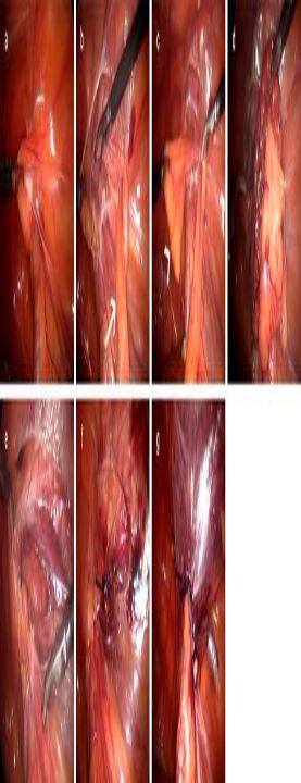
Citation: Lee SR, Park P (2018) Laparoscopic Treatment for Lipoma in the Inguinal Canal without Hernia: Intracorporeal Lipoma Excision and Suture
Repair of the Deep Inguinal Ring. J Clin Case Rep 8: 1065. doi: 10.4172/2165-7920.10001065
Page 2 of 3
presented as mean ± standard deviation and categorical variables as
frequency and percentage. The Shapiro–Wilk test was used to assess the
normality of continuous variables. Continuous variables were analyzed
using the t-test or Wilcoxon rank sum test, whereas categorical variables
were analyzed using the Fisher’s exact test or χ2 test. A probability (p)
value of ≤ 0.05 was considered statistically significant.
Results
Patient characteristics are shown in Table 1. There was no
conversion to open surgery and no difference in age between males
and females, although the body mass index was higher in males than
in females (24.8 ± 3.06 (19.5-33.7) kg/m2 vs. 21.7 ± 2.33 (18.1-25.0) kg/
Figure 2: Laparoscopic view of the deep inguinal ring. (A) Bulging of an indirect m2, p=0.004). The laterality of the IL was more common on the right
area (white arrow) without a hernia sac. (B) Bulging of a direct area (black side in both sexes, but there was no significant difference between
arrow) without a hernia sac. sexes. Inguinal pain was more frequent in female than in male patients
(90.9% vs. 74.3%), but this difference was not statistically significant.
The location of the IL tended to be in an indirect area more commonly
in male than in female patients (91.4% vs. 63.6%, p=0.046). IL was
separated with preperitoneal fat in 21 (60%) males and 6 (54.5%)
females (Figure 4). The mean surgical duration was approximately 31
min, with no significant difference between males and females. The
mean postoperative hospital stay was also similar between males and
females, with no significant difference in complication rates between
groups (one IH in one male patient). After conservative treatment, the
IH subsided with no recurrence to date.
Discussion
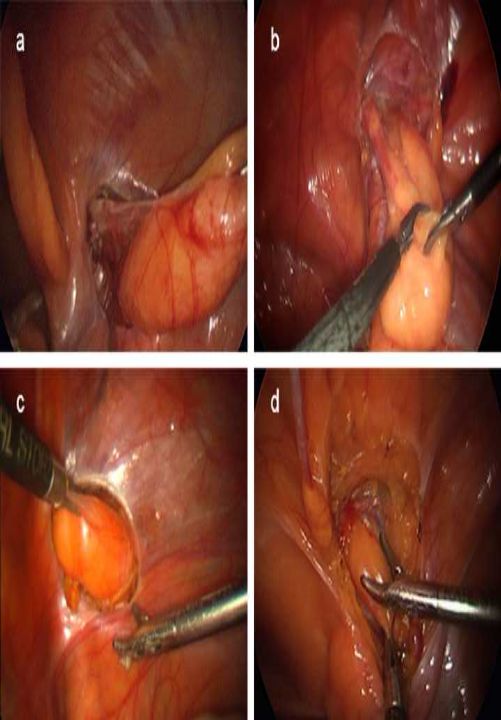
Figure 3: Surgical procedure. (A) Closed processus vaginalis. (B) First
dissection of an inguinal lipoma (IL; arrow) connected to preperitoneal fat. The IL primarily manifests as an inguinal bulge with symptoms similar
deep inguinal ring was observable after IL dissection. (C) Second dissection of to those of IH. The prevalence of IL was reportedly 75% among male
the IL (arrowhead) separated from the preperitoneal fat. (D) Full excision of the autopsy cases, although the symptoms were not confirmed [4]. In the
IL. (E) The deep inguinal ring after complete IL excision. (F) Suture repair of the
deep inguinal ring. (G) Peritoneum closure with continuous sutures.
present study, hyperechoic lesions were observed in the inguinal canal
of the hernia protruding from the greater omentum. Although IH was
the inguinal area revealed a peritoneum protruding into the abdominal
Variables Male (n=35) Female (n=11) p-value*
cavity (Figure 2). An incision of the peritoneum was performed using
48.7 ± 13.9 (19-
a dissector to confirm IL. After complete IL excision, the deep inguinal Age (years, range)
76)
50.5 ± 13.2 (34-70) 0.706
ring was closed using sutures with a nonabsorbable multifilament (silk 24.8 ± 3.06 (19.5- 21.7 ± 2.33 (18.1-
BMI (kg/m2, range) 0.004
1-0). The technical details of the suture are as follows: initially, sutures 33.7) 25.0)
were placed while avoiding the cord vessel and vas deferens and tied at Laterality
the bottom of the defect wall. Next, they were sutured in the upward Right 19 (54.3%) 7 (63.6%)
direction until the top of the defect wall, then continuously sutured Left 10 (28.6%) 4 (36.4%) 0.430
in the opposite direction and intracorporeally tied at the initial knot Both 6 (17.1%) 0 (0.0%)
(Figure 3). The peritoneum was closed with continuous absorbable Symptom
sutures (Vicryl 1-0). Inguinal bulging only 9 (25.7%) 1 (9.1%)
0.410
Bulging with pain 26 (74.3%) 10 (90.9%)
Follow-up protocols Location of lipoma
Eating and drinking were permitted after a 2-h observation period. Indirect 32 (91.4%) 7 (63.6%)
0.046
In accordance with the protocol of the institution, the patient was Direct 3 (8.6%) 4 (36.4%)
discharged once comfortable with daily activities, such as walking Connection to preperitoneal fat
and eating. Telephone interviews were conducted on day 2 to check Yes 14 (40.0%) 5 (45.5%) 1
the postoperative status, such as pain, wound state, possible symptoms No 21 (60.0%) 6 (54.5%) -
of hematoma, and seroma. The outpatient follow-up routine included Surgical duration 31.2 ± 11.8 (15-
31.4 ± 12.9 (17-55) 0.915
(minutes) 60)
physical examination at 7 days and 6 months and annual telephone
12.1 ±15.5 (3.33- 12.9 ± 12.9 (3.83-
follow-ups thereafter. The telephone follow-ups in this study were Hospital stay (hour)
67.6) 40.7)
0.321
conducted until June 2017. The mean follow-up period was 26.2 ± Complication
15.7 (range, 6-57) months for males and 28.8 ± 16.4 (8-55) months for Seroma 0 0 1
females. Hematoma 1 (2.9%) 0 -
Statistical analysis Follow-up period (months) 26.2 ± 15.7 (6-57) 28.8 ± 16.4 (8-55) 0.595
*Categorical variables were analyzed using the χ² test or Fisher’s exact test.
All statistical analyses were performed using R 3.3.2 software (R Continuous variables were analyzed using the independent samples t-test.
Development Core Team, Vienna, Austria). Continuous variables are Table 1: Patient characteristics.
J Clin Case Rep, an open access journal
Volume 8 • Issue 1 • 10001065
ISSN: 2165-7920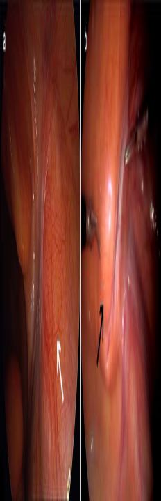
Citation: Lee SR, Park P (2018) Laparoscopic Treatment for Lipoma in the Inguinal Canal without Hernia: Intracorporeal Lipoma Excision and Suture
          Repair of the Deep Inguinal Ring. J Clin Case Rep 8: 1065. doi: 10.4172/2165-7920.10001065

                                                                                                                                                              Page 3 of 3

                                                                                      performed after IL resection. The current trend in surgical treatment is
                                                                                      to ensure minimal invasiveness and avoid recurrence. As IL occupies
                                                                                      the inguinal canal even when not accompanied by IH, it may appear
                                                                                      as an IH defect with no sac. Therefore, posterior wall repair must be
                                                                                      performed. Posterior wall repair using laparoscopic surgery with mesh
                                                                                      implantation and tissue repair and mesh implantation using open
                                                                                      surgery have been reported. However, tissue repair by laparoscopic
                                                                                      surgery has not been reported. The posterior wall repair method used
                                                                                      in this study was the same as Marcy repair in open surgery. In this
                                                                                      cohort, there has been no IL recurrence or IH development to date.

 Figure 4: Multiple characteristics of inguinal lipoma (IL). (A) IL connected to
                                                                                          As a limitation, this was a retrospective study conducted at a single
 preperitoneal fat in an indirect area in a male patient. (B) IL separated from the   center. In addition, no comparison was made with open surgeries.
 preperitoneal fat at an indirect area in a male patient. (C) IL separated from the   Laparoscopic excision was not difficult because the IL was located in
 preperitoneal fat at an indirect area in a female patient. (D) IL separated from     the direct IH defect or deep inguinal ring. Furthermore, the follow-up
 the preperitoneal fat at a direct area in a female patient.
                                                                                      period was relatively short; thus, longer follow-up terms are required to
                                                                                      obtain the IL recurrence or IH occurrence rates.
preoperatively diagnosed, the external compression of the inguinal
area with bulging of the IL was laparoscopically confirmed [5]. A                     Conclusion
misdiagnosis of IL can lead to IH recurrence and IL enlargement; thus,
current recommendations include the removal of IL with or without an                      In conclusion, when a patient presents with a symptom of an
associated IH [3,6]. Fatty tissues occupying the preperitoneal space can              inguinal bulge, the possibility of IL should be considered. In this study,
give rise to symptoms. One author described “true” IL as the one not                  46 IL patients without IH were successfully treated using laparoscopy
connected to extraperitoneal fat and confined to the inguinal canal [1],               by complete IL excision and intracorporeal suturing of the deep
while another described it as preperitoneal fat that projects through the             inguinal ring as posterior wall repair.
internal inguinal ring [2]. In this study, IL was defined as a protrusion             References
of preperitoneal fat or its separation from the retroperitoneal tissues.              1. Fawcett AN, Rooney PS (1997) Inguinal cord lipoma. Br J Surg 84: 1169.
In this study, 21 males and 6 females had an IL separated from the
preperitoneal fat. All ILs with direct defects were separated from the                2. Nyhus LM, Bombeck CT, Klein MS (1991) Textbook of Surgery: The Biological
                                                                                         Basis of Modern Surgical Practice (Sabiston DC eds), Philadelphia, USA.
preperitoneal fat. The prevalence of IL not associated with IH, reported
in previous studies, was greater than that in the present study (13%-16%              3. Bittner R, Arregui ME, Bisgaard T, Dudai M, Ferzli GS, et al. (2011) Guidelines
                                                                                         for laparoscopic (TAPP) and endoscopic (TEP) treatment of inguinal hernia
vs. 4.2%) [7,8]. In this study, the primary symptom in all patients was                  [International Endohernia Society (IEHS)]. Surgical endoscopy 25: 2773-2843.
a round movable inguinal mass, and 26 (74.3%) males and 10 (90.9%)
                                                                                      4. Heller CA, Marucci DD, Dunn T, Barr EM, Houang M, et al. (2002) Inguinal
females complained of pain. The incidence of pain in the present study                   canal “lipoma”. Clin Anat 15: 280-285.
was similar to that in previous reports (78% vs. 77%) [7].
                                                                                      5. Yang XF, Liu JL (2016) Laparoscopic repair of inguinal hernia in adults. Ann
    IL is difficult to distinguish from IH through physical examination.                 Transl Med 4: 402.
Preoperative ultrasonography showed a round movable mass                              6. Felix E, Scott S, Crafton B, Geis P, Duncan T, et al. (1998) Causes of recurrence
composed of dense fat similar to a herniated omentum. Pelvic                             after laparoscopic hernioplasty. A multicenter study. Surgical Endoscopy 12:
computed tomography (CT) is reportedly useful to accurately diagnose                     226-231.
IL. However, as a drawback, pelvic CT is not applicable to all patients               7. Lilly MC, Arregui ME (2002) Lipomas of the cord and round ligament. Ann Surg
due to cost concerns [9]. In this study, one female patient was diagnosed                235: 586-590.
with preoperative IL by pelvic CT (Figure 1), IL was confirmed in 63%                 8. Nasr AO, Tormey S, Walsh TN (2005) Lipoma of the cord and round ligament:
(29/46) of cases by preoperative sonography, and 35% (16/46) cases                       An overlooked diagnosis? Hernia. J Hernias Abdominal Wall Surg 9: 245-247.
were indistinguishable from those of IH. In a previous report, 55% of                 9. Fataar S (2011) CT of inguinal canal lipomas and fat-containing inguinal
cases were preoperatively diagnosed by pelvic CT [7].                                    hernias. J Med Imaging Radiat Oncol 55: 485-492.

    IL is often found during IH surgery. Both open and laparoscopic                   10. Carilli S, Alper A, Emre A (2004) Inguinal cord lipomas. Hernia. J Hernias
                                                                                          Abdominal Wall Surg 8: 252-254.
surgeries have been applied for herniorrhaphy [8,10]. Most surgeons
agree that IL is not accompanied by IH, but its treatment is based on                 11. Yener O, Demir M, Yigitbasi R, Yilmaz A (2013) Missed lipoma of the spermatic
that of IH [11]. Therefore, we agree that posterior wall repair should be                 cord. Prague Med Rep 114: 5-8.

 J Clin Case Rep, an open access journal
                                                                                                                                          Volume 8 • Issue 1 • 10001065
 ISSN: 2165-7920
You can also read