Rheumatology Pearls and Updates - Shriram Lokare, MD UnityPoint Health Rheumatology August 1, 2019
←
→
Page content transcription
If your browser does not render page correctly, please read the page content below
Rheumatology Pearls and
      Updates
           Shriram Lokare, MD
     UnityPoint Health Rheumatology

            August 1, 2019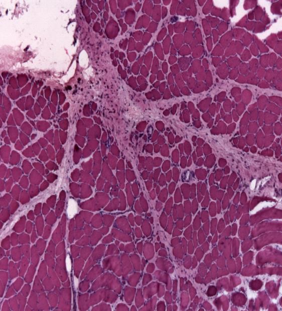
Disclosures
None

References
. ACR practice guidelines
. Primer on Rheumatic diseases
. Kelley’s textbook of Rheumatology
. ACR Image bank
. National Osteoporosis Foundation
Diagnosis and management of Rheumatological diseases remain challenging both
for Primary Care Physicians and Rheumatologists.

Rheumatological diseases are rare but still significantly prevalent.

Musculoskeletal disorders cause 30% of visits to Physicians in the US.

Rheumatological diseases are responsible for increased work and functional
disability.

Rheumatologic diseases are high cost. More than $150 billion every year.
Early diagnosis and referral can result in significantly better outcomes.

One of the least focused areas of our educational curriculums.
Rheumatology rotations are not mandatory.
Educational Objectives
. Approaching rheumatological diseases. Recognizing patterns.

. Appropriate laboratory testing and understanding limitations of labs.

. Overview of therapeutic plan. Different treatment options.

. Developing comprehensive care plan and a team approach.
We will be reviewing...
Rheumatoid Arthritis

Seronegative Spondyloarthropathy

SLE

Myositis

Polymyalgia Rheumatica

Giant Cell Arteritis

Osteoporosis

Gout , Pseudogout

Soft tissue rheumatism

Rheumatoid Arthritis
The most common autoimmune inflammatory arthritis in adults (1%).
Women/men= 2/1
Onset - insidious or sudden.

Common symptoms-
. joint pains, joint swelling, morning stiffness > 30-60 min
. tingling in the hands
. feeling ill, worsening fatigue, anorexia, weight loss.

Labs
ESR, CRP - may be elevated in upto 40-60% patients at presentation
RF – seen in up to 40- 50 %
Anti CCP Antibody- more specific, high risk of erosive and polyarticular
disease, higher risk of extra-articular disease.

Other labs- leucocytosis, thrombocytosis, elevated alkaline
phosphatase, decreased serum albumin
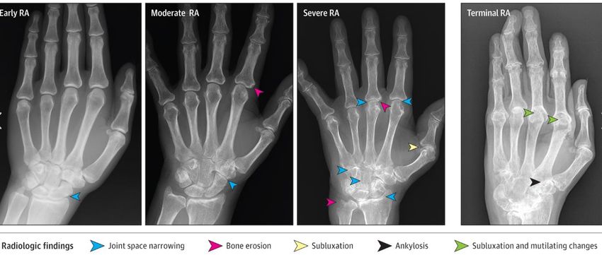
Xrays
RA Treatment Guidelines
– Early referral to a Rheumatologist
– Rapid control of symptoms with short-term low-dose glucocorticoids
– DMARDs therapy as soon as possible with early aggressive
treatment approach
– Advance to biologics or escalate treatment if no response
– Multidisciplinary approach
– Treat to Target therapy
– Monitoring for comorbid conditions, complications and drug toxicities
Non- steroidal DMARD Medications
Seronegative Spondyloarthropathy
Initial work up of a patient with Rheumatologic disease
CBC with differential
CMP
ESR
CRP
Hepatitis B/C serologies
TB quantiferon gold test
RF and Anti CCP antibody
ANA
Serum CPK
Gout
Excess body burden of uric acid.
Spectrum of clinical and pathologic features due mostly to tissue
deposition of monosodium urate monohydrate crystals (MSU).
Hyperuricemia = serum uric acid > 6.8 or 7.0 mg/dl.
Typically presentation: acute and episodic
Can be chronic

Prevalence in the US estimated at 3.9% of adults (8.3 million people)
Risk factors
HTN
Obesity
Metabolic syndrome
DM2
CKD
Diuretics
Psoriasis
Treatment of Gout

Acute Gout - NSAIDs, Steroids, Colchicine

UA lowering agents - allopurinol, febuxostat, probenecid, Losartan
   Serum UA Target < 6 mg/dl
   Tophaceous gout < 5 mg/dl

Refractory gout- Pegloticase (Krystexxa)
Pseudogout
Due to deposition of calcium pyrophosphate dihydrate in the cartilage.

Can be misdiagnosed as inflammatory arthritis or cellulitis.

Often associated with osteoarthritis.

Associated diseases-
 1.   Hyperparathyroidism
 2.   Hemochromatosis
 3.   Hypothyroidism
Clinical features of Pseudogout

Joint involvement - MCP, wrists, elbows, hips, knees, shoulders.

Diagnostic clues -
1.     sudden onset symptoms, mainly pain, swelling, erythema
2.     lack of morning stiffness
3.     distribution of involved joint (s)
4.     typical appearance on x-rays , e.g. chondrocalcinosis
5.     Synovial fluid- rod shaped crystals on polarized microscopy
Treatment of Pseudogout

. NSAIDs
. Colchicine.
. Steroids- orally or intraarticular

. Treat the underlying cause if it was identified.
Polymyalgia Rheumatica
Age ≥ 50

Bilateral upper extremity pain and or pelvic girdle pain > 2 weeks with
morning AM stiffness > 45 min.
ESR and CRP are often moderately high.

16-21% of patients may have GCA. These patients may have
unilateral or bilateral headaches, jaw claudication, myalgias, fatigue,
fever, weight loss, and sometimes acute vision loss.
PMR Treatment
Start treatment with prednisone- 12.5 mg to 25 mg daily.
Decrease prednisone by 2.5 mg to 5 mg every 2 weeks. Assess
response by symptoms and lab improvement.
Once the symptoms are under remission at prednisone 10 mg dose,
then decrease the dose gradually by 1 mg every 4 weekly.

Some patients can remain symptoms free after the steroids are
discontinued. More than 50% would have a recurrence.

Consider steroid sparing agents- Methotrexate, Azathioprine
Giant Cell Arteritis
Usually age > 50 yr. More in women.

Most common presentations -
1. Temporal arteritis. (severe headaches, jaw claudication, vision
   changes)
2. Elderly patient with myalgia, fatigue, involuntary weight loss
3. Patient with PMR with recurrence of symptoms at prednisone 10-
   15 mg/day or persistent ESR/CRP.
4. Incidental finding on a CT scan
Investigations
ESR, CRP - often high.
Studies have shown that 25% of all patients with positive TA biopsy
had normal ESR.

Temporal artery biopsy is the gold standard.
Can be done with 1- 4 days of steroid initiation without significant
effect on biopsy results.
Bilateral TA biopsies are recommended to increase the yield. Skip
lesions are common.
At least 2 cm of arterial sampling is recommended.
Recommend Pathologist to do internal elastic lamina staining.
GCA therapy
. Depending on the severity of disease.
. For temporal arteritis- Initially IV Methylprednisolone 1000 mg for 3-5
days.
. Transition to prednisone 1 mg /kg/ day (max dose of 80 mg/ day).
. Decrease prednisone by 10% every 1-2 weeks.
. Steroid sparing agents - Actemra (FDA approval). Methotrexate,
Imuran, Cellcept have been used as well.
. Aspirin 81-325 mg per day , if no contraindications.
ANA
Auto-antibodies against antigens in the cell nucleus.
Diseases associated with ANA
SLE, Sjogrens, MCTD, Scleroderma, Myositis
R.A, Psoriatic arthritis, ANCA vasculitis
Hashimoto’s disease, Grave’s disease
IBD
Infections
Cancers
Medications- NSAIDs, minocycline, hydralazine
Healthy individuals 3-15%. More so, beyond age 65
SLE

Female : Male- 9:1

Usually around menarche, pregnancies.

Aggressive disease in African – American patients.
SLICC diagnostic criteria
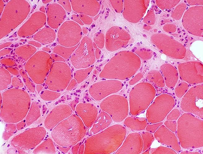
Inflammatory Myositis
Clinical features-
1.    Symmetric proximal muscle weakness. Often painless.
2.    Insidious or acute
3.    Low grade fevers, fatigue can be present
4.    Rashes of Dermatomyositis.
5.    Associated symptoms may include- inflammatory arthritis,
     Raynaud’s, GERD, dysphagia, shortness of breath.
Diagnosis-
1.   Elevated CPK, Aldolase, LDH, AST/ALT
2.   MRI evidence of myositis
3.   EMG
4.   Muscle biopsy
Dermatomyositis Polymyosits Inclusion body
myositisDifferential diagnosis of Myositis Asymptomatic CPK elevation, can be seen in upto 30% people Infections- viral Severe hypothyroidism Dyselectrolytemia - hypokalemia Medications- statins, steroids, colchicine, diuretics Metabolic myopathies Muscular dystrophies
Treatment of Myositis
Steroids - careful long term use advised as it can cause steroid induced myopathy.

Methotrexate

Azathioprine

Mycophenolate mofetil

Cyclosporine

IVIG

Plasmapheresis

Rituximab

Cyclophosphamide
Osteoporosis

About 10 million with osteoporosis in the US. About 43 millions have low BMD.

Estimated lifetime risk of a fracture from osteoporosis -

 1. 50% in Caucasian women
 2. 20% in men
Hip fracture- 8-36% increased mortality with in 1 year.

Vertebral fracture- 5 fold increased risk of future fractures.
Diagnosis
.

The FRAX tool is a fracture risk assessment tool.

 Based on the patient's age, body mass index (BMI), tobacco use, alcohol use, presence of RA, use of glucocorticoids, prior history of
fracture, parental history of hip fracture and presence of secondary causes of osteoporosis.

The FRAX tool can be applied to patients with osteopenia (T-score between -1.0 and -2.5) who have not been taking medications for
osteoporosis (except for Calcium and Vitamin D) and age > 50 yr.
Risk factors for low BMD
. Post menopausal
. Diet limited in calcium, vitamin D
. Alcohol > 3 drinks per day
. Smoking- active or passive
. Low BMI
. Low activity / immobilization
. Medications- steroids, Warfarin, Omeprazole, Phenytoin,
Furosemide, Pioglitazone, Anastrozole, Orlistat
. Ethnicity- Caucasian, Asian
Osteoporosis treatment
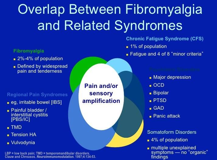
Fibromyalgia
                -   4% of US population

                -   Chronic widespread
                    pain, TENDERNESS
                -   Increased pain
                    sensitivity

               Frequently associated
               symptoms include-
                - Chronic fatigue
                - Poor nonrestorative
                   sleep
                - Memory issues
                - IBS
                - Interstitial cystitis
Labs to be ordered for evaluating Fibromyalgia CBC CMP ESR, CRP CPK TSH Hep B/C serologies Serum Vitamin D Other labs as pertinent- RF, CCP antibody, ANA, Lyme
Treatment of Fibromyalgia
. Targeting the cause, if identifiable- depression, poor sleep, snoring, arthritis, etc
. Education about their disease, sleep hygiene, exercises.
. Stressing the importance of patient participation in symptom management.
. Shared responsibility between different providers.
. CBT- relaxation training, goal setting, distraction strategy, problem solving.
. Referrals- sleep study, P.T. , Psychologist, Psychiatrist, Pain clinic, Chiropractors,
Acupuncture, Massage therapy.
. FMLA/ letters to employers to request accommodation
Meds - NSAIDs, muscle relaxants, Elavil, Cymbalta, Neurontin, Lyrica.

Establish a realistic expectation when controlling pain with
medications.

Refrain from using narcotics on a long term basis.
Vaccinations
Flu vaccines - regular vs sequential vs high dose flu vaccine

Pneumovax 23 - can be given every 5 years
Prevnar 13

Shingrix - this is being increasingly recommended by Rheumatologists

Live vaccinations- can be considered after consultation with a
Rheumatologist or Infectious disease. May need DMARD meds to be
Management of infections in Rheumatologic
diseases
Contact the Rheumatologist.

Most DMARD medications except steroids can be held while infections
are being treated.

Sometimes, patient may in fact need higher dose of steroids if they are
steroid dependent.
PreOp evaluation
Perioperative use of DMARD medications needs to be carefully weighed with both
patient and the Orthopedic surgeon.

Patient with chronic steroid use - may need stress dosing of steroids. Many
anesthesiologist would also opine on this and can be involved in decision making.

Patient with neck pain or long standing inflammatory arthritis - please get x-ray of
cervical spine with 5 views.
Contraception
Patients (and their partners, if possible) need to be made aware of the need for
double contraception (contraceptive pills /IUD and barrier contraception).
Reasses any conception plans and contraception methods at every visit.

Estrogen containing contraceptives can be used but need to weighed carefully,
especially in SLE, APS. Involve Rheumatologist in discussion as well.

Most DMARDs need to be discontinued at least 3 months prior to
conception.
If a patient is on Arava- cholestyramine chelation therapy is highly recommended.
In case of an unexpected pregnancy while on DMARDs, a referral to high
risk OB need to be done.
Pregnancy
RA activity can lessen during pregnancy. Lupus disease activity can flare up.
Medications that can be used- Plaquenil, Steroids, Imuran, Sulfasalazine.
Most pregnancies are uneventful. But, a high risk OB referral is recommended.
Sjogrens/ SLE - need close and frequent monitoring by OB, high risk OB and
Rheumatologist.
   . Anti SSA/ SSB antibodies- risk of fetal heart block, neonatal lupus
   . HELLP syndrome
   . ITP
   . Antiphospholipid syndrome- involve Hematology as well
   . if having significant anemia- involve Hematology as well
Non-pharmacological aspects of care

Exercises - strength, conditioning, balance, improved tolerance to pain
Smoking cessation.
Limited alcohol use.
Dietary changes.
Emotional support. Assessing depression.
Education of family members when they accompany patients.
Some important clinical findings..
DLE
SLE
Relapsing polychondritis
Sjogren’s syndrome
Baker’s cyst
Rheumatoid nodules
Calcinosis cutis
Edematous phase of scleroderma
Keratoderma blenorrhgaicum- Reactive arthritis
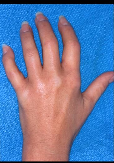
Dactylitis
Nail pitting changes in psoriatic arthritis
Periungal erythema- scleroderma, myositis
Mechanic’s hands- Anti synthetase syndrome
(myositis, inflammatory arthritis, scleroderma)
Dermatomyositis
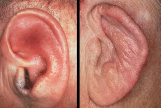
Fluoroquinolone related tendinitis
Plaquenil related eye toxicity
Atypical fracture from prolonged bisphosphonate therapy
Injection site reaction from biologic medications
Stevens Johnsons syndrome from Sulfasalazine
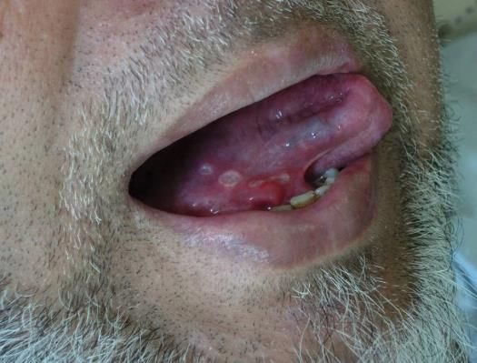
Mucosal ulcerations are suspicious when they are
recurrent, idiopathic, long lasting ulcers at atypical
sites.
Erythema nodosum- IBD related arthritis
Advantage of DECT- Dual energy Ct scan
                      This 70 year-old man with longstanding
                      seropositive rheumatoid arthritis was
                      evaluated for right index metacarpophalangeal
                      (MCP) swelling.

                      Because of a remote history of gout, a dual
                      energy CT scan was obtained and showed
                      monosodium urate deposits (green pixelation)
                      in multiple MCP and interphalangeal (IP)
                      joints. Also seen were carpal collapse and
                      MCP and IP subluxations consistent with
                      advanced rheumatoid arthritis.

                      The patient's uric acid at the time of the study
                      was 2.9 mg/dl.
Questions?
Thank you.
Joint involvement in different arthritis
You can also read