Objective Evaluation of the Longevity of A Calcium Hydroxylapatite-Based Filler (Radiesse)
←
→
Page content transcription
If your browser does not render page correctly, please read the page content below
Journal of Clinical and Cosmetic Dermatology
          Sci Forschen
                 Open HUB for Scientific Researc h   ISSN 2576-2826 | Open Access

RESEARCH ARTICLE                                                                                                                         Volume 5 - Issue 2

Objective Evaluation of the Longevity of A Calcium Hydroxylapatite-Based
Filler (Radiesse®)
Maria Angelo-Khattar*
Overseas Director, American Academy of Anti-aging Medicine, Dubai, UAE

*Corresponding author: Maria Angelo-Khattar, Overseas Director, American Academy of Anti-aging Medicine, Dubai, UAE, Tel: 971506245494; E-mail:
mkhattar@younatagroup.com

Received: 19 Apr, 2021 | Accepted: 15 May, 2021 | Published: 22 May, 2021

Citation: Angelo-Khattar M (2021) Objective Evaluation of the Longevity of A Calcium Hydroxylapatite-Based Filler (Radiesse®). J Clin Cosmet
Dermatol 5(2): dx.doi.org/10.16966/2576-2826.163
Copyright: © 2021 Angelo-Khattar M. This is an open-access article distributed under the terms of the Creative Commons Attribution License,
which permits unrestricted use, distribution, and reproduction in any medium, provided the original author and source are credited.

Abstract
Background: Calcium hydroxylapatite (CaHA) based soft-tissue filler (Radiesse®, Merz) is categorised as a biodegradable collagen-stimulating implant.
It is comprised of a suspension of 25-45 micron diameter microspheres of CaHA(30%) in a carboxymethylcellulose (CMC) gel carrier (70%) and is one
of the most well researched soft-tissue fillers. However, great discrepancies exist in the literature regarding the duration of clinical effect with the
implant, ranging from only 6 to 24 months.
Objective: This is a retrospective chart analysis of four patients injected with CaHA filler for volume augmentation in the malar region of the face.
Objective volume calculations were performed with the Canfield Vectra 3D Imaging System three and five months post-implantation, with the view
to determine the longevity of the volumizing effect of the filler substance.
Discussion: CaHA based filler is believed to afford immediate volume restoration due to the CMC gel component and a long-term action due to
neocollagenesis induced by the CaHA microspheres. The CMC gel is known to dissipate within 6-8 weeks, only to be replaced by new collagen
induced by the CaHA particles. Thus soft-tissue formation is believed to lead to a sustained volumizing effect.
Results of this retrospective study clearly show that the volume replacement afforded by the filler was not sustained, with 65% and up to 96%
reduction in volume at five months.
Conclusion: The volumizing effect of CaHA based filler is shown to have limited longevity, despite the fact that it is a proven biostimulant. It is
possible that the CaHA microspheres are degraded relatively swiftly in interstitial fluid and therefore cannot sustain their biostimulating effect.

Keywords: Calcium hydrxylapatite; Radiesse®; Biostimulant; Poly-L-lactic acid; Vectra 3D

Introduction                                                                    between 6-12 months, since they are metabolized by endogenous
                                                                                hyaluronidase enzymes. The hyaluronic acid filler’s longevity depends
   Soft tissue fillers, originally developed for the filling of rhytids and
                                                                                upon the extent of cross-linkage and the concentration and particle
folds, are currently used for the global restoration of facial volume
                                                                                size of each product. These medical devices are replacement fillers
and optimization of facial contours as in chin, tear trough, cheek, lip,
                                                                                and simply restore lost volume but do not induce long-term collagen-
and jawline correction. Recently several fillers have proven to be of
                                                                                stimulating effects [5,6].
value in the correction of non-facial areas. They are of great benefit in
hand rejuvenation, rejuvenation of the décolleté and neck, reduction               The non-biodegradable products are generally not approved in
of cellulite dimpling, buttock augmentation, volumizing of the labia            most countries as they are associated with severe side effects and
majoré, and post-liposuction deformity correction [1,2].                        persistent granulomas that can occur several years after implantation
                                                                                [4].
    The current plethora of soft-tissue filler substances essentially
comprises three general categories; the replacement biodegradable                  The quest for longer-lasting biodegradable substances has resulted
fillers (Hyaluronic acid products), the permanent non-biodegradable             in the development of substances that provide longer-term correction
fillers (ex. Polymethylmethacrylate, polyacrylamide gel, silicone), and         by inducing new soft tissue formation. This category of biodegradable
biodegradable collagen-stimulating substances. There is no doubt that           collagen-stimulating substances includes three types of implantable
hyaluronans are the most widely used soft-tissue fillers worldwide              medical devices; poly-l-lactic acid (PLLA), calcium hydroxyapatite
[3,4]. This is despite the fact that they have limited longevity,               (CaHA), and polycaprolactone-based fillers. Typically, these are

J Clin Cosmet Dermatol | JCCD                                                                                                                          1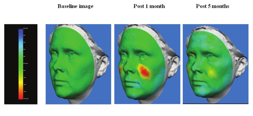
Sci Forschen
         Open HUB for Scientific Researc h
                                                                                                          Journal of Clinical and Cosmetic Dermatology
                                                                                                                                    Open Access Journal

particulate substances between 25 to 45 microns in size that illicit         importing the baseline and the 3 and 5 month images into the Vectra
an inflammatory reaction resulting in encapsulation of the particles         mirror analysis 3D software. The mid-face region was selected in each
and the prevention of particle migration. They ultimately lead to            case and highlighted as the area to be measured. Change in mid-face
neocollagenesis, and the induction of new soft tissue formation by           volume was computed by registering each the post-treatment images
these biostimulating substances results in long-lasting cosmetic             against the baseline image. All volume measurements were recorded
correction, in certain cases persisting well over two years [7,8].           in millilitres.
   CaHA-based filler (Radiesse®, Merz) is comprised of a suspension             Furthermore, qualitative volume visualization of the degree of
of CaHA microsphere ranging from 25 to 45 micron in diameter, in a           contour change was made possible with a colour distance map (See
carboxymethyl cellulose gel carrier [9]. It is one of the most extensively   figure 2).
studied fillers currently used for soft tissue augmentation. However,
great discrepancies exist in the literature regarding the duration           Results
of clinical effect with the implant, ranging anywhere from only 6            Qualitative data
months to 24 months [9-13]. The vast majority of these studies have
employed subjective evaluation scales such as the global aesthetic              Figure 2 is representative of colour distance maps indicating a
improvement scale and the wrinkle severity rating scale, which rely on       qualitative change in volume over time. The colour scale is shown
the investigator’s and patient’s evaluation of the results.                  whereby green is baseline and any increase in volume manifests
                                                                             incrementally as yellow and red.
   A review of the literature revealed only one objective study whereby
3D surface scanning was employed for the quantification of soft tissue          Figure 2 represents a 45-year-old patient who had been injected
changes in three female subjects treated with CaHA filler. Volume            with a total of 1.5ml of CaHA filler on both malar areas. The yellow and
quantifications were made immediately post injection, 2 weeks and            intense red colours are indicative of volume increase from baseline,
                                                                             seen at one month post treatment. The decline in colour intensity at
six months after injection with an Artec MHT surface scanner (Artec
                                                                             the fifth month is a clear indication that the volume was not sustained.
Group, San Diego, California). The authors concluded that the filler
persisted for six months after treatment. However qualitative data           Quantitative data
from the colour coding of the heat maps showed a clear decline in
                                                                                Table 1 shows the volume of CaHA filler injected on each side of
volume at six months [14].
                                                                             the face and the total volume, for each of the patients. The volume
   Three-dimensional (3D) imaging systems for the face and body are          remaining at 3 months and 5 months are also shown. The residual
currently employed for surgical planning and research. The Canfield          volume at 5 months, computed as a percentage of total filler remaining,
3D stereophotogrammetric camera and software (Vectra; Canfield               was only a fraction of the original volume, between 4% and 35%.
Scientific, Fairfield, NJ) is being increasingly used in computerized
volume calculations post filler injections, breast augmentation and          Discussion
cryolipolysis [15-18]. The system has been shown to be reliable and             The CaHA-based filler has gained great popularity as both a
reproducible and the 3D imaging system measurements show a linear            volumizing agent and a treatment modality for skin rejuvenation
association with Magnetic Resonance Imaging [19].                            in non-facial skin such as on the neck, decolletage, inner arms, and
Materials and Methods                                                        hands. In certain cases, the bio-stimulant is used concomitantly with
                                                                             microfocused ultrasound for optimal effects [20-24].
   This is a retrospective chart review of four female subjects, aged
28-50 years, treated with CaHA filler on the malar area of the face,            The mechanism of action of the soft-tissue filler ultimately depends
between january to june 2015. The subjects retrospectively studied           upon the activation of fibroblasts by the CaHA microspheres to elicit
were randomly selected. Ethical approval to analyse the patient data         collagen production. Histological and electron microscopic studies in
retrospectively was awarded by the Clinic Director based on consent          human volunteers have demonstrated new collagen deposition around
given by the patients to analyse and publish the data (Figure 1).            the microspheres for up to six months [25,26]. Immunohistochemical
                                                                             analysis [23,25] demonstrated a significant increase in both collagen
Injection technique                                                          type I and type III at 4 and 7 months post-treatment. A more recent
   In our clinics, all patients that receive filler injections in the        study using picrosirius red staining and polarized light microscopy,
infraorbital area are pre-treated with an infraorbital block (2%             2 months after implantation, mainly newly formed type III collagen
xylocaine+1:80,000 adrenaline). The CaHA filler is then injected onto        was identified [27]. Hence, there is no doubt that CaHA in a collagen
the supra-periosteal plane in the malar area on both sides of the face,      stimulating agent. Nonetheless, several studies have demonstrated
with a 22 G × 50 mm blunt-tipped cannula.                                    limited clinical efficacy [14] with the filler and, our results using
Imaging                                                                      quantitative Vectra 3D evaluation have also shown a clear decline in
                                                                             volume over five months. Hence it may be concluded that the collagen
   The Vectra 3D Imaging system (Canfield Imaging Systems,                   stimulating effects of the CaHA filler are not long-lasting.
Fairfield, N.J) is used as part of standard of care in our clinics. The
system contains six cameras and captures images in 180 degrees.                  Data from studies on the longevity of another collagen stimulating
Standardized full face 2D and 3D images are generated. The high              agents, Poly-L-Lactic acid (PLLA), shows that in contrast to CaHA
resolution 3 D images produced by the Vectra software can be used            filler, the volumizing action of PLLA is between 2 to 3 years [28-32].
for both qualitative volume visualisation and quantitative volume               PLLA is a biodegradeable and biocompatible polyhydroxyacid that
measurements.                                                                does not augment the skin directly but has an indirect effect due to
   Standardized full-face 2D and 3D digital surface imaging were taken       neocollagenesis [33]. When injected into the tissue, the particles of
of the four patients at baseline and then at 3 months and 5 months after     PLLA degrade over time, only to be replaced by the patients’ own
the injection session. Data from the four patients were analysed by          collagen, a process that persists for up to 25 months [34,35].

Citation: Angelo-Khattar M (2021) Objective Evaluation of the Longevity of A Calcium Hydroxylapatite-Based Filler (Radiesse®). J Clin
                                                                                                                                                  2
Cosmet Dermatol 5(2): dx.doi.org/10.16966/2576-2826.163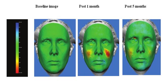
Sci Forschen
Open HUB for Scientific Researc h
Journal of Clinical and Cosmetic Dermatology
Open Access Journal
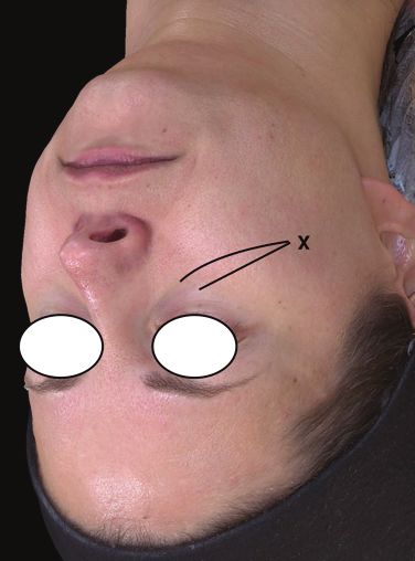
Figure 1: Site of injection of Calcium hydroxylapatite filler in the mid face. A 22 G, 50 mm length cannula was inserted at point X on the zygomatic
arch and the filler was deposited on the supraperiosteal plane.
Figure 2: Frontal and profile colour distance maps of 45-year old patient injected with a total volume of 1.5 cc of CaHA filler both malar areas.
Table 1: The volume of CaHA filler injected on each side of the face and the total volume for each of the patients.
Volume CaHA filler Volume % Residual % Decrease in
Total Volume CaHA Total Volume at Total Volume at
Patient injected on right side CaHA filler injected volume at 5 volume at 5
filler injected 3 months 5 months
(cc) on left side months months
AP 0.45 0.35 0.80 0.39 0.02 4 96
GE 0.35 0.45 0.80 0.20 0.06 7.5 92.5
JK 0.55 0.45 1.00 0.49 0.35 35.0 65
KK 0.40 0.30 0.70 0.24 0.07 10 90
Citation: Angelo-Khattar M (2021) Objective Evaluation of the Longevity of A Calcium Hydroxylapatite-Based Filler (Radiesse®). J Clin
3
Cosmet Dermatol 5(2): dx.doi.org/10.16966/2576-2826.163Sci Forschen
Open HUB for Scientific Researc h
Journal of Clinical and Cosmetic Dermatology
Open Access Journal
A study using 3-D digital imaging showed that subjects treated 11. Tzikas TL (2008) A 52-month summary of results using calcium
with PLLA had significant increases from baseline in mid-face hydroxylapatite for facial soft tissue augmentation. Dermatol Surg
volume at the end of the trial, six months after the first injection 34 Suppl 1: S9-S15.
session [36]. 12. Sadick SN, Katz EB, Roy D (2007) A multicenter, 47-month study
Over 29 types of collagen are present in the human body with of safety and efficacy of calcium hydroxylapatite for soft tissue
types I and III being predominantly found in the skin. The collagen augmentation of nasolabial folds and other areas of the face.
Dermatol Surg 33: S122-S126.
fibres, which are the most abundant components of the extracellular
matrix, are of high tensile strength and impart strength and resilence 13. Jacovella PF (2008) Use of calcium hydroxylapatite (Radiesse®) for
to the skin [37].The efficacy of biostimulating agents in inducing neo- facial augmentation. Clin Interv Aging 3: 161-174.
collagenesis and new soft tissue formation is contingent upon on the 14. Simunovic F, Schlager S, Montanari M, Iblher N (2017) Prospective
persistence of the biostimulant in the tissue to allow for long-term 3D analysis of facial soft tissue augmentation with calcium
fibroblast stimulation. hydroxylapatite. J Cosmet Laser Ther 19: 283-289.
Hence, the limited volumizing action of CaHA may be explained 15. Lowe P, Lowe NJ, Patnaik R (2011) Three-dimensional digital surface
by the limited longevity of the microspheres in the body, possibly imaging measurement of the volumizing effect of injectable poly-L-
due to the swift degradation of the CaHA microspheres in interstitial lactic acid for nasolabial folds. J Cosmet Laser Ther 13: 87-94.
fluid.
16. Killaars RC, Preuβ MLG, de Vos NJP, Van Berlo CCJLY, Lobbes MBI, et
Conclusion al. (2020) Clinical Assessment of Breast Volume: Can 3D Imaging Be
the Gold Standard? last Reconstr Surg Glob Open 8: e3236.
Calcium hydroxylapatite soft tissue filler is a proven biostimulant.
However, objective volume assessments performed with the Canfield 17. Garibyan L, Sipprell WH, Jalian HR, Sakamoto FH, Avram M, et al.
Vectra 3D analysis system, showed that the volume augmentation with (2014) Three-dimensional volumetric quantification of fat loss
the filler is not sustained but on the contrary, a significant decline is following cryolipolysis. Lasers Surg Med 46: 75-80.
seen within five months of injection. 18. Landau MJ, Kim JS, Gould DJ, Patel KM (2018) Vectra 3D Imaging
for Quantitative Volumetric Analysis of the Upper Limb: A Feasibility
Disclosure
Study for Tracking Outcomes of Lymphedema Treatment. Plast
The author reports no conflicts of interest in this work. Reconstr Surg 141: 80e-84e.
References 19. De Menzes M, Rosati R, Ferrario VF, Sforza C (2010) Accuracy and
reproducibility of a 3-dimensional stereophotogrammetric imaging
1. Goldberg DJ, Bass LM, Fitzgerald R, Graivier MH, Lorenc ZP (2018)
system. J Oral Maxillofac Surg 68: 2129-2135.
Expanding Treatment Options for Injectable Agents. Aesthet Surg J
38: S1-S7. 20. Amselam M (2015) Radiesse(®): a novel rejuvenation treatment for
2. Jabbar A, Arruda S, Sadick N (2017) Off Face Usage of Poly-L-Lactic the upper arms. Clin Cosmet Investig Dermatol 9: 9-14.
Acid for Body Rejuvenation. J Drugs Dermatol 16: 489-494. 21. De Almeida AT, Figueredo V, Da Cunha ALG, Casabona G, De Faria
3. Rohrich RJ, Bartlett EL, Dayan E (2019) Practical Approach and Safety JRC, et al. (2019) Consensus Recommendations for the Use of
of Hyaluronic Acid Fillers. Plast Reconstr Surg Glob Open 7: e2172. Hyperdiluted Calcium Hydroxyapatite (Radiesse) as a Face and Body
Biostimulatory Agent. Plast Reconstr Surg Glob Open 7: e2160.
4. Funt D, Pavicic T (2013) Dermal fillers in aesthetics: an overview
of adverse events and treatment approaches. Clin Cosmet Investig 22. Casabona G, Marchese P (2017) Calcium Hydroxylapatite Combined
Dermatol 6: 295-316. with Microneedling and Ascorbic Acid is Effective for Treating Stretch
Marks. Plast Reconstr Surg Glob Open 5: e1474.
5. Keen MA (2017) Hyaluronic Acid in Dermatology. 15: 441-448.
23. Yutskovskaya YA, Kogan EA (2017) Improved Neocollagenesis and
6. Lee W, Hwang SG, Oh W, Kim CY, Lee JL, et al. (2020) Practical Skin Mechanical Properties After Injection of Diluted Calcium
Guidelines for Hyaluronic Acid Soft-Tissue Filler Use in Facial
Hydroxylapatite in the Neck and Décolletage: A Pilot Study. J Drugs
Rejuvenation. Dermatol Surg 46: 41-49.
Dermatol 16: 68-74.
7. Breithaupt A, Fitzgerald R (2015) Collagen Stimulators: Poly-L-Lactic
24. Casabona G, Teixeira DN (2018) Microfocused ultrasound in
Acid and Calcium Hydroxyl Apatite. Facial Plast SurgClin North Am
combination with diluted calcium hydroxylapatite for improving
23: 459-469.
skin laxity and the appearance of lines in the neck and décolletage.
8. Melo FD, Nicolau P, Piovano L, Lin SL, Fernandes TB, et al. (2017) J Cosmet Dermatol 17: 66-72.
Recommendations for volume augmentation and rejuvenation of
the face and hands with the new generation polycaprolactone- 25. Berlin AL, Hussain M, Goldberg DJ (2008) Calcium hydroxylapatite
based collagen stimulator (Ellansé®). Clin Cosmet Investig Dermatol filler for facial rejuvenation: a histologic and immunohistochemical
10: 431-440. analysis. Dermatol Surg 34: S64-S67.
9. Loghem JV, Yutskovskaya YA, Werschler WP (2015) Calcium 26. Marmur ES, Phelps R, Goldberg DJ (2004) Clinical, histologic
hydroxylapatite: over a decade of clinical experience. J Clin Aesthet and electron microscopic findings after injection of a
Dermatol 8: 38-49. calciumhydroxylapatite filler. J Cosmet Laser Ther 6: 223-226.
10. Emer J, Sundaram H (2013) Aesthetic applications of calcium 27. Zerbinati N, Calligaro A (2018) Calcium hydroxylapatite treatment of
hydroxylapatite volumizing filler: an evidence-based review and human skin: evidence of collagen turnover through picrosirius red
discussion of current concepts: (part 1 of 2). J Drugs Dermatol 12: staining and circularly polarized microscopy. Clin Cosmet Investig
1345-1354. Dermatol 11: 29-35.
Citation: Angelo-Khattar M (2021) Objective Evaluation of the Longevity of A Calcium Hydroxylapatite-Based Filler (Radiesse®). J Clin
4
Cosmet Dermatol 5(2): dx.doi.org/10.16966/2576-2826.163Sci Forschen
Open HUB for Scientific Researc h
Journal of Clinical and Cosmetic Dermatology
Open Access Journal
28. Lacombe V (2009) Sculptra: a stimulatory filler. Facial Plast Surg 25: 34. Vleggar D (2005) Facial volumetric correction with injectable poly-L-
95-99. lactic acid. Dermatol Surg 31: 1511-1517.
29. Lorenc ZP (2012) Techniques for the optimization of facial and 35. Narins RS, Baumann L, Brandt FS, Fagien S, Glazer S, et al. (2010)
nonfacialvolumization with injectable poly-l-lactic acid. Aesthetic A randomized study of the efficacy and safety of injectable poly-L-
Plast Surg 36: 1222-1229. lactic acid versus human-based collagen implant in the treatment of
nasolabial fold wrinkles. J Am Acad Dermatol 62: 448-462.
30. Butterwick K, Lower NJ (2009) Injectable poly-L-lactic acid for
cosmetic enhancement: learning from the European experience. J 36. Lowe P, Lowe N, Patnaik R (2011) Three-dimensional digital surface
Am Acad Dermatol 61: 281-293. imaging measurement of the volumizing effect of injectable poly-L-
lactic acid for nasolabial folds. J Cosmet Laser Ther 13: 87-94.
31. Levy RM, Redbord KP, Hanke CW (2008) Treatment of HIV lipoatrophy
37. Lin J, Shi Y, Men Y, Wang X, Ye J, et al. (2020) Mechanical Roles in
and lipoatrophy of aging with poly-L-lactic acid: a prospective 3-year
Formation of Oriented Collagen Fibers. Tissue engineering part B:
follow-up study. J Am Acad Dermatol 59: 923-933.
Reviews 26: 2.
32. Lowe NJ, Mazwell, CA, Lowe P, Shah A, Patnaik R (2009) Injectable
poly-l-lactic acid: 3 years of aesthetic experience. Dermatol Surg 35
Suppl 1: 344-349.
33. Gogolweskin S, Jovanovic M, Perren SM, Dilton JG, Hughes MK
(1993) Tissue response and in vivo degradation of selected
polyhydroxyacids: polylactides (PLA), poly(3-hydroxybutyrate)
(PHB), and poly(3-hydroxybutyrate-co-3-hydroxyvalerate) (PHB/VA).
J Biomed Mater Res 27: 1135-1148.
Citation: Angelo-Khattar M (2021) Objective Evaluation of the Longevity of A Calcium Hydroxylapatite-Based Filler (Radiesse®). J Clin
5
Cosmet Dermatol 5(2): dx.doi.org/10.16966/2576-2826.163You can also read