In vivo confocal scanning laser microscopy of pigmented Spitz nevi: Comparison of in vivo confocal images with dermoscopy and routine histopathology
←
→
Page content transcription
If your browser does not render page correctly, please read the page content below
In vivo confocal scanning laser microscopy of
         pigmented Spitz nevi: Comparison of in vivo
        confocal images with dermoscopy and routine
                        histopathology
  Giovanni Pellacani, MD,a Anna Maria Cesinaro, MD,b Costantino Grana, PhD,c and Stefania Seidenaria
                                             Modena, Italy

     Background: Spitz nevus is a benign melanocytic lesion sometimes mistakenly diagnosed clinically as
     melanoma.

     Objective: Our aim was to evaluate in vivo reflectance-mode confocal scanning laser microscopy (CSLM)
     aspects of globular Spitz nevi and to correlate them with those of surface microscopy and histopathology.

     Methods: A total of 6 Spitz nevi, with globular aspects on epiluminescence observation, were imaged with
     CSLM and subsequently excised for histopathologic examination.

     Results: A close correlation among CSLM, epiluminescence, and histopathologic aspects was observed.
     Individual cells, observed in high-resolution confocal images, were similar in shape and dimension to the
     histopathologic ones. Lesion architecture was described on reconstructed CSLM images. Melanocytic nests
     corresponded to globular cellular aggregates at confocal microscopy and to globules at epiluminescence
     observation. Melanophages were clearly identified in the papillary dermis both by confocal microscopy
     and histopathology.

     Conclusion: In vivo CSLM enabled the identification of characteristic cytologic and architectural aspects of
     Spitz nevi, correlated with histopathology and epiluminescence microscopy observation. ( J Am Acad
     Dermatol 2004;51:371-6.)

T       he epithelioid and/or spindle cell nevus, also             the globular aspect and/or the presence of a rim of
        called ‘‘Spitz nevus,’’ is an acquired, usually            large globules, are present. Because the differentia-
        benign, melanocytic tumor. Its alarming clini-             tion between Spitz nevus and melanoma may be
cal presentation may sometimes lead to its being                   sometimes very difficult, histopathology is usually
mistakenly diagnosed as melanoma. Using epi-                       performed for the definitive diagnosis. Typical his-
luminescence microscopy (ELM), characteristic                      topathologic aspects, which help the pathologist to
features of different types of pigmented skin lesions              distinguish between Spitz nevi and melanomas, have
have been identified.1-3 In such instances, ELM has                been identified8-11 as architectural patterns and cy-
been shown to improve diagnostic accuracy of Spitz                 tologic features.12
nevi,4-7 in particular when typical findings, such as                 In vivo reflectance-mode confocal scanning laser
                                                                   microscopy (CSLM) is a novel technique providing
                                                                   instantaneous visualization of skin structures at a cel-
From the Departments of Dermatology,a Pathology,b and Com-         lular-level resolution.13,14 Its recent application in
   puter Engineering, University of Modena and Reggio Emila.       experimental dermatology for the characterization of
Supported by a grant of the Fondazione Cassa di Risparmio di
  Modena.
                                                                   melanocytic lesions enabled the identification of
Conflicts of interest: None identified.                            specific features associated with melanoma, and
Accepted for publication December 5, 2003.                         dysplastic and benign nevi, tightly correlated with
Reprint requests: Giovanni Pellacani, Department of Dermatology,   conventional histopathologic examination.15-19
   University of Modena and Reggio Emilia, Via del Pozzo 71,          The aim of this study was to evaluate in vivo CSLM
   41100 Modena, Italy. E-mail: pellacani.giovanni@unimo.it.
0190-9622/$30.00
                                                                   aspects of globular Spitz nevi or Spitz nevi with
ª 2004 by the American Academy of Dermatology, Inc.                peripheral globules. The implementation of software
doi:10.1016/j.jaad.2003.12.041                                     for image reconstruction of CSLM pictures enabled

                                                                                                                      371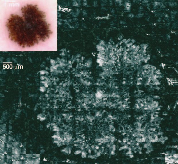
372 Pellacani et al                                                                                J AM ACAD DERMATOL
                                                                                                        SEPTEMBER 2004

                                                                 Windows (Microsoft Corp., Redmond, Wash). The
                                                                 digitized images offer a spatial resolution of 768 3
                                                                 576 pixels and a resolution of 16 million colors.
                                                                    For CSLM imaging, confocal images were ac-
                                                                 quired by means of a near-infrared reflectance con-
                                                                 focal laser scanning microscope (Vivascope 1000,
                                                                 Lucid Inc, Henrietta, NY).14 After acquiring the ELM
                                                                 image, the adapter ring was filled with water and the
                                                                 arm of the CSLM with the 30x water immersion
                                                                 objective lens (numeric aperture of 0.9) was placed
                                                                 onto it. The instrument uses a diode laser at 830 nm
                                                                 with a maximum power of 35 mW at tissue level,
                                                                 enabling visualization of skin structures at a maxi-
                                                                 mum depth of 350 mm. Images have a spatial reso-
                                                                 lution of 0.5 to 1.0 mm in the lateral dimension and 4
                                                                 to 5 mm in the axial dimension. Each image corre-
                                                                 sponds to a horizontal plane of skin section at
                                                                 a selected depth with an effective field of view of
Fig 1. Reconstructed confocal scanning microscopy image          475 3 350 mm, with a resolution of 640 3 480 pixels
of Spitz nevus, used for evaluation of architectural features.   and 255 colors. An automated stepper was used to
Lesion appears more refractive in comparison with sur-           obtain a grid of 16 contiguous horizontal images at
rounding skin. Inset, Corresponding epiluminescence im-          a selected depth, constructing a montage image with
age as observed with polarized light and 50-fold
                                                                 an in vivo field of view of 1.9 3 1.4 mm (block
magnification.
                                                                 image). A sequence of 30 block images was acquired
                                                                 for each lesion at dermoepidermal junction level and
the description of the architecture of the lesion.               mounted by means of a software developed by us to
Together with cytologic features, the former was                 obtain a field of view of 7.60 3 6.65 mm (re-
used to establish a correlation between ELM images               constructed image) (Fig 1). Sequences of confocal
and histopathologic sections.                                    sections, beginning at the stratum corneum and into
                                                                 the papillary dermis, were recorded at areas of
                                                                 interest on the border and inside the lesion.
MATERIALS AND METHODS
Patients                                                         Image description
   This study included 6 Spitz nevi in as many                      For ELM images, traditional dermoscopic features
patients, with an average patient age of 29.8 years.             were described for each lesion.21
Before biopsy, all lesions were recorded using both                 CSLM images of Spitz nevi were described con-
ELM and CSLM. To have an exact correspondence                    sidering both the overall aspect of the lesion, eval-
between ELM and CSLM images, the CSLM adapter                    uated on the reconstructed image with a field of 7.60
ring was first positioned onto the skin and centered             3 6.65 mm, and the cytologic features, evaluated on
around the lesion. Subsequently, ELM and CSLM                    the single images with the best resolution (475 3 350
images were acquired. All lesions were then excised              mm). For the overall aspect, the symmetry, border
and underwent histopathologic examination for di-                cutoff, and cell and structure uniformity were taken
agnostic confirmation.                                           into account. According to previous reports con-
                                                                 cerning normal skin or melanocytic lesions observed
Evaluation methods                                               with CSLM, cytologic features were evaluated at
   ELM imaging was performed with a digital vid-                 different depth. The standard confocal features of
eomicroscope (VMS-110A, Scalar Mitsubishi, Tama-                 the epidermis and of the dermis were described.14-18
shi, Tokyo, Japan) using 20-, 50-, and 200-fold                  Moreover, the presence within the superficial layers
magnification, positioning the probe onto the CSLM               of ovoid highly refractive structures without nuclei
adapter ring. The instrument has been described                  was reported. At the basal layer, cells clustered into
elsewhere.20 The images were digitized by means of               globular aggregates were described15 also consider-
a Matrox Orion frameboard (Matrox Electronic                     ing their size, shape, brightness, and homogeneity
Systems, Ltd, Dorval, Quebec, Canada) and stored                 throughout the lesion. The cell dimension was
by an image acquisition program (VideoCap 8.09,                  compared with keratinocytes of the surrounding
DS-Medica, Milan, Italy), which runs under Microsoft             healthy skin.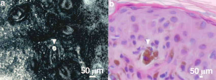
J AM ACAD DERMATOL                                                                       Pellacani et al 373
VOLUME 51, NUMBER 3

   The traditional histopathologic description of       Table I. In vivo confocal scanning laser microscopy
both architectural patternsesuch as lesion symmetry;    features observed in 6 globular Spitz nevi
border cutoff; aspect and distribution of the nests;                                             Present
epidermal hyperplasia and flogistic infiltrate; and
cytologic features, such as cell type and aspect,       Overall aspect
presence of pagetoid infiltration, number of mitoses,     Symmetric silhouette                      6
cell maturation with increasing depth, and cell           Abrupt borders                            5
uniformity from one side of the lesion to the other-      Cell/structure uniformity                 6
                                                        Cytologic features
were described.
                                                          Stratum corneum/granular
                                                             layer/ spinous layer
RESULTS                                                      Honeycombed                            0
ELM features                                                    appearance
   All the 6 Spitz nevi were symmetric lesions with          Bright refractive                      4
a diameter ranging between 4 to 8 mm, characterized             particles
by a globular appearance. In 5 cases a rim of large          Single round cells                     1
globules was present at the periphery of the lesion.         Ovoid refractive                       2
The central portion was characterized by darkly              structures
pigmented globules in 3 cases and light brown in          Basal layer
the remaining 3, associated with gray-bluish areas in        Small bright cells                     6
                                                             Large bright cells          6 (round, 6; spindle
4 lesions.
                                                                                            or dendritic, 4)
                                                            Globules (aggregates         6 (diameter range:
CSLM ASPECTS                                                     of clustered cells)        140-240 mm)
   Evaluation of lesion architecture on the                   Central globules                      6
reconstructed image. As shown in Table I, Spitz               Peripheral globules                   5
nevi markedly differed from the control skin.             Dermal papillae
Melanin present in nevus cells represented a strong         Plump bright cells           6 (abundant in 4 cases)
source of contrast, rendering the pigmented skin            Dark canalicular                       6
lesion lighter than the surrounding skin (Fig 1). All         structures
lesions presented a symmetric silhouette, with cell
and structure uniformity. Sharp borders were ob-
served in 5 Spitz nevi with the globular rim.
   Evaluation at cellular level. The normal             tion (Fig 4). All lesions examined presented contig-
honeycombed pattern, constituted by polygonal           uous dense nonconfluent aggregates, with variable
low refractive cells with a mean diameter of 22         diameter ranging between 140 to 420 mm, homo-
mm, was always observable in superficial layers,        geneously distributed inside the lesion. Well-de-
whereas bright refractive particles were noticed in     marcated, highly refractive aggregates formed by
4 out of 6 lesions. Moreover, few individual cells,     large homogeneous cells were observed at the
round in shape and with bright cytoplasm and dark       periphery of the 5 Spitz nevi with a globular
nucleus, with a mean diameter of 35 mm, were            rim. Plump, bright cells with ill-defined borders,
observed in one lesion. In two cases ovoid highly       corresponding to melanophages,16 were present in
refractive structures with a diameter of approxi-       different amounts in all lesions (Fig 3, C ). In 4 cases
mately 50 mm were observed at the basal layer or        a great amount of plump cells was observed,
immediately upward (Fig 2). Small bright cells were     correlating with the presence of ELM gray-bluish
found at the dermoepidermal junction, correlating       areas and of a great histopathologic inflammatory
with melanocytes and pigmented keratinocytes            infiltrate rich in melanopahges. Small canalicular
seen by conventional histopathology, frequently         structures within the papillary dermis, corre-
with intercalated large brighter cells that were        sponding to capillaries, were evident in all cases.
round to oval (all cases) and spindle or dendritic         Histopathologic examination. All lesions
(4 cases) in shape (Fig 3). Oval to round polygonal     were symmetric and well circumscribed. Epidermal
aggregates with well-defined borders, composed          hyperplasia with elongated rete ridges was observed
by clustered cells, frequently large in size and        only in one case. Usually large nests, homogeneous
highly refractive, were observed in all lesions,        in size, predominantly at the dermoepidemal junc-
corresponding in structure and dimension to the         tion, were observed in all Spitz nevi. No permeation
pigmented globules of the ELM observation and to        of the epidermis was reported. Although a flogistic
melanocytic nests at the histopathologic examina-       infiltrate was present in all lesions, it was marked and
374 Pellacani et al J AM ACAD DERMATOL
SEPTEMBER 2004
Fig 2. Large ovoid homogeneously bright structure in suprabasal layers (a) (arrowhead)
corresponding to large coarse pigment conglomerates inside epidermis at histopathologic
examination (arrowhead) (b).
Fig 3. Cytologic aspects: round to oval large brighter cells (a), spindle/dendritic cells (b) (both
corresponding to Spitz nevus melanocytes), and plump bright cells (c) with ill-defined borders
inside dermal papillae, corresponding to melanophages (arrowhead).
Fig 4. Globular structures: correlation among pigmented globules with 200-fold epilumines-
cence microscopy (in vivo horizontal plane image) (a), refractive globular structures with in
vivo confocal scanning laser microscopy (CSLM) (in vivo horizontal plane image) (b), and
melanocytic nests in routine histology (ex vivo vertical plane image) (c). With CSLM (b),
globular structures appeared as oval to round polygonal aggregates with well-defined borders,
composed by clustered cells, frequently large in size and highly refractive.
rich in melanophages in 4 out of 6 cases. Cytologic in 4 lesions. In two cases, large coarse pigment
features consisted predominantly of large epithelioid conglomerates inside the epidermis were present.
cells, intensely pigmented in 4 cases. Poorly
pigmented spindle cells predominated only in one DISCUSSION
case. Cell uniformity and maturation was observed in In vivo reflectance-mode CSLM is a novel tech-
all cases. Transepidermal melanin loss was reported nique that enables the in vivo study of the skin atJ AM ACAD DERMATOL Pellacani et al 375
VOLUME 51, NUMBER 3
a nearly histopathologic resolution, producing melanocytic cells. Although detection of the
pictures of horizontal planes of the skin. Confocal pagetoid spread of melanocytes within the epider-
images of normal skin and of keratinocytic and mis is an important clue for melanoma diagnosis,
inflammatory skin lesions with detailed correlation this feature is also observable in Spitz nevi, which
to histopathologic sections have been previously are usually characterized by the presence of spo-
reported.22-25 Because melanin represents a strong radic cells only in suprabasal layers. Moreover, the
source of contrast, the use of this technique for the presence in two Spitz nevi of ovoid homoge-
characterization of pigmented skin lesions seems neously bright structures in basal and suprabasal
particularly interesting. The appearance of melano- layers appeared correlated with large coarse pig-
cytes, pigmented keratinocytes, and melano- ment conglomerates inside the epidermis at the
phages,16 together with the features of common histopathology (Fig 2).
and atypical nevi,15 and of melanomas,15,17-19 have In conclusion, in globular Spitz nevi, CSLM al-
been described. Because dermoscopy enables the lowed the in vivo recognition of characteristic histo-
visualization of subsurface structures that can be pathologic aspects, with an excellent correlation
correlated with specific histopathologic aspects,26-28 with the corresponding ELM features. The im-
the capability of CSLM in identifying cytologic and plementation of a program for the composition of
architectural features with a tight correlation to an overall CSLM image enabled the evaluation of the
histopathology may improve diagnostic accuracy, architectural aspects of the lesions and the correla-
especially for pigmented skin lesions characterized tion of ELM features with the exactly corresponding
by unspecific features, such as in situ melanoma confocal structures. Single cells or cellular aggregates
versus atypical nevus or lentigo maligna versus can be imaged for the cytologic description of the
lentigo maligna melanoma.19 lesion and for the correlation with histopathology.
Spitz nevi may represent a diagnostic pitfall both Thus, CSLM may represent the missing link between
for dermatologists and for pathologists. Histo- the ELM technique and the histopathologic exami-
pathologic criteria useful for distinction between nation.
melanomas and Spitz nevi have been identified and
defined.9-12 ELM description of Spitz nevi enables
REFERENCES
the subdivision of these lesions into 3 main 1. Pehamberger H, Steiner A, Wolff K. In vivo epiluminescence
categories: globular; starburst; and atypical.6 The microscopy of pigmented skin lesions, I: pattern analysis of
aim of our study was to describe Spitz nevi of the pigmented skin lesions. J Am Acad Dermatol 1987;17:571-83.
globular type by CSLM. With specific software for 2. Steiner A, Binder M, Schemper M, Wolff K, Pehamberger H.
image reconstruction of CSLM images, we were Statistical evaluation of epiluminescence microscopy criteria
for melanocytic pigmented skin lesions. J Am Acad Dermatol
able to describe the overall aspect and the archi- 1993;29:581-8.
tecture of Spitz nevi. This approach also enabled 3. Kenet RO, Kang S, Kenet BJ, Fitzpatrick TB, Sober AJ, Barnhill
the exact correlation with the corresponding ELM RL. Clinical diagnosis of pigmented lesions using digital
and histopathologic features. On the whole, con- epiluminescence microscopy: grading protocol and atlas. Arch
focal images of our globular Spitz nevi were Dermatol 1993;129:157-74.
4. Steiner A, Pehamberger H, Binder M, Wolff K. Pigmented Spitz
characterized by aggregated globules. They were nevi: improvement of the diagnostic accuracy by epilumines-
uniform in size and shape, and tended not to cence microscopy. J Am Acad Dermatol 1992;27:697-701.
become confluent, in accordance to histopathol- 5. Pellacani G, Martini M, Seidenari S. Digital videomicroscopy
ogy. All Spitz nevi with peripheral globules pres- with image analysis and automatic classification as an aid for
ented refractive polygonal aggregates at the diagnosis of Spitz nevus. Skin Res Technol 1999;5:266-72.
6. Argenziano G, Scalvenzi M, Staibano S, Brunetti B, Piccolo D,
periphery, corresponding to melanocytic nests at Delfino M, et al. Dermatoscopic pitfalls in differentiating
the margin of lesion in the histopathologic sections. pigmented Spitz naevi from cutaneous melanoma. Br J
In confocal images, superficial layers presented Dermatol 1999;141:788-93.
a normal honeycombed appearance. In the 4 Spitz 7. Pellacani G, Cesinaro AM, Seidenari S. The morphological
nevi characterized by dark pigmentation, bright features of Spitz nevus as observed by digital videomicros-
copy. Acta Derm Venereol 2000;80:117-21.
granular particles between the meshes of the 8. Requena L, Sanchez Yus E. Pigmented spindle cell naevus. Br J
honeycombed pattern were observable, probably Dermatol 1990;123:757-63.
corresponding to transepidermal melanin loss at 9. Ackerman AB, Magana-Garcia M. Naming acquired melano-
the histopathologic examination. Few individual cytic nevi. Am J Dermatopathol 1990;12:193-209.
cells, round to oval in shape, with bright granular 10. Sau P, Graham JH, Helwig EB. Pigmented spindle cell nevus:
a clinicopathologic analysis of ninety-five cases. J Am Acad
cytoplasm and dark eccentric nucleus, were ob- Dermatol 1993;28:565-71.
served in the suprabasal layers in one case, 11. Piepkorn M. On the nature of histologic observations: the case
corresponding to pagetoid infiltration of nevo- of the Spitz nevus. J Am Acad Dermatol 1995;32:248-54.376 Pellacani et al J AM ACAD DERMATOL
SEPTEMBER 2004
12. Elder D, Elenitsas R. Benign pigmented lesions and malignant 20. Seidenari S, Burroni M, Dell’Eva G, Pepe P, Belletti B. Comput-
melanoma. In: Elder D, Elenitsas R, Jaworsky C, Johnson B, erized evaluation of pigmented skin lesion images recorded
editors. Lever’s histopathology of the skin. 8th ed. Philadelphia: by a videomicroscope: comparison between polarizing mode
Lippincott-Raven; 1997. observation and oil/slide mode observation. Skin Res Technol
13. Rajadhyaksha M, Grossman M, Esterowitz D, Webb RH, 1995;1:187-91.
Andersson RR. In vivo confocal scanning laser microscopy of 21. Argenziano G, Soyer HP, Chimenti S, Talamini R, Corona R, Sera
human skin: melanin provides strong contrast. J Invest F, et al. Dermoscopy of pigmented skin lesions: results of
Dermatol 1995;104:946-52. a consensus meeting via the Internet. J Am Acad Dermatol
14. Rajadhyaksha M, Gonzalez S, Zavislan JM, Andersson RR, Webb 2003;48:679-93.
RH. In vivo confocal scanning laser microscopy of human skin 22. Gonzalez S, Rajadhyaksha M, Rubinstein G, Andersson RR.
II: advances in instrument and comparison with histology. Characterization of psoriasis in vivo by reflectance confocal
J Invest Dermatol 1999;113:101-13. microscopy. J Med 1999;30:337-56.
15. Langley RG, Rajadhyaksha M, Dwyer PJ, Sober AJ, Flotte TJ, 23. Gonzalez S, Gonzalez E, White MW, Rajadhyaksha M, Ander-
Andersson RR. Confocal scanning laser microscopy of benign sson RR. Allergic contact dermatitis: correlation of in vivo
and malignant melanocytic skin lesions in vivo. J Am Acad confocal imaging to routine histology. J Am Acad Dermatol
Dermatol 2001;45:365-76. 1999;40:708-13.
16. Busam KJ, Charles C, Lee G, Halpern AC. Morphological 24. Aghassi D, Anderson RR, Gonzalez S. Confocal laser micros-
features of melanocytes, pigmented keratinocytes, and me- copy of actinic keratoses in vivo: a preliminary report. J Am
lanophages by in vivo confocal scanning laser microscopy. Acad Dermatol 2000;43:42-8.
Mod Pathol 2001;14:862-8. 25. Gonzalez S, Tannous Z. Real-time, in vivo confocal reflectance
17. Busam KJ, Hester K, Charles C, Sachs DL, Antonescu CR, microscopy of basal cell carcinoma. J Am Acad Dermatol 2002;
Gonzalez S, et al. Detection of clinically amelanotic malignant 47:869-74.
melanoma and assessment of its margins by in vivo confocal 26. Yadav S, Vossaert KA, Kopf AW, Silverman M, Grin-Jorgensen
scanning laser microscopy. Arch Dermatol 2001;137:923-9. C. Histopathologic correlates of structures seen on dermo-
18. Busam KJ, Charles C, Lohmann CM, Marghoob A, Goldgeier M, scopy (epiluminescence microscopy). Am J Dermatopathol
Halpern AC. Detection of intraepidermal malignant melanoma 1993;15:297-305.
in vivo by confocal scanning laser microscopy. Melanoma Res 27. Soyer HP, Kenet RO, Wolf IH, Kenet BJ, Cerroni L. Clinicopath-
2002;12:349-55. ological correlation of pigmented skin lesions using dermos-
19. Tannous ZS, Mihm MC, Flotte TJ, Gonzalez S. In vivo exam- copy. Eur J Dermatol 2000;10:22-8.
ination of lentigo maligna and malignant melanoma in situ, 28. Ferrara G, Argenziano G, Soyer HP, Corona R, Sera F, Brunetti
lentigo maligna type by near-infrared reflectance confocal B, et al. Dermoscopic and histopathologic diagnosis of
microscopy: comparison of in vivo confocal images with equivocal melanocytic skin lesions: an interdisciplinary study
histologic sections. J Am Acad Dermatol 2002;46:260-3. on 107 cases. Cancer 2002;95:1094-100.You can also read