Polyploidy in Spitz Nevi: A Not Uncommon Karyotypic Abnormality Identifiable by Fluorescence in Situ Hybridization
←
→
Page content transcription
If your browser does not render page correctly, please read the page content below
ORIGINAL STUDY

    Polyploidy in Spitz Nevi: A Not Uncommon Karyotypic
         Abnormality Identifiable by Fluorescence in
                       Situ Hybridization
   Anjeli Krishnan Isaac, MD, Terakeith Lertsburapa, MD, Jyoti Pathria Mundi, MD, Mary Martini, MD,
                              Joan Guitart, MD, and Pedram Gerami, MD

                                                                             Hence, further studies elucidating the biologic basis for this
Abstract: Fluorescence in situ hybridization (FISH) often reveals            unique subset of nevi are necessary.
imbalanced chromosomal gains in melanoma, whereas Spitz nevi                        Recently, molecular techniques such as comparative
typically have a normal complement of chromosomes. However,                  genomic hybridization (CGH) and fluorescence in situ
there may be a subset of Spitz nevi that are perfectly tetraploid by         hybridization (FISH) have emerged as adjunctive diagnostic
FISH analysis, and these cases may be confused diagnostically with           tools in dermatopathology. With these techniques it has
melanoma. This study evaluates 41 cases of Spitz nevi that were              become evident that in addition to their unique clinical and
histologically confirmed to be benign. Four of these lesions dem-            histologic features, Spitz nevi also have unique molecular
onstrated polyploidy by FISH. Three of the 4 cases were from the             features.5,6 Although 95% of melanomas show evidence of
same patient, a 17-year-old woman; 1 lesion was from the wrist,              clonal chromosomal abnormalities by CGH, such changes are
whereas the other 2 were from the buttocks. The other case was from          only rarely seen in nevi. The primary exception to this rule is
a 14-year-old man from the ankle. All 4 cases that were polyploid            Spitz nevi in which 10%–20% contain an isolated clonal gain
were confirmed using a probe for the X chromosome. This article              in 11p.5 Additionally, previous studies using multivariate DNA
highlights the importance of polyploidy as a feature of some benign          cytometry have suggested the presence of polyploidy in
Spitz nevi.                                                                  approximately 10% of Spitz nevi.7
Key Words: polyploidy, Spitz nevi, fluorescence in situ hybridiza-                  FISH is emerging as a diagnostic tool in melanocytic
tion, melanoma                                                               neoplasms. This assay relies upon the recognition of clonal
                                                                             chromosomal gains or losses in melanomas, which are not
(Am J Dermatopathol 2010;32:144–148)                                         identified in nevi. Melanomas frequently have clonal
                                                                             chromosomal gains, but these are most often imbalanced
                                                                             gains.8 However, a subset of Spitz nevi may be perfectly
                                                                             tetraploid. It is critical to recognize the possibility of
                                                                             tetraploidy in Spitz nevi to avoid misdiagnosis as melanoma
                        INTRODUCTION                                         when using FISH as a diagnostic adjunct. In this study, we
       More than 60 years after Sophie Spitz’s original                      used FISH to study the incidence and extent of tetraploidy in
description of Spitz nevi as benign juvenile melanoma in                     41 Spitz nevi. Additionally, we reviewed the clinical and
1948, the subject of Spitz nevi continues to be an enigma to                 histologic features of tetraploid Spitz nevi.
clinicians and pathologists. Despite our improved familiarity
with the clinical and histologic features of Spitz nevi, there
continues to be significant discordance between even expert                              MATERIALS AND METHODS
dermatopathologists in the diagnosis of a subset of Spitz nevi                      After approval from the Northwestern University
as some cases may have conflicting histopathologic features                  Internal Review Board, 41 cases of Spitz nevi were identified
making them difficult to distinguish from melanoma.1–3                       from Northwestern University’s dermatopathology depart-
Furthermore, in some of these ambiguous cases, in which                      ment. These cases had varying degrees of cytologic atypia, but
a histologic diagnosis of benign Spitz nevi was favored by an                all histologic cases were evaluated by 2 dermatopathologists
expert dermatopathologists, the lesion resulted in metastases.4              (P.G. and J.G.), and only cases, which were unequivocally
                                                                             benign Spitz nevi, were included in the study. As part of the
                                                                             initial search method, 10 cases of Spitz tumor were also
From the *Department of Dermatology, Northwestern University, Feinberg
    School of Medicine, Chicago, IL.                                         identified, which were excluded from the study because of the
M. Martini serves on the board of Dove/Unilever and is an investigator for   presence of conflicting histopathologic features.
    Electro-Optical Sciences, Inc.                                                  Five-micrometer-thick sections were obtained from
P. Gerami served as a consultant for Abbott Molecular Laboratories and has   formalin-fixed paraffin-embedded tissue and mounted on
    received honoraria.
Reprints: Pedram Gerami, MD, 676 North St., Clair St, Suite 1600, Chicago,
                                                                             SuperFrost Plus positively charged slides. The slides were
    IL 60611 (e-mail: pgerami@nmff.org).                                     baked overnight at 56°C and stored at room temperature. The
Copyright Ó 2010 by Lippincott Williams & Wilkins                            sections were then deparaffinized in 3 changes, each for 5

144   | www.amjdermatopathology.com                                              Am J Dermatopathol  Volume 32, Number 2, April 2010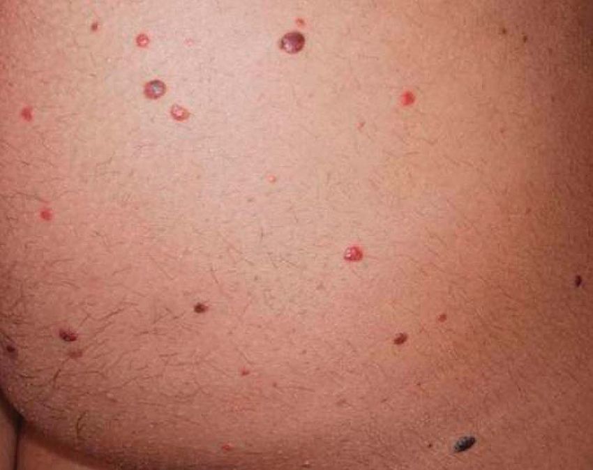
Am J Dermatopathol  Volume 32, Number 2, April 2010                                                       Polyploidy in Spitz Nevi

minutes of Hemo-De solvent and clearing agent. Specimens            common particularly in large spitzoid cells. Additionally, in
were then rinsed for 1 minute for 2 cycles in absolute ethanol      any cases in which a significant number of tetraploid cells
and then incubated in 13saline sodium citrate pH 6.3 at 80°C        were identified, further validation was performed with
for 35 minutes. This was followed by a 1-minute water rinse.        FISH for the X chromosome. The presence of 2 copies
The specimens were then digested in protease 1 (Abbott              of the X chromosome in the Spitz nevi from males or 4 in
Molecular, Inc., 02J08-32, 32-801260; 4 mg protease/mL, 0.2         those from females were used to confirm our impression
N HCl). Digestion time at 15 minutes consistently resulted in       of tetraploidy.
high-quality specimens of benign lesions. In melanomas, 15
minutes digestion occasionally resulted in excessive back-
ground and need to be varied between 13 and 15 minutes for                                   RESULTS
optimal specimens. A 3-minute water rinse was then                         Forty-one Spitz nevi cases were studied from 38
performed followed by dehydration in 70%, 85%, and 100%             patients. The patient’s ages ranged from 23 months to age
ethanol, each for 1 minute and then dried.                          57. Of the 41 cases, 31 were women and 10 were men. One
      The ThermoBrite codenaturation/hybridization oven             patient had multiple lesions and was diagnosed as having
(Abbott Molecular) was set at 73°Cfor 5 minutes denaturation,       agminated Spitz nevi. The most common sites of involvement
and at 37°C for 16–18 hours hybridization. The slides were          were the lower extremities followed by the upper extremities,
then placed on oven surface and 10 mL of the Vysis LSI              but the head and neck, back, groin, and buttock areas were also
RREB1/LSI MYB/CEP 6 probe solution and 10 mL of the                 represented in the sample set. Histologically, all cases were
Vysis LSI CCND1 SpectrumGreen probe solution were added.            considered unequivocally benign Spitz nevi by both derma-
A coverslip was applied, the edges were sealed with rubber          topathologists. The average clinical follow-up period was
cement, and the oven was started. After hybridization, the          approximately 2 years, ranging from 1 to 3 years. In this
rubber cement was removed and the slides were placed at room        period, no lesions recurred or showed evidence of aggressive
temperature, 23SSC/0.3% NP40, for 2–10 minutes to remove            behavior.
the coverslips. The slides were then immersed in 23SSC/0.3%                Among 41 Spitz nevi, FISH analysis with our probe
NP40 at 73°C for 2 minutes and allowed to dry in the dark.          set using the definitions as determined above, 4 cases were
4#-6-Diamidino-2-phenylindole 1 antifade solution (Abbott           identified as having a significant number of polyploid cells
Molecular) and a coverslip were then applied.                       (Fig. 1). In these 4 nevi, 11–16 cells examined showed
      Slides were evaluated with an epifluorescence micro-          balanced gains in 6p25, 6q23, 11q13, and CEP 6, with all cells
scope equipped with DAPI, Aqua, Gold, GreenV2, and Red              having 3 or 4 identifiable copies of each chromosomal segment
single bandpass filter sets (Abbott Molecular). All cases were      (Fig. 2). The 4 cases were from 2 patients, and the follow-up
studied with probes targeting the following 4 loci: (1) ras         time for these cases was approximately 2.5 years with no
responsive element-binding protein-1 (RREB1) (chromo-               evidence of recurrence or aggressive behavior. Three of the 4
some 6p25), (2) myeloblastosis (MYB) (chromosome 6q23),             polyploid nevi were from 1 patient, a 17-year-old woman who
(3) cyclin D1 (chromosome 11q13), and (4) CEP 6                     had agminated Spitz nevi (Fig. 3). One of the lesions was from
(centromeric portion of chromosome 6). Five total reviewers         the right wrist, whereas the other 2 were from the buttock.
including 2 attending dermatopathologists, 1 research fellow,       These Spitz nevi were all considered benign. They were
and 2 dermatology residents were involved in the enumeration        symmetrical melanocytic lesions with both epithelioid and
process. The reviewers were blinded to the histologic               spindle melanocytes arranged in vertically oriented nests and
diagnosis. The protocol used involved quick scanning under          fascicles. These had prominent lavender nucleoli and open
each of the 4 filters to detect any areas with gains or losses in   chromatin with abundant cytoplasm, and there was maturation
any of the DNA loci being studied [$63% of enumerated               of the dermal component. Kamino bodies were also noted in
cells showing abnormalities with RREB1 (gains/losses),              some of the lesions (Fig. 4). Interestingly, some of her other
average MYB signals $2.5 per nucleus, average cyclin D1             Spitz nevi not included in this study consisted entirely of
signals .2.5 per nucleus, or MYB/CEP 6 loss of $31%].               normal diploid cells.
When a nest or area was identified, a minimum of 10 cells                  The last polyploid Spitz nevus was from a 14-year-old
from the section were evaluated, and at least 3 separate nests      man, whose histology was read as an atypical Spitz nevus on
or areas were identified per specimen. Therefore, 30 countable      the ankle. The sections demonstrated a broad compound
cells per case were enumerated. Cells with overlapping              melanocytic proliferation of epithelioid and spindle cells
borders were not included because of potential falsely              arranged in nests and fascicles along the dermal–epidermal
elevated signals.                                                   junction and within the dermis. The nests at the junction had
      The criteria used that would select a cell as highly          clefting and rare Kamino bodies. There were focal areas with
likely to be polyploid were as follows: (1) For a given cell,       confluent single-cell suprabasal spread. Some of the dermal
3 or 4 copies of the chromosomal loci targeted needed to            nests showed prominent crowding of nuclei and some
be identified with the probes RREB1 (6p25), MYB (6q23),             expansile features. Moderate nuclear atypia was noted.
and cyclin D1 (11q13) and (2) nine or more cells of the                    All 4 cases that were polyploid were confirmed by use of
30 enumerated per case had to be polyploid in order for             a probe for the X chromosome, which showed 4 copy numbers
the case to be considered as having significant numbers             for women and 2 copy numbers for men in the same regions, in
of polyploid cells. In the first criteria, identification of only   which copy number gains were identified with the original
3 copies was considered acceptable because truncation is            probe set.

q 2010 Lippincott Williams & Wilkins                                                     www.amjdermatopathology.com |        145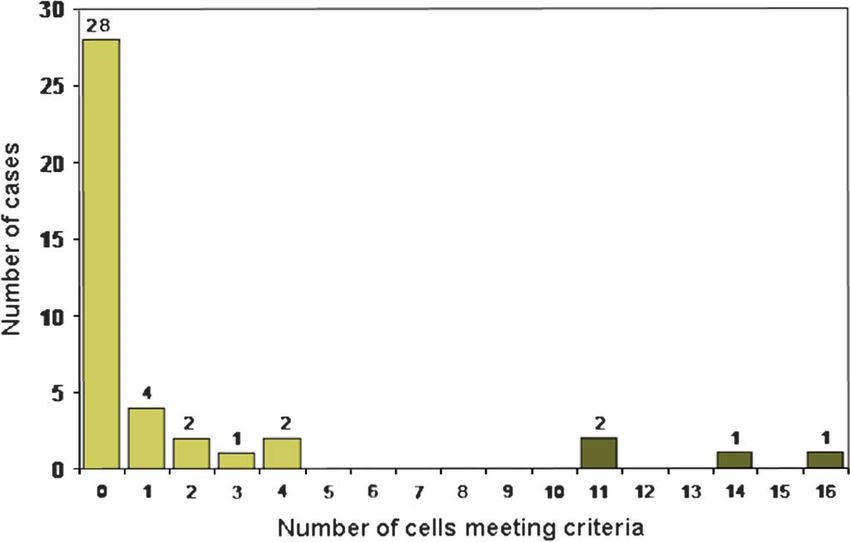
Isaac et al Am J Dermatopathol Volume 32, Number 2, April 2010
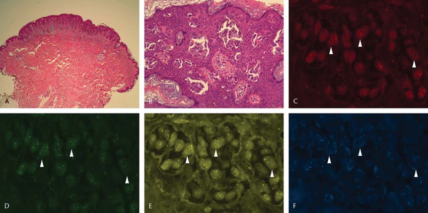
FIGURE 1. A, Low-power magnification (34) of polyploid Spitz nevus. B, High-power magnification (320) of polyploid Spitz nevus.
Polyploidy demonstrated by balanced gains in RREB1 (6p25; C), MYB (6q23; D), cyclin D1 (11q13; E), and CEP 6 (centromeric
portion of chromosome 6; F).
DISCUSSION a finding not seen in melanomas.5 In our study using FISH, we
For decades Spitz nevi have been appreciated for their showed that 4 of 41 Spitz nevi demonstrated polyploidy.
unique histologic features, which can closely resemble Polyploidy exists although is extremely rare in melanoma both
melanoma. With the emergence of CGH and FISH as tools by our own observations and in the literature.12 In melanoma,
for studying melanocytic neoplasms, unique cytogenetic imbalanced gains are more common. Recognizing the
features of Spitz nevi are also becoming evident. Previous presence of polyploidy in Spitz nevi is highly significant to
studies have shown that although the vast majority of benign avoid making a definitive diagnosis of melanoma based on the
nevi lack chromosomal abnormalities, approximately 10%– presence of chromosomal gains by FISH. In this study, we
20% of Spitz nevi may show an isolated gain in 11p. This is
FIGURE 2. Criteria to select cell as polyploidy: (1) For each cell,
at least 3 copies of the chromosomal locus targeted by the
probes RREB1, MYB and cyclin D1 were required and (2) 9 or FIGURE 3. Numerous pink, brown, and black papules in a
more cells of 30 total cells per case had to show these increased 17-year-old woman with agminated Spitz nevi (3 of 4
copy numbers. polyploid cases).
146 | www.amjdermatopathology.com q 2010 Lippincott Williams & WilkinsAm J Dermatopathol Volume 32, Number 2, April 2010 Polyploidy in Spitz Nevi
cytokinesis, or chromosome segregation, and mutations in cell
cycle checkpoint regulators, such as p53, may be the root
cause of the polyploid state.9,10 Conversely, melanoma cells
tend to show aneuploidy with imbalanced chromosomal gains
and losses.7,11 Again, although melanomas with tetraploidy
have been identified, it seems to be much less common than
aneuploidy.12
The significance of polyploidy as a whole, however, is
controversial; although there are data suggesting that poly-
ploidy can trigger cellular senescence, other studies suggest
that it acts as an intermediary in the development of cancer.13,14
Investigators have shown that polyploid cells in general may
be more sensitive to growth arrest secondary to DNA damage.
There may, in fact, be a G1 checkpoint that prevents polyploid
intermediates from producing aneuploid cells.15 If so, this
may be an important mechanism of growth arrest of polyploid
Spitz nevi. Conversely, polyploidy has also been identified
FIGURE 4. Polyploid Spitz nevus, 310 magnification. Both in a number of precancerous or intermediate states in the
epithelioid and spindle melanocytes arranged in vertically development of cancer, including esophageal adenocarcinoma
oriented nests and fascicles. Maturation of the dermal (intermediate state, Barrett’s esophagus), colorectal carcinoma
component was noted. (intermediate state, ulcerative colitis), and breast cancers.
Several in vivo studies have supported this possibility.15 In this
theory, polyploidy may represent an intermediate state of
defined a case as having a significant number of polyploid cells genomic instability, after which cells pass into an aneuploid
as those cases with 9 or more of the 30 enumerated cells and a more tumorigenic state.16 In fact, there have been studies
having gains because in our previously established criteria for suggesting that tetraploid cancer cells are resistant to DNA
melanoma, cases with 9 or more cells with gains in damage–induced apoptosis, further suggesting an association
RREB1/6p25 were considered malignant.8 between polyploidy and tumorigenicity.17
Although these 4 cases would have met our previously In Spitz nevi, we believe that the polyploid state likely
determined criteria for malignancy, recognizing them as does induce cellular senescence, and that this can be seen as
polyploid would allow one to circumvent this. In addition, part of an entirely benign lesion rather that it representing an
when using FISH for the diagnosis of Spitz nevus versus intermediate or precancerous state. This is supported by the
melanoma, one should consider the possibility of polyploidy. fact that polyploidy in Spitz nevi is far more common than the
Using the 4-probe panel targeting RREB1/6p25, MYB/6q23, development of melanoma from a Spitz nevus, which is a rare
CEP 6, and cyclin D1/11q13, specific patterns should raise event. In conclusion, our study highlights a novel finding of
concern for polyploidy. Specifically, most melanomas show polyploidy seen in certain cases of benign Spitz nevi.
imbalances in chromosome 6 with greater copy numbers of the Polyploidy may be more common among cases of agminated
short-arm RREB1/6p25 relative to the long-arm MYB/6q23. Spitz nevi and Spitz nevi, which have an atypical spitzoid
In fact, most melanomas demonstrate loss of MYB/6q23.8 In epithelioid cell component. More research needs to be done to
polyploidy, one finds uniform gains in all probes being studied, further characterize polyploid Spitz nevi and to determine if
including 6p25, 6q23, 11q13, and CEP 6. Then polyploidy can the biologic behavior of polyploid Spitz nevi varies from that
be further confirmed by use of a probe for the X chromosome, of other Spitz nevi.
which in polyploidy cases should show 2 copy numbers for
men or 4 for women. Although we definitively found
polyploidy in 2 of 38 (5%) of our patients, the true number ACKNOWLEDGMENT
is likely a bit higher because cells may be truncated, all the The authors would like to thank the Irene D. Pritzker
gene copies may not be identified, or they may have not quite Foundation and the Dermatology Foundation for their support
met our strict criteria for polyploidy. of this work.
Typically, human cells are diploid, containing 2
homologous sets of chromosomes. Polyploidy, in turn, is the REFERENCES
state of having a chromosome complement that is a multiple of 1. Dahlstrom JE, Scolyer RA, Thompson JF, et al. Spitz naevus: diagnostic
the haploid number, and tetraploidy refers to exactly twice the problems and their management implications. Pathology. 2004;36:
452–457.
number of diploid chromosomes. The molecular significance 2. Crotty KA, Scolyer RA, Li L, et al. Spitz naevus versus spitzoid
of polyploidy is not fully resolved, but the underlying melanoma: when and how can they be distinguished? Pathology. 2002;34:
mechanism seems to be a failure to link DNA replication 6–12.
with normal mitotic division. Hence, although in most cases, 3. Busam KJ, Pulitzer M. Sentinel lymph node biopsy for patients with
diagnostically controversial spitzoid melanocytic tumors? Adv Anat
DNA replication is closely followed by cell division, Pathol. 2008;15:253–262.
polyploidy results when cell division is halted after DNA 4. Barnhill RL. Childhood melanoma. Semin Diagn Pathol. 1998;15:
replication. It can arise from failure of spindle assembly, 189–194.
q 2010 Lippincott Williams & Wilkins www.amjdermatopathology.com | 147Isaac et al Am J Dermatopathol Volume 32, Number 2, April 2010
5. Bastian BC, Wesselmann U, Pinkel D, et al. Molecular cytogenetic 11. Gattuso P, Reddy V, Solans E, et al. Is DNA ploidy of prognostic
analysis of Spitz nevi shows clear differences to melanoma. J Invest significance in stage I cutaneous melanoma? Surgery. 1990;108:702–708.
Dermatol. 1999;113:1065–1069. 12. Satoh S, Hashimoto-Tamaoki T, Furuyama J, et al. High frequency of
6. Wettengel GV, Draeger J, Kiesewetter F, et al. Differentiation between tetraploidy detected in malignant melanoma of Japanese patients by
Spitz nevi and malignant melanomas by interphase fluorescent in situ fluorescence in situ hybridization. Int J Oncol. 2000;17:707–715.
hybridization. Int J Oncol. 1999;14:1177–1183. 13. Erenpreisa J, Cragg MS. Cancer: a matter of life cycle? Cell Biol Int. 2007;
7. Vogt T, Stolz W, Glässl A, et al. Multivariate DNA cytometry dis- 31:1507–1510.
criminates between Spitz nevi and malignant melanomas because large 14. Andreassen PR, Martineau SN, Margolis RL. Chemical induction of
polymorphic nuclei in Spitz nevi are not aneuploid. Am J Dermatopathol. mitotic checkpoint override in mammalian cells results in aneuploidy
1996;18:142–150. following a transient tetraploid state. Mutat Res. 1996;372:181–194.
8. Gerami P, Jewell SS, Morrison LE, et al. Fluorescence is situ hybridization 15. Andreassen PR, Lohez OD, Lacroix FB, et al. Tetraploid state induces
(FISH) as an ancillary diagnostic tool in the diagnosis of melanoma. Am J p53-dependent arrest of nontransformed mammalian cells in G1. Mol Biol
Surg Pathol. 2009;33:1146–1156. Cell. 2001;12:1315–1328.
9. Ganem NJ, Pellman D. Limiting the proliferation of polyploid cells. Cell. 16. Margolis RL. Tetraploidy and tumor development. Cancer Cell. 2005;8:
2007;131:437–440. 353–354.
10. Thorpe PH, Gonzalez-Barrera S, Rothstein R. More is not always 17. Castedo M, Coquelle A, Vitale I, et al. Selective resistance of tetraploid
better: the genetic constraints of polyploidy. Trends Genet. 2007;23: cancer cells against DNA damage-induced apoptosis. Ann N Y Acad Sci.
263–266. 2006;1090:35–49.
148 | www.amjdermatopathology.com q 2010 Lippincott Williams & WilkinsYou can also read