Cumulative radiation exposure during current scoliosis management
←
→
Page content transcription
If your browser does not render page correctly, please read the page content below
DANISH MEDICAL JOURNAL
Cumulative radiation exposure during
current scoliosis management
Ari Demirel, Peter Heide Pedersen & Søren Peter Eiskjær
ABSTRACT taining adequate image quality for correct treatment of
Introduction: Patients undergoing scoliosis management our patients. The primary aim of this study was to evalu- Original Article
are exposed to repeated radiological imaging. Previous ate the frequency and type of radiographic examinations Spine Section,
studies have shown an increase in incidence of cancer to which scoliotic patients are exposed at our institution Orthopaedics
among these patients. The primary aim of this study was to Department, Aalborg
and to estimate the total cumulative radiation dose to
evaluate the radiographic examinations and cumulative University Hospital,
which a typical scoliotic patient is subjected. A second- Denmark
radiation dose to which scoliotic patients are exposed.
ary aim was via a survey sent out to nine international
A secondary aim was to compare in-house algorithms of
spine-centres inviting them to provide information on Dan Med J
scoliosis management and radiographic follow-up to 2020;67(2):A06190366
scoliosis assessment and follow-up to compare our in-
international spine centres and current consensus literature.
house algorithms and the current consensus literature.
Materials and methods: A single-centre retrospective
review evaluating type and frequency of radiographic
imaging and total cumulative radiation exposure to patients Methods
treated for scoliosis. Inclusions: patients followed for A single-centre retrospective review of medical charts
idiopathic scoliosis in the years 2013-2016. A survey asking on patients treated for idiopathic scoliosis was per-
for information on management and radiological follow-up formed. Ethical approval for the study was not needed
algorithms was sent to a number of international spine according to the Regional Committee on Health Ethics.
centres for comparison with the in-house algorithm. Approval for establishing a database was obtained as
Results: Patients who underwent surgery received an required under Danish law.
approximately ten-fold higher median cumulative radiation Inclusions: all patients aged 0-18 years of age, who
dose than those treated conservatively. A variety of were treated either surgically or conservatively, at our
radiological follow-up algorithms among eight spine centres institution in the years 2013-2016 were included.
was observed. Braced patients and patients followed only by radio
ConclusionS: Cumulative radiation dose during scoliosis logical observation of curve progression were gathered
treatment varies substantially depending on radiographic in the same group and termed conservative. Patients
follow-up protocol, intraoperative and ancillary imaging. By with neuromuscular disease or any type of severe syn-
using low-dose X-ray systems in combination with a low- dromic disease were excluded.
dose protocol for intraoperative navigation, it is possible to Medical records and the Picture Archiving and
keep exposure to patients at a minimum while still providing Communication System were scrutinised to retrieve in-
optimal care.
formation on the number and types of radiographic im-
Funding: none.
aging to which patients were subjected. Included were
TRIAL REGISTRATION: not relevant.
all imaging in relation to the assessment, follow-up and
treatment of idiopathic scoliosis used at our institution.
This included conventional digital full-spine scoliosis
Scoliosis patients are exposed to repeated radiological radiographs (CR), low-dose full-spine stereo-radiog
imaging during assessment, treatment and follow-up. raphy and CT, including both intraoperative navigation
Previous studies have shown a correlation between in- (O-arm), ancillary CT and PET-CT.
creased risk of cancer and exposure to ionizing radiation The number and projections of full-spine CR were
during scoliosis follow-up [1-3]; an increase in breast registered as separate exposures, e.g., coronal and lat-
cancer mortality, in particular, has been of concern. In eral imaging of the spine was counted as two expos
recent years, considerable efforts have gone into optim ures. In the case of splicing/stitching of full-spine im-
isation of X-ray equipment and imaging protocols in or- ages, each separate exposure was counted. The same
der to reduce exposure of our patients to ionizing radi method was used to quantify the number and projec-
ation [4, 5]. As of today, no known lower threshold of tions of EOS images.
amount of ionizing radiation has been established that
might lead to radiation-induced cancer. We therefore Estimation of cumulative radiation dose
need to limit the use of ionizing radiation, while main- The total cumulative radiation dose to the scoliotic pa-
Dan Med J 67/2 / February 2020 1DANISH MEDICAL JOURNAL
erative scan protocol (70 kVp/20 mA) introduced by
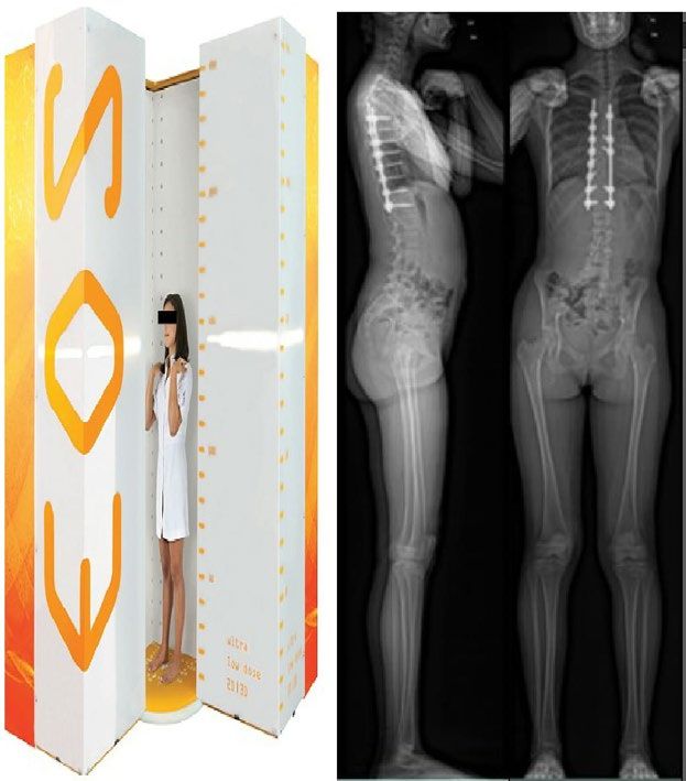
FIGURE 1 / Illustration of the EOS low-dose scanner and the outcome. Petersen et al in 2012 [9], whereby the CT dose from
the O-arm was reduced by nearly 90% compared with
the default protocol.
Algorithms for scoliosis follow-up at our institution
Follow-up for scoliosis at our institution comprises clin-
ical examination and biplane full-spine imaging. Since
the autumn of 2014, EOS low-dose stereo-radiography
has been the first choice for full-spine radiography. CR
biplane imaging has been used solely in cases where
EOS was not available. Figure 1 illustrates the EOS
low-dose scanner, a system that has been described to
markedly reduce radiation dose exposure to patients.
The system has previously been described in detail
[10]. Figure 2 illustrates the follow-up and treatment
algorithms at our institution.
Post-operative radiographs are performed for all
scoliosis cases: before discharge from hospital and at
six months, one year and two years post-operatively.
Consensus survey for scoliosis follow-up at interna-
tional spine centres
A survey was forwarded to nine international ortho
paedic spine centres, in Denmark, Norway, Sweden,
Spain, UK, France and the US, dealing with the assess-
ment and treatment of scoliosis, using a web-based sur-
vey tool. All centres served a population larger than
one million. Four of these centres were high-volume
centres performing more than 100 surgeries for scoli
osis annually.
The survey included questions about the practice of
tients was estimated by calculating the theoretical radiological follow-up of scoliotic patients to illustrate
amount of full-body absorbed radiation dose in terms the similarities or differences among centres and to
of effective dose in mSv. In order to calculate the cumu- compare the answers with current international con-
lative radiation dose from CR and EOS, the number of sensus guidelines [11-13]. The questionnaire used can
images divided by two (coronal and lateral planes) was be found at the following link as referred [14].
multiplied by effective dose references for CR and EOS
stereo-radiography for full-spine examinations. The Results
reference doses for full-spine examinations were esti- Final inclusions
mated based on phantom dosimetry [6]. Reference Demographics for the 61 patients included in this study
doses: CR anterior-posterior-lateral (APL) projections are shown in Table 1. Six patients underwent more
(0.545 mSv) and EOS standard-dose APL projections than one surgery; three were managed initially with
(0.220 mSv). All doses were calculated according to growing rod systems and afterwards with final correc-
the International Commission of Radiologic Protection tion surgery with posterior spinal fusion. Another three
(ICRP), ICRP-103 approach [7]. patients underwent additional revision surgeries; two
Effective doses for the O-arm 3D cone-beam CT and cases owing to progression of curves adjacent to fusion,
ancillary CT and PET-CT were calculated based on dose and one patient owing to implant failure (screw loosen-
length product (and CT conversion factors according to ing and rod breakage).
the Danish Health Authority, Institute of Radiation
Protection (SIS) [8]. Effective doses for intra-operative Radiological exposure
2D fluoroscopy using the O-arm were calculated by us- Radiologic imaging and exposure have been expressed
ing dose area product values and X-ray conversion fac- in terms of the number of images and radiation doses
tors according to the Danish Health Authority, SIS. At (effective dose) to patients from all modalities during
our institution, we routinely use the low-dose intraop- assessment, treatment and follow-up, Table 1. In 66%
2 Dan Med J 67/2 / February 2020DANISH MEDICAL JOURNAL
(73/111) of the intraoperative scans used for safe in- tients for radiographic controls every three months; the
strumentation, the O-arm low-dose protocol was used. other half every six months. Surgically treated patients
The dose from a single low-dose scan (70 kVp/20 mA) were seen anywhere from one to four times for radio-
was found to be 0.45 mSv; for comparison, a default graphic follow-up over a period ranging from six
scan of 120 kVp/40mA was 4.02 mSv. months to two years post-operatively. Five centres saw
Patients who underwent surgery received an ap- patients every six months after instituted brace treat-
proximately ten-fold higher median cumulative radi ment; three centres once annually. A variety of radio-
ation dose (excluding ancillary CT and PET-CT) than graphic systems and techniques were used at the differ-
those treated conservatively. ent centres.
Ancillary radiological imaging from CT and PET-CT
on average resulted in an approximate 100% increase Discussion
of the total dose from all routine imaging (CR, EOS and To the best of our knowledge, this retrospective study
intra-operative O-arm-based navigation and fluoros- represents the first assessment of the cumulative radi
copy), Table 2. ation dose from CR, EOS stereo-radiography and the O-
Approximately 25% (39.04 mSv/161.82 mSv) of arm for a typical patient undergoing current manage-
the total intraoperative radiation dose from the O-arm ment for idiopathic scoliosis. Patient demographics and
was due to 2D fluoroscopy. magnitude of perioperative X-ray/EOS acquisitions
The mean/median weight and height at the time of were comparable to those reported in other studies [1,
surgery were 54.9/54 kg (range: 42-80 kg) and 2, 16-18], as was the mean number of levels fused.
166.2/165 cm (range: 147-184 cm), respectively.
These values are directly comparable to the female do- Exposures and radiation dose
simetry phantom from which the reference doses were As expected and previously shown by Presciutti et al
estimated; weight 55 kg and height 160 cm [15]. [17], the patients who underwent surgery were ex-
posed to substantially more radiographic imaging than
Survey the conservative group, and thus received a higher
Eight out of nine international spine centres, each serv- level of absorbed radiation dose. The large difference in
ing a population of one million to more than ten mil- observed absorbed radiation dose between the two
lion, completed our survey regarding treatment and ra- groups was mainly due to intraoperative as well as an-
diological follow-up algorithms among patients with cillary imaging. Apart from this, the combined observa-
adolescent idiopathic scoliosis. The usual interval be- tion time for the conservative group was shorter. This
tween preoperative imaging varied among the centres. was – in part – caused by the fact that a part of the con-
Half (3/6) of those who answered this question saw pa- servative group was assessed only once or just a few
Juvenile/adolescent idiopathic scoliosis (Cobb angle > 10°) FIGURE 2 / Algorithms for
scoliosis follow-up at our institution.
3-9 years 10-18 years
Cobb angle ≤ 20° Cobb angle ≥ 45° Cobb angle ≤ 20° Cobb angle ≥ 45°
Radiographs every 6-12 Radiographs every 6-12
months until curve months until curve
progression or maturity progression or maturity
Cobb angle 25°-44° Cobb angle 25°-44°
Brace treatment Brace treatment
Progression to Progression to
Cobb angle ≥ 45° Cobb angle ≥ 45°
Surgery with magnetlcally controlled rods Scollosls surgery
Dan Med J 67/2 / February 2020 3DANISH MEDICAL JOURNAL
times, resulting in a low number of total follow-up im-
TABLE 1 / Demographics and radiation exposure..
ages.
Treatment Intraoperative imaging from O-arm CT and fluoros-
conservative surgery both groups copy comprised roughly 50% of the total amount of ab-
Demographics sorbed radiation dose in the surgical group. In the
Patients, n: study by Presciutti et al [17], intraoperative imaging
Males 6 11 17 accounted for 78% of the total accumulated dose. The
Females 13 31 44 reason why our percentage was lower may very well be
Total 19 42 61 the low-dose intraoperative scan protocols used at our
Age, yrs, median (range):
institution. However, our intraoperative dose exposure
At initial assessment 15 (5-18) 14 (3-17) 14 (3-18)
would have been even lower if a higher degree of ad-
At final assessment 15 (9-19) 17 (14-20) 17 (9-20)
herence to the low-dose protocol had been observed.
Time of follow-up, mo.s, median (range) 9 (0-52) 38 (13-163)
The fact that just one ancillary CT or PET-CT re-
Cobb angle, °, median (range):
sulted in a two-fold increase of total cumulative radi
Initial assessment 19 (10-50) 45 (10-80)
Just prior to surgery 52 (36-82) ation dose is a very disturbing finding seen in relation
Final assessment 23 (12-65) 16 (4-30) to the overall dose assessment of these patients. This
Fused vertebrae, n, mean - 11 once again emphasises the importance of keeping the
Radiation exposure, median (range) number of CTs at a bare minimum.
14.5 (2-57)/4.1
CR: spine X-rays, n/radiation dose, mSv 4 (0-20)/1.1 (0-5.5)a
(0.6-15.5) Survey and consensus
Biplanar EOS imaging, nb /radiation dose, 2 (0-17)/ 10.5 (0-26)/ Our survey showed that most of the spine centres
mSv 0.58 (0-2.4)a 1.3 (0-3.1)
agreed on surgical technique and on avoiding post-op-
O-arm 3D scans, n /radiation dose, mSv -
b
2 (1-4)/3.8 (0.9-10.0)
erative CT. However, the survey also illustrated some
O-arm 2D fluoroscopy time, sec/radiation 33.7 (20.3-136.0)/
dose, mSv
-
0.9 (0.4-3.5)
of the discrepancies among centres as to how often to
Radiation dose combined: CR, EOSc, O-arm, assess and for how long to follow patients with idio-
1.1 (0.2-7.2)a 10.3 (3.8-20.4) pathic scoliosis, as well as a discrepancy in choice of ra-
mSv
Additional CT and PET-CT, n d
0 a
1 (1-2) diographic systems. Roughly half of the centres used a
Radiation dose, mSv 0a 11.9 (0.6-20.1) follow-up algorithm similar to our algorithm, which is
Total radiation dose, all modalities, mSv 1.1 (0.2-7.2)a 10.8 (3.8-35.9) in line with the consensus guidelines of Kleuver et al
CR = conventional radiographs. [11] and Knott et al [12]. Implementation of interna-
a) Braced and observational. tional consensus guidelines on follow-up algorithms for
b) Total number of coronal and lateral images.
c) Low-dose stereo-radiography. scoliosis vary depending on the department, local trad
d) A total of 6 patients had additional imaging owing to various reasons explained in the results section. ition and on which consensus guideline was used.
In fact, there is still a lack of clear international con-
sensus as to how often and how many X-rays are
needed in the course of scoliosis treatment. A review of
TABLE 2 / The magnitude of radiation dose from ancillary CT and PET-CT. recent literature gives no clear picture of this [11-13,
Radiation dose, Factor of increase in Part of total 18]. There is agreement as to the needs for at least one
Patient ID Modality mSV total radiation doseb radiation dose, %c coronal and lateral radiograph during or after surgery,
13 CT cervical spine 5.38 2.0 50 but not as to the timing of subsequent follow-up inter-
27 CT thoraco-lumbar spinea 17.03 1.9 48 vals or the duration of patient follow-up.
34 PET-CT full spine and CT 20.13 3.1 67 Only one in eight centres used post-operative CT
lumbar spinea routinely; likewise, one in eight used intraoperative
38 CT lumbar spine 6.70 1.7 42
navigation. At our institution, we use intraoperative
45 CT cervical spine 0.57 1.1 6
navigation for all scoliosis surgery based on studies
46 CT thoraco-lumbar spinea 18.03 2.1 53
showing significantly less screw misplacements than
CR = conventional radiographs.
a) Metal artefact reducing software technique. for freehand technique and or fluoroscopy-based in-
b) (CR, EOS, O-arm and ancillary CT and PET-CT)/(ancillary CT and PET-CT). strumentation [19, 20].
c) (Ancillary radiation dose)/(total radiation dose from all modalities).
Limitations
Our study has some limitations. The size of the study
population was limited and thus not well suited for
statistical analysis, e.g., on various possible correla-
tions. Because of the small number of patients, we com-
bined braced patients and observation patients in the
4 Dan Med J 67/2 / February 2020DANISH MEDICAL JOURNAL
same group for comparison with the surgical group. 8. Danish Health Authority. CT referencedoser. Indsamling og vurdering
af patientdoser ved CT. Copenhagen: Danish Health Authority, 2015.
This comparison provides an overview of the variation http://sundhedsstyrelsen.dk/da/udgivelser/2015/~/media/5FFD01914
of doses between the groups, but it does not reflect very F8A430A8407C9B46D94AA6E.ashx (17 Nov 2019).
9. Petersen AG, Eiskjær S, Kaspersen J. Dose optimisation for intraopera-
well what the typical dose is for braced patients speci tive cone-beam flat- detector CT in paediatric spinal surgery. Pediatr
Radiol 2012;42:965-73.
fically. 10. Després P, Beaudoin G, Gravel P et al. Physical characteristics of a
low-dose gas microstrip detector for orthopedic x-ray imaging. Med
Phys 2005;32:1193-204.
Dose reduction 11. De Kleuver M, Lewis SJ, Germscheid NM et al. Optimal surgical care for
There are several ways to reduce radiation exposure. adolescent idiopathic scoliosis: an international consensus. Eur Spine
J 2014;23:2603-18.
The simplest is to reduce the number of X-rays and 12. Knott P, Pappo E, Cameron M et al. SOSORT 2012 consensus paper: re-
avoid CTs, while optimising radiological equipment is a ducing x-ray exposure in pediatric patients with scoliosis. Scoliosis
2014;9:4.
continuously ongoing process. In fact, little might be 13. Fletcher ND, Glotzbecker MP, Marks M et al. Development of consen-
sus-based best practice guidelines for postoperative care following
gained from routine imaging unless the patient has un- posterior spinal fusion for adolescent idiopathic scoliosis. Spine (Phila
expected symptoms. Garg et al [18] found that only Pa 1976) 2017;42:E547-E554.
14. www.dropbox.com/s/mlladixj2dwy8bm/SurveyMonkey_160922409-2.
2.9/1,000 spine X-rays led to revision surgery. Thus, it pdf?dl=0.
may very likely be possible to lower the number of 15. ATOM dosimetry phantoms. Size and age-related dose calculations
features. Virginia: CIRS, 2015.
spine radiographs without affecting the quality of treat- 16. Law M, Ma W-K, Lau D et al. Cumulative radiation exposure and associ-
ment. ated cancer risk estimates for scoliosis patients: Impact of repetitive
full spine radiography. Eur J Radiol Elsevier Ireland Ltd; 2016;85:625-8.
17. Presciutti SM, Karukanda T, Lee M. Management decisions for adoles-
Conclusions cent idiopathic scoliosis significantly affect patient radiation expos
ure. Spine J 2014;14:1984-90.
The magnitude of cumulative radiation during scoliosis 18. Garg S, Kipper E, La Greca J et al. Are routine postoperative radiog
raphs necessary during the first year after posterior spinal fusion for
treatment varies substantially depending on the radio- idiopathic scoliosis? A retrospective cohort analysis of implant failure
graphic follow-up protocol and on intraoperative and and surgery revision rates. J Pediatr Orthop 2014;35:33-8.
19. Ughwanogho E, Patel NM, Baldwin KD et al. Computed tomography-
ancillary imaging used. Future studies are needed to guided navigation of thoracic pedicle screws for adolescent idio-
elucidate the clinical consequences of a lowered or an pathic scoliosis results in more accurate placement and less screw
removal. Spine (Phila Pa 1976) 2012;37:473-8.
elevated frequency of X-ray monitoring. Such studies 20. Baky FJ, Milbrandt T, Echternacht S et al. Intraoperative computed
may also lay the foundation for future consensus guide- tomography-guided navigation for pediatric spine patients reduced
return to operating room for screw malposition compared with free-
lines on radiographic follow-up. hand/fluoroscopic techniques. Spine Deform 2019;7:577-5.
By using low-dose X-ray systems such as EOS ste-
reo-radiography in combination with low-dose protocol
for intraoperative navigation, it is possible to keep pa-
tient exposure at a minimum, balancing potential risks
of adverse effects such as screw misplacement and radi-
ation-induced cancer while still providing optimal care.
One ancillary CT may double the total cumulative full-
body absorbed radiation dose.
CORRESPONDENCE: Ari Demirel. E-mail: ari.demirel@hotmail.com
ACCEPTED: 2 December 2019
CONFLICTS OF INTEREST: none. Disclosure forms provided by the authors
are available with the full text of this article at Ugeskriftet.dk/dmj
LITERATURE
1. Doody MM, Ronckers CM, Land CE et al. Cancer mortality among
women frequently exposed to radiographic examinations for spinal
disorders. Radiat Res 2010;174:83-90.
2. Simony A, Carreon LY, Jensen KE et al. Incidence of cancer and infertil-
ity, in patients treated for adolescent idiopathic scoliosis 25 years
prior. Eur Spine J 2015;24:S740.
3. Pace N, Ricci L, Negrini S. A comparison approach to explain risks re-
lated to X-ray imaging for scoliosis, 2012 SOSORT award winner. Scoli-
osis 2013;8:11.
4. Deschênes S, Charron G, Beaudoin G et al. Diagnostic imaging of spinal
deformities: reducing patients radiation dose with a new slot-scan-
ning X-ray imager. Spine (Phila Pa 1976) 2010;35:989-94.
5. Dietrich TJ, Pfirrmann CW, Schwab A et al. Comparison of radiation
dose, workflow, patient comfort and financial break-even of standard
digital radiography and a novel biplanar low-dose X-ray system for up-
right full-length lower limb and whole spine radiography. Skeletal Ra-
diol. 2013;42:959-67.
6. Pedersen PH, Petersen AG, Østgaard SE et al. EOS Micro-dose Protocol:
first full-spine radiation dose measurements in anthropomorphic
phantoms and comparisons with EOS standard-dose and conven-
tional digital radiology. Spine (Phila Pa 1976) 2018;43:E1313-E1321.
7. The International Commission on Radiological Protection. The 2007
Recommendations of the International Commission on Radiological
Protection. ICRP Publication 103. Ann ICRP 2007;37.
Dan Med J 67/2 / February 2020 5You can also read