Scholars Journal of Applied Medical Sciences
←
→
Page content transcription
If your browser does not render page correctly, please read the page content below
Scholars Journal of Applied Medical Sciences
Abbreviated Key Title: Sch J App Med Sci
ISSN 2347-954X (Print) | ISSN 2320-6691 (Online)
Journal homepage: https://saspublishers.com                                                                  Radiological Sciences

Paranasal Sinuses in the Evaluation of Sinusitis using Computed
Tomography: Cross Sectional Study
Syed Faizan Haider Naqvi1*, Nosheen Arshad2, Muhammad Ahmad Naeem2, Nazeeha Waseem1, Narjis Batool2, Abid
Ali3
1
  Medical Imaging Doctor, Department of Radiological Sciences and Medical Imaging, the University of Lahore, Gujrat, Pakistan
2
  Lecturer, Department of Radiological Sciences and Medical Imaging, the University of Lahore, Gujrat, Pakistan
3
  Associate Professor, Faculty of Allied Health Sciences, the University of Lahore, Gujrat, Pakistan

DOI: 10.36347/sjams.2021.v09i04.024                               | Received: 18.03.2021 | Accepted: 25.04.2021 | Published: 29.04.2021
*Corresponding author: Syed Faizan Haider Naqvi

    Abstract                                                                                              Original Research Article

    Background: Recent advances in the understanding the pathophysiology paranasal sinuses have revolutionized the
    surgical management of chronic and recurrent sinusitis. Paranasal sinus is air-filled spaces in the interior skull and
    facial bones. Symptoms of a common cold include nasal discharge, nasal obstruction, headache, and nasal allergy in
    the majority of patients. CT is a superlative modality to describe the sinus anatomy along with soft tissue structure. As
    compared to sinus radiographs, computed tomography scanning has improved the imaging of paranasal sinus anatomy.
    Objective: To determine paranasal sinuses in evaluation of sinusitis using computed tomography. Material Methods:
    From October 2020 to March 2021, a cross-sectional analysis was performed at Aziz Bhatti Shaheed (DHQ) Teaching
    Hospital Gujrat and Azeem ultrasound and diagnostic center Gujrat. Data of 100 patients was collected through
    convenient sampling technique adults and children with sinusitis were included in the study data of patients with
    recent cold associated with irritated nasal discharge and headache along with congestion was collected from the
    patients. For data processing, the Statistical Kit for the Social Sciences (spss) is used. Results: Out of 100 patients 50
    were male & 50 were female majority of the patients were in 25-40 age group. 12 out of 100 had nasal obstructions, 27
    patients were presented with headache, 27 patients had the history of facial pain. Maxillary sinuses are mostly
    involved in study 13 had bilateral maxillary sinusitis 13% left maxillary right sphenoidal and ethmoidal sinusitis
    followed by ethmoid sinus and frontal sinus. Mucosal thickening (mild 76 percent, moderate 20 percent) is the most
    often observed mass and symptom of sinusitis. The final result came out 5 % PAN sinusitis. Conclusion: Pathologies
    in paranasal sinuses is frequently found on computed tomography imaging and has to be cured or monitored
    consequently. The best modality for diagnosing and preparing therapy for clinically current sinusitis is computed
    tomography.
    Keywords: Para nasal sinuses, sinusitis, computed tomography.
Copyright © 2021 The Author(s): This is an open-access article distributed under the terms of the Creative Commons Attribution 4.0 International
License (CC BY-NC 4.0) which permits unrestricted use, distribution, and reproduction in any medium for non-commercial use provided the original
author and source are credited.

                                                                             nasal sinuses. The pathological lesions of the para nasal
INTRODUCTION                                                                 sinuses vary from inflammation to cancerous types,
          Para nasal sinuses are hollow air-filled cavities                  which may be benign or malignant however; sinusitis is
in human facial bones, which acts an essential role                          usually described as nasal and Para nasal sinus mucosal
which includes reducing skull weight, humidifying and                        inflammation of the mucous membrane that lines the
warming of inhaled air, pressure control within the                          sinuses which resulting in symptoms like nasal
nasal cavity, also trapping dust particles [1]. There are                    discharge, sore throat, nasal obstruction, cough,
four combined Para nasal sinuses; are maxillary, frontal,                    headache, nasal allergy [4]. Earlier studies have
and sphenoid sinuses as well as ethmoid cells by                             revealed that people are prone to several diseases of
abundant inter and intra-individual similarities [2].                        Para nasal sinuses due to different causes such as
Three essential components make up the para nasal                            allergy, extreme exposure to cold, tobacco smoking,
sinus: thin natural mucus secretions, normally working                       alcohol intake, trauma, and infections by a pathogen
hair-like cilia that exchange mucus out of sinuses, and                      such as a virus, bacteria, and fungi [1]. Sinusitis is a
an open sinus drainage opening known as the sinus                            swelling of the nasal sinus, it is also well-known as
ostium [3]. The word sinusitis mentions a collection of                      rhino sinusitis, and is a common medical problem in the
conditions described by swelling of the mucosa of Para
    Citation: Syed Faizan Haider Naqvi et al. Paranasal Sinuses in the Evaluation of Sinusitis using Computed Tomography: Cross
                                                                                                                                       605
    Sectional Study. Sch J App Med Sci, 2021 Apr 9(4): 605-611.
Syed Faizan Haider Naqvi et al; Sch J App Med Sci, Apr, 2021; 9(4): 605-611
ear, nose, and throat ENT department. The maxillary sinus abnormalities is linked with environmental
sinus is one of four nasal sinuses held in cheekbones. Its pollution, which is common due to numerous oil and
shapes similar to the pyramid and each hold three gas industrial activities in the area [1]. The goal of the
cavities [5]. The analysis of acute sinusitis is made current study was to estimate the CT findings in three
through medical conditions also depend on the different areas i.e. nasal septum, nasal turbinate’s, and
occurrence and array of indications that can distinguish Para nasal sinuses, in patients booked for rhinoplasty
between acute sinusitis and a simple virus-related URI [13]. Computed tomography executes a very important
[6]. Acute sinusitis is a small period of inflammation of role to value pathologies in strenuous especially in
the membrane of the nose and surrounding sinus is ethmoid in sphenoid sinuses [9]. Essentially, CT scan
mostly due to cold-causing infection or it may be non- proved to be an excellent imaging modality because it
infection [5]. Rhino sinusitis is a unique disease accurately diagnosed and differentiated benign and
affecting people worldwide with a significantly bad malignant lesions, as well as delineated their genesis,
effect on the quality of life [7]. The presence of at least appearance, expansion, and presence. With the unique
2 out of four cardinal symptoms (facial pain, ability of CT to image soft bone tissue, direct coronal
compression, drainage, nasal obstacle) for at least 12 scans and sagittal reconstruction, the lesions occupying
repeated weeks is well established in maximum the space [5]. The management and results of smell
guidelines [8]. Sinusitis is now well recognized as complaint of conductive damage are dissimilar to
primarily a psychiatric diagnosis. A physical sensorineural damage [14]. The prevalence of these
examination may help differentiate sinusitis from a findings is needed to determine their clinical relevance
minor upper respiratory tract infection as patient records and to guide its management [15].
suggest sinusitis [3].
Imaging technologies used in nose study and
MATERIAL AND METHOD
From October 2020 to March 2021, a cross-
Para nasal sinuses (PNS) play a significant role in the
sectional analysis was performed at Aziz Bhatti
treatment of multiple pathologies. Recent or innovative
Shaheed (DHQ) Teaching Hospital Gujrat and Azeem
imaging technology such as computed tomography and
ultrasound and diagnostic center Gujrat. Data of 100
MRI plays a vital role to analyze Para nasal sinuses
patients was collected through convenient sampling
more accurately than conventional X-rays [9].
technique adults and children with sinusitis were
Computed tomography has shown huge progress since
included in the study data of patients with recent cold
the initial house field CT images obtained at the end of
associated with irritated nasal discharge and headache
1970 [4]. On a point that preoperative worksheet in the
along with congestion was collected from the patients.
estimation of Sino nasal CT scan boosts the protection
For data processing, the Statistical Kit for the Social
also the ability of nose as well as Para nasal sinus
Sciences (SPSS) is used.
surgical treatment [10]. Additionally in the physical
sets, the multislice CT scans are organized that reduced
patient distress due to smaller as well as less breath RESULTS
holds also reducing the requirement of lethargy for Out of 100 patients 50 were male & 50 were
agitated patients [11, 14]. This study meant to estimate female majority of the patients were of 25 to 40 age
also concludes the incidence of the structural group.12 out of 100 had nasal obstructions, 27 patients
differences must be measured to escape the difficulties were presented with headache, 27 patients had the
that might take place during the invasive process [12]. history of facial pain. Maxillary sinuses are mostly
Conventional x-rays of Para nasal sinuses PNS is still involved in study 13 had bilateral maxillary sinusitis
the simplest and cheapest method of diagnosing the 13% left maxillary right sphenoidal and ethmoidal
PNS pathologies but because of the superimposition of sinusitis followed by ethmoid sinus and frontal sinus.
structure in x-rays and inadequate diagnostic Mucosal thickening (mild 76 percent, moderate 20
information, it cannot be used as a guide for endoscopic percent) is the most often observed mass and symptom
sinus surgery [1]. The high prevalence of Para nasal of sinusitis. Final result came out 5 % PAN sinusitis.
Table-1: Clinical findings
Symptoms Frequency Percent Valid Percent Cumulative Percent
Fever , headache 1 1.0 1.0 51.0
Fever , nasal obstruction 1 1.0 1.0 52.0
Fever , nasal stiffness 1 1.0 1.0 53.0
Fever ,pressure or pain in sinus 2 2.0 2.0 55.0
Fever facial tenderness 1 1.0 1.0 56.0
Fever, facial tenderness 1 1.0 1.0 57.0
Headache , facial pain 27 27.0 27.0 84.0
Nasal discharge , fever 12 12.0 12.0 96.0
Nasal discharge, fever, facial swallowing 2 2.0 2.0 98.0
Nasal obstruction 2 2.0 2.0 100.0
Total 100 100.0 100.0
© 2021 Scholars Journal of Applied Medical Sciences | Published by SAS Publishers, India 606Syed Faizan Haider Naqvi et al; Sch J App Med Sci, Apr, 2021; 9(4): 605-611
Results indicate that headache and facial pain are 27% nasal discharge and fever are 12%, pressure and pain are
2%.
Fig-1
Fig-2: Age Distribution
Results indicate that majority of cases are between 25 to 40 years and most of them are 30year old (7%), 40 year
old (7%) and 25 year old (5%).
Table-3: CT PNS Findings Mucosal Thickening
Severity Frequency Percent Valid Percent Cumulative Percent
mild 76 76.0 76.0 76.0
mild 3 3.0 3.0 79.0
moderate 20 20.0 20.0 99.0
normal 1 1.0 1.0 100.0
Total 100 100.0 100.0
This result indicate that mild mucosal thickening is 76% and moderate mucosal thickening is 20%
© 2021 Scholars Journal of Applied Medical Sciences | Published by SAS Publishers, India 607Syed Faizan Haider Naqvi et al; Sch J App Med Sci, Apr, 2021; 9(4): 605-611
Table-4: Impression
Frequency Percent
Valid LT mastioditis , LTsphenoid sinusitis 1 1.0%
bilateral ethmoidal and left sphenoidal sinusitis 1 1.0%
bilateral ethmoid sinusitis 1 1.0%
bilateral ethmoid sinusitis and LF sphenoidal sinusitis 1 1.0%
bilateral ethmoidal sinusitis 1 1.0%
bilateral inferior turbinate thickening 1 1.0%
bilateral maxillary and ethmoidal sinusitis 1 1.0%
bilateral maxillary sinusitis 13 13.0%
bilateral maxillary sinusitis ,LT ethmoidal sinusitus 1 1.0%
bilateral maxillary sinusitis ,LT sphenoidal and ethmoidal 1 1.0%
sinusitis
bilateral maxillary sinusitis and left ethmoidal sinusitis 1 1.0%
ethmoidal sinusitis 1 1.0%
ethmoidal sinusitis and maxillary sinusitis 1 1.0%
extensive fungal sinusitis 4 4.0%
extensive pan sinusitis 1 1.0%
extensive sinusitis involving left side sinuses 1 1.0%
extensive sinusitis involving left side sinuses with 1 1.0%
polypolidial
Fungal sinusitis 2 2.0%
LT ethmoidal and LT sphenoidal sinusitis 1 1.0%
LT ethmoidal sinusitis 1 1.0%
LT frontal and bilateral sinusitis 2 2.0%
LT maxillary , RT sphenoidal and ethmoidal sinusitis 1 1.0%
LT maxillary sinusitis 13 13.0%
LT maxillary sinusitis ,polyp sinus 1 1.0%
LT maxillary sinusitis, acute sinusitis 1 1.0%
LT nasal polyps 1 1.0%
LT spheniodal sinusitis 5 5.0%
maxillary sinusitis 1 1.0%
nasal polyposis 1 1.0%
nasal polyposis and sinusitis 1 1.0%
pan sinusitis 5 5.0%
RT acute sphenoidal sinusitis 1 1.0%
RT ethmoidal fungal sinusitis, RT sphenoid chronic sinusitis 1 1.0%
RT ethmoidal sinusitis 1 1.0%
RT frontal sinusitis 1 1.0%
RT frontal , ethmoidal, bilateral maxillary, RT sphenoidal 1 1.0%
sinusitis
RT inferior turbinate thickening 1 1.0%
RT mastiodititis 1 1.0%
RT maxillary ,frontal ,ethmoidal sinusitis 1 1.0%
RT maxillary and ethmoidal sinusitis 1 1.0%
RT maxillary sinusitis 5 5.0%
RT maxillary sinusitis, LT sinunasal polyp 1 1.0%
RT maxillary sinusitis 3 3.0%
© 2021 Scholars Journal of Applied Medical Sciences | Published by SAS Publishers, India 608Syed Faizan Haider Naqvi et al; Sch J App Med Sci, Apr, 2021; 9(4): 605-611
RT maxillary acute sinusitis 1 1.0%
RT nasal polyp and sinusitis 2 2.0%
RT sphenoid sinusitis 5 5.0%
Sinusitis 5 5.0%
sphenoidal sinusitis 1 1.0%
Total 100 100.0%
Frequency Percent Valid Percent Cumulative Percent
sphenoidal sinusitis 1 1.0 1.0 100.0
Total 100 100.0 100.0
Result indicate that mostly seen patients with these patients who had sinusitis. These patients were
bilateral maxillary sinusitis 13% left maxillary sinusitis presented with a history of cough, headache, and facial
13% pain it is critical to confirm that if chronic rhino
sinusitis is described based on subjective knowledge; an
empirical result may be obtained with CT. The
evaluation of sinusitis between male and female ratio is
50%. According to the study conducted by author
Michael promise Ogolodom et al., sinusitis
was the most common paranasal sinuses disease in this
study, while osteoma was the least common.
The most often affected sinuses were the maxil
lary sinuses [1]. This study conducted by author ohood
A. Mohammed et al., 2019. Preoperative identification
of the anatomical difference of the paranasal sinuses is
crucial. These differences were discovered to differ by
area and nation. In our research, we discovered that all
Fig-3: Gender patients with rhinosinusitis have one or more of the
Graphical representation shows those patients anatomical variations listed.
of sinusitis on CT scan finding present 50% male and
50% female patients. The most common anatomical variation is a de
viated nasal septum (93.11 percent), followed by aggern
asi cell d(93.11 percent) (51.02 percent ) Junaid Iqbal et
al., 2017 [2]. Computed Tomography Evaluation of
Anatomical Variations of the Paranasal Sinuses Region
of Rhinosinusitis was the subject of a research project.
Of the 120 patients, 49 (41%) were female and 71 (59%
) were male. The patients were 35.211.61 years old on a
verage. 33percent (27.5%) were between the ages of 20
and 30, 35 percent (29%) were between the ages of 30 a
nd 40, 29 percent (24%) were between the ages of 40 an
d 50, and the remaining 23 percent (19%) were over 40.
Sinusitis caused by fungus on both sides was marginall
y more common [2]. According to this study conducted
by author Alia Ahmad et al., 2016 In this study, 55
patients ranging in age from 20 to 55 years old were
included. 33 (60%) of the patients were male, while the
remaining 22 (40%) were female. 12 (21.82%) patients
were between the ages of 20 and 29, 13 (23.64%)
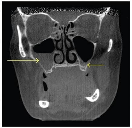
Fig-4: Showing CT scan image arrows indicating Bilateral patients were between the ages of 30 and 39, 18
maxillary sinusitis (coronal view) (32.72%) patients were between the ages of 40 and 49,
and 12 (21.82%) patients were over the age of 49. By
clinically standard x-ray observation, 26 (47.27 percent)
DISCUSSION of the 55 patients had acute sinusitis, while 29 (52.73
percent) had chronic sinusitis [3].
The aim of the research was to use CT to
confirm the diagnosis of paranasal sinusitis. A history
of 100 patients was collected and compared among
© 2021 Scholars Journal of Applied Medical Sciences | Published by SAS Publishers, India 609Syed Faizan Haider Naqvi et al; Sch J App Med Sci, Apr, 2021; 9(4): 605-611
The pathologies in this study were seen in peo 4. Verma J, Tyagi S, Srivastava M, Agarwal A.
ple aged 16 to 30.The oldest were between the ages of 0 Computed tomography of Para nasal sinuses for
and 15, while the youngest were between the ages of 0 early and proper diagnosis of nasal and sinus
and 15.Sinusitis has been the most common PNS. For pathology. Int J Otorhinolaryngol Head Neck Surg.
several years, I worked in pathology. The diagnosis of 2016;2(2):70.
chronic rhino sinusitis is focused mostly on major and 5. Gohar A, Tariq I, Saeed M, Waqar M, Mazhar R,
minor symptoms in these age ranges, with care Daniel S, et al. Frequency of Computed
administered based on the diagnosis. Patients who Tomography Para nasal sinuses in the Evaluation
follow the symptoms-based definitions of chronic rhino of Sinusitis. Journal of Health and Medical
sinusitis are advised to administer antibodies for several Sciences. 2019;2(4).
weeks. This project was undertaken to provide a 6. DeMuri GP, Eickhoff JC, Gern JC, Wald ER.
slandered description of chronic rhino sinusitis, as well Clinical and virological characteristics of acute
as to make a diagnosis based on endoscopy or CT scans. sinusitis in children. Clinical Infectious Diseases.
Endoscopy was used to assess the function of 2019;69(10):1764-70.
endoscopy in the diagnosis of chronic, non-polyp, 7. Chakraborty P, Ragni J. Radiologic variations of
unoperated rhino sinusitis. My research showed that the nose and Para nasal sinuses: ACT-based study.
pathologies such as maxillary sinusitis are commonly J Med Sci Clin Res. 2016;4:10536-41.
detected on Computed tomography imaging and must 8. Ahmmed SU, Khan MNI, Hossain MZ, Mridha
be treated or monitored appropriately. Computed MKI, Bhuiyan AP, Ahmed KSU. Study of
tomography should be used to diagnose and schedule Prevalence of Concha Bullosa, Nasal Septal
therapy for sinusitis that is clinically present. The Deviation, and Sinusitis based on CT Findings.
commonest Para nasal sinuses frequency in this study Bangladesh Journal of Otorhinolaryngology.
was sinusitis these findings are keeping with the study 2020;26(1):18-23.
done. 9. Ahmed A, Malik G, Rauf M. Comparison between
Imaging Technologies (X-Ray with Compute
Tomography Scan) Of Para Nasal Sinuses (PNS) in
CONCLUSION Sinusitis Patients. Pakistan Postgraduate Medical
Pathologies in paranasal sinuses is frequently Journal. 2016;27(3):68-71.
found on computed tomography imaging and has to be 10. Alshaikh N, Aldhurais A. Anatomic variations of
cured or monitored consequently. The best modality for the nose and Para nasal sinuses in Saudi
diagnosing and preparing therapy for clinically current population: computed tomography scan analysis.
sinusitis is computed tomography. For diagnosing The Egyptian Journal of Otolaryngology.
paranasal sinus pathologies CT imaging shows an 2018;34(4):234-41.
accurate use in diagnosis of acute sinusitis and a 11. Dhong H-J, Jung J-Y, Park JH. Diagnostic
sufficient limit in the diagnosis of chronic sinusitis. accuracy in sinus fungus balls: CT scan and
Without waiting for culture results, the imaging operative findings. American journal of rhinology.
modality can be used to diagnose certain infections 2000;14(4):227-32.
quickly. 12. Güngör G, Okur N. Evaluation of Para nasal sinus
Variations with Computed Tomography. surgery.
Financial Support and Conflict of Interest: No 2019;2(5):6.
financial support and we declared that there is no 13. Abbasi M, Izadi P, Yarmohammadi M. Computed
conflict of study in this research. Tomographic Findings of Nasal and Para nasal
sinuses in Patients Scheduled for Rhinoplasty in
REFERENCES Shahid Mostafa Khomeini Hospital during 2011-
1. Ogolodom MP, Ugwu AC, Ohagwu CC, 13. ENT Updates. 2019;9(3):199-205.
Chukwuemeka E, Joseph TCO, Egbeyemi OO. 14. Naeini AS, Karimi-Galougahi M, Raad N,
Patterns and prevalence of Para nasal sinuses Ghorbani J, Taraghi A, Haseli S, Mehrparvar G,
diseases among patients referred for Para nasal Bakhshayeshkaram M. Paranasal sinuses computed
sinuses computed tomography in Port Harcourt tomography findings in anosmia of COVID-19.
Rivers State, Nigeria. International Journal of American Journal of Otolaryngology. 2020 Nov
Medical and Health Research. 2018;4(11):71-5. 1;41(6):102636.
2. Guidugli GA, Gibelli DM, Michaela C, Oliva AG, 15. Bakheet E, Bakheet TM. Prevalence of Anatomical
Barni L, Sartori P, Sforza C. Volume, asymmetry Abnormalities of Nose & Para nasal sinuses in
and reciprocal relationships between paranasal Cases of Rhinogenic Headache among Sohag
sinuses: a 3D segmentation study on head CT- University Students. Sohag Medical Journal.
scans. Stoma Edu J. 2020;7(1):20-7. 2020;24(1):140-9.
3. Hussein AO, Ahmed BH, Omer MAA, Manafal 16. Ogolodom MP, Ugwu AC, Ohagwu CC,
MF, Elhaj AB. Assessment of clinical, X-Ray, and Chukwuemeka E, Joseph TCO, Egbeyemi OO.
CT in the diagnosis of Para nasal sinus diseases. Int Patterns and prevalence of paranasal sinuses
J Sci Res. 2014;3:7-11. diseases among patients referred for paranasal
© 2021 Scholars Journal of Applied Medical Sciences | Published by SAS Publishers, India 610Syed Faizan Haider Naqvi et al; Sch J App Med Sci, Apr, 2021; 9(4): 605-611
sinuses computed tomography in Port Harcourt 18. Ahmed A, Malik G, Rauf M. Comparison between
Rivers State, Nigeria. International Journal of Imaging Technologies (X-Ray with Compute
Medical and Health Research. 2018;4(11):71-5. Tomography Scan) Of Paranasal Sinuses (PNS) in
17. Iqbal J, Rashid S, Darira J, Shazlee MK, Ahmed Sinusitis Patients. Pakistan Postgraduate Medical
MS, Fatima S. Diagnostic accuracy of CT scan in Journal. 2016 Jun 1;27(3):68-71.
diagnosing paranasal fungal infection. J Coll
Physicians Surg Pak. 2017 May 1;27(5):271-74.
© 2021 Scholars Journal of Applied Medical Sciences | Published by SAS Publishers, India 611You can also read