Alterations of Uterine Blood Flow During the Follicular Phase in Patients With Recurrent Implantation Failure: A Doppler Ultrasonographic Study
←
→
Page content transcription
If your browser does not render page correctly, please read the page content below
http://www.ijwhr.net                                                                                                         doi 10.15296/ijwhr.2021.40

    Open Access                                                                                                                      Original Article
                                              International Journal of Women’s Health and Reproduction Sciences
                                              Vol. 9, No. 3, July 2021, 217–221
                                              ISSN 2330- 4456

Alterations of Uterine Blood Flow During the Follicular
Phase in Patients With Recurrent Implantation Failure: A
Doppler Ultrasonographic Study
                     ID                              ID                         ID                      ID                      ID
Marjan Amini1 , Mahnaz Ranjkesh2 , Saba Nikanfar3 , Amir Fattahi4 , Laya Farzadi1 , Kobra
Hamdi1* ID

    Abstract
    Objectives: The dynamics of blood flow in the endometrium plays a crucial role during the implantation process. This study aimed
    to assess the uterine perfusion during the follicular phase in patients with a history of recurrent implantation failure (RIF) and healthy
    fertile women using the transvaginal ultrasound color Doppler method.
    Materials and Methods: To this end, 50 patients with RIF and 50 age-matched healthy fertile women were recruited in this case-
    control study. The transvaginal color Doppler ultrasonography was used to evaluate the pulsatility index (PI) and resistance index (RI)
    of the uterine, arcuate, and sub-endometrial arteries during the follicular phase in both groups.
    Results: The RI and PI of both right and left uterine arteries were higher in the RIF group compared to the fertile women (P
Amini et al
Key Messages PI= PSV- EDV/mean maximum flow velocity, RI= PSV-
EDV/ PSV
►► The uterine blood flow of RIF patients is significantly
altered in the follicular phase.
The alterations of uterine blood flow in the follicular
phase were investigated for all participants.
Al-Zahra Hospital of Tabriz, Iran. RIF was defined as a
Statistical Analysis
failure of pregnancy after the transfers of 1-2 high-quality
Due to time and financial constraints, the convenience
embryo(s) during at least three consecutive IVF cycles.
sampling method was applied for the sample selection,
The decision about the type of protocol or doses of drugs
and the sample size was calculated using the PASS 11.0
was made by a gynecologist according to the ovarian
software (PASS, Kaysville, UT, USA). In this regard, beta
response to the doses of the applied drugs during previous
and type I error were set to 0.20 (80% power) and 0. 05,
cycles. However, 150-400 IU/day of gonadotropins in
respectively. Data are expressed as the mean ± standard
combination with human menopausal gonadotropin
deviation (SD). After analyzing the data distribution by
(75-150 IU) were generally administered for 10-12 days
Kolmogorov-Smirnov test, variables were evaluated by
during the gonadotropin-releasing hormone antagonist
the Student’s t test or the Mann-Whitney U test for data
protocol. When the follicles reached a mean size of >17
with a normal distribution or skewed ones, respectively.
mm, ovulation was induced by 5000 or 10 000 IU doses
A P value lower than 0.05 was considered statistically
of human chorionic gonadotropin. After 36 hours,
significant, and all statistical analyses were performed
the cumulus‐oocyte complexes were retrieved by the
using SPSS software, version 16.0 (Chicago, IL, USA).
transvaginal ultrasound‐guided puncture of the follicles.
After checking the maturity of the denuded oocytes,
Results
metaphase II oocytes underwent the ICSI procedure. After
This study recruited 50 RIF and 50 healthy women. The
the development of embryos, high-quality blastocysts
mean age of women in the RIF and control groups was
(1-2 cases) based on Veeck and Zaninovic’s criteria were
32.72 ± 5.46 and 31.84 ± 4.21 years, respectively (P = 0.342).
selected for transfer (11). In this regard, the early formation
Furthermore, the groups did not differ regarding the day
of an expanded and eccentric cavity with a distinct layer
of the follicular phase in which the ultrasonography was
of trophectoderm and inner cell mass was considered
performed (4.87 ± 4.14 in RIF women vs. 7.14 ± 3.84 in
for the characterization of a good quality blastocyst.
the control group, P=0.103), and the mean duration of
Women of the control group were ovulating females who
infertility was 8.26 ± 4.60 years in the RIF group.
experienced regular menstrual cycles and had no history
Embryo factors such as sperm and oocyte quality,
of pregnancy failure. The inclusion criteria for all women
chromosomal anomalies in parents, and the number and
were having 20-40 years of age with a body mass index of
developmental stage of embryos can play a role in RIF.
˂30 kg/m2, no history of alcohol consumption or special
Furthermore, according to the guidelines of our fertility
diet, and being a nonsmoker. The patients with diabetes,
center, the number and developmental stage of embryos
dyslipidemia, uterine anomalies, uterine myomas,
were approximately the same in most cases. Moreover,
uterine adenomyosis, disorders of ovulation (including
patients with uterine anomalies and autoimmune diseases
prolonged oligomenorrhea, premature ovarian failure,
were excluded from our study. However, this study did
and hypothalamic amenorrhea), autoimmune diseases,
not assess all the possible reasons for RIF. Our results
hypertension, and coagulation disorders were excluded
demonstrated that the total number of embryo transfer
from the study. The participants were enrolled in the
cycles and the total number of transferred embryos were
study after obtaining a signed informed consent form.
2.96± 1.24 and 10.36± 8.21, respectively.
All patients underwent Doppler transvaginal ultrasound
Table 1 presents the means (±SD) of PI and RI for
to assess PI and RI of the right and left uterine, arcuate
uterine, arcuate, and sub-endometrial arteries in RIF
uterine, and sub-endometrial arteries.
patients and healthy women. The results of Doppler
ultrasound examinations showed that the PI and RI of
Transvaginal Doppler Ultrasound
the right and left uterine arteries significantly increased
Transvaginal color Doppler ultrasonography was
in the RIF group in comparison with the control group.
performed for both RIF patients and control women
Moreover, the indices of pulsatility and resistance for
using the Samsung WS-80 ultrasound system (Samsung
sub-endometrial uterine arteries significantly elevated in
Medison Company Ltd, Seoul, South Korea) equipped
women with a history of RIF compared to the control group
with a 4- to 8-MHz transvaginal probe. The calculation
(PAmini et al
Table 1. Mean ± SD of PI and RI for Uterine, Arcuate, and Sub-endometrial
Arteries in RIF and Control Groups
Study Group Control Group
Uterine Arteries (n=50) (n=50) P Value
Mean (±SD)
Right uterine arteries
PI 2.96± 0.61 1.87± 0.55 ˂0.001*
RI 0.92± 0.10 0.73± 0.14 ˂0.001*
Left uterine arteries
PI 3.05± 0.69 1.88± 0.40 ˂0.001*
RI 0.89± 0.06 0.74± 0.14 ˂0.001*
Arcuate arteries
PI 1.87± 0.61 1.72± 0.65 0.229
RI 0.74± 0.12 0.65± 0.17 0.005*
Sub-endometrial arteries
PI 1.478± 0.87 0.71± 0.25 0.001*
RI 0.67± 0.20 0.53± 0.06 ˂0.001*
was significantly higher in the study group compared to
the fertile women. However, the PI of arcuate arteries did
not represent a statistically significant difference between
the groups (P = 0.229).
Discussion
The dynamics of blood flow in reproductive tissues has an
essential role in the endometrial growth and implantation,
as well as the maturation of follicles and their subsequent
conversion to corpus luteum (12-14). Given the important
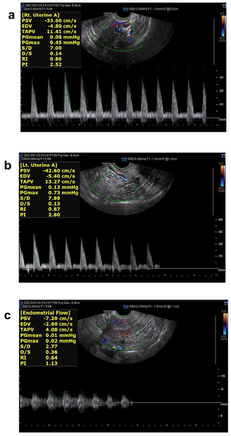
role of blood flow dynamics, alterations in blood supply Figure 1. Doppler Examination of (a) Right Uterine Arteries, (b) Left Uterine
(e.g., an increase in uterine artery resistance) may be Arteries, and (c) Endometrial Arteries in RIF Woman. Note. PI: Pulsatility
index; RI: Resistance index. PI and RI were computed using equations PI
responsible for diminished endometrial receptivity and
= (PSV-EDV)/TAPV and RI = (PSV-EDV)/PSV. RIF: Recurrent implantation
thus RIF. Several studies have examined the role of the failure; Rt. uterine: Right uterine arteries; Lt. uterine: Left uterine arteries; PSV:
luteal phase in pregnancy loss (10,15) while less attention Peak systolic velocity; EDV: End diastolic velocity; TAPV: Timed averaged
has been paid to the importance of the follicular phase in peak velocity; PGmean: Mean pressure gradient; PGmax: Maximum pressure
gradient; S/D: Systolic/diastolic ratio.
these cases. However, it has been demonstrated that any
defect in the follicular phase of the menstrual cycle could
be associated with the poor quality of the oocyte and may
compromise the quality of the embryo, and consequently, impaired uterine perfusion and decreased vascularity have
the pregnancy outcome (14). On the other hand, been reported during the mid-luteal phase of the menstrual
researchers rarely confirmed that the quality of oocyte can cycle in patients with RPL and recurrent miscarriage,
be a consequence of the vascularity and endocrinology of respectively. Impaired uterine perfusion showed adverse
the follicular phase. Therefore, the present research aimed effects on reproductive functions (17-20). It has been
to investigate vascularity in the follicular phase in both demonstrated that patients with RIF have a higher uterine
RIF and healthy fertile women. artery PI in comparison with the examined infertile
Our findings revealed vascularity variations in the control cases during the mid-luteal phase before their first
uterus of women with RIF in comparison to the healthy IVF trial (21). In this regard, individuals with successful
women during the follicular phase. In this regard, higher implantation during the IVF procedure represented a low
resistance was found in the uterine arteries of patients with PI (22). Moreover, Abdel-Razik et al reported an increased
RIF compared to the control group. Based on our results, PI, along with the higher RI of the uterine arteries in
the RI and PI of the right and left uterine arteries and patients with unexplained recurrent abortion (23). In
sub-endometrial arteries were higher in the study group contrast, some studies demonstrated that the mean of the
compared to the controls. Our results are in line with the PI of left and right uterine arteries in different days of the
findings by Pattinaja et al, indicating a higher PI of uterine menstrual cycle was not significantly different between
arteries in women with a history of at least two embryo the women with pregnancy failure after embryo transfer
transfers during IVF treatment compared to control and those with successful pregnancy (24,25). Additionally,
women with normal reproduction (16). Furthermore, Prakash et al (14) reported no significant difference in the
International Journal of Women’s Health and Reproduction Sciences, Vol. 9, No. 3, July 2021 219Amini et al
ultrasound measurements of patients with a history of the follicular phase could give useful information about
pregnancy loss compared to the control group during the endometrial receptivity. Moreover, the growing body of
follicular phase, which is inconsistent with our results. It evidence regarding the critical role of sufficient uterine
should be noted that in the above-mentioned study, luteal perfusion in pregnancy success makes it more important
phase defects among approximately 30% of patients and a in the clinical approach. However, to validate our findings
small sample size, especially for the control group could and define the mechanisms involved in the relationship
be the possible reasons for such a controversy. In addition, between RIF and defective uterine artery blood flow in the
the assessment of Doppler indices between days 8 and 9 of follicular phase, further studies with a larger sample size
the cycle could be another explanation for the discrepant are necessary.
results with the present study.
The results of the present study showed that the PI and Conclusions
RI of sub-endometrial arteries were higher in women with In general, using transvaginal ultrasound examinations, it
RIF compared to the controls. These findings conform to was revealed that the PI and RI of the right and left uterine
those of a previous study showing that RPL patients had arteries and sub-endometrial uterine arteries, along with
an increased RI of sub-endometrial blood flow compared the RI of the arcuate arteries were significantly higher in
to normal controls (26). This is also consistent with patients with a history of RIF compared to their control
reports of NG et al (27) regarding higher sub-endometrial counterparts. These findings highlight the increased
vascularity in women with a successful live birth in blood flow resistance and reduced uterine perfusion in
comparison with those who experienced miscarriages. RIF patients. In this respect, evaluating uterine perfusion
Two other studies linked implantation failure in IVF cycles early in the follicular phase may force physicians to better
with a decrease (28) or lack (29) of blood flow in the sub- support the luteal phase or prevent embryo transfer.
endometrium. Furthermore, it has been represented that Finally, using Doppler sonography may be a useful non-
women with a successful IVF procedure had lower PI and invasive method for evaluating patients with implantation
RI of uterine and arcuate arteries compared to those with or pregnancy loss.
an unsuccessful IVF (30). Similarly, Yalti et al (31) found
that women who conceived with intrauterine insemination Authors’ Contribution
Study conception or design: KH; Acquisition of data: MR, MA, and LF;
had a lower uterine artery PI compared with those who
Analysis and interpretation of data: SN; Drafting of the manuscript: SN and
did not conceive at all. This study was conducted during MA; Critical revisions: AF, KH, MR, and LF.
the follicular phase before human chorionic gonadotropin
administration. Thus, blood flow impedance in uterine Conflict of Interests
The authors declare that they have no conflict of interests.
arteries may be an indicator of pregnancy success possibly
due to the association of the vascularity of endometrium Ethical Issues
and sub-endometrium with the development of the The study received approval from the Ethics Committee of Tabriz University
placenta during pregnancy (29,32). of Medical Sciences (IR.TBZMED.REC.1399.679).
The ischemia of the endometrium and sub-
Financial Support
endometrium could be considered as one of the causative This study was supported by Women’s Reproductive Health
factors involved in RPL (26). In this regard, patients with Research Center, Department of Obstetrics and Gynecology, Faculty
miscarriage demonstrated significantly lower endometrial of Medicine, Tabriz University of Medical Sciences, Tabriz, Iran.
and sub-endometrial vascularity compared to pregnant
Acknowledgments
women with live birth (27). However, due to the lack of
The authors would like to thank the staff of Al-Zahra Hospital for their help
a difference between the role of endometrial and sub- in conducting this research.
endometrial blood flow in getting pregnant, Chien et al
proposed examining these two areas by color Doppler References
1. Ly KD, Aziz N, Safi J, Agarwal A. Evidence-based management of infertile
(28). Further studies on vascular changes, especially in
couples with repeated implantation failure following IVF. Curr Womens
pregnancy loss, may provide a broader understanding of Health Rev. 2010;6(3):200-18. doi:10.2174/157340410792007073
the pathophysiology and etiology of these conditions. 2. Simon A, Laufer N. Repeated implantation failure: clinical
Transvaginal Doppler ultrasound is a non-invasive approach. Fertil Steril. 2012;97(5):1039-1043. doi:10.1016/j.
fertnstert.2012.03.010
method for measuring uterine vascularity (33) and can
3. Valdes CT, Schutt A, Simon C. Implantation failure of endometrial
predict adverse pregnancy outcomes in high-risk women origin: it is not pathology, but our failure to synchronize the developing
based on serum screening for pregnancy complications embryo with a receptive endometrium. Fertil Steril. 2017;108(1):15-18.
(34). Thus, finding markers to show the endometrial doi:10.1016/j.fertnstert.2017.05.033
developmental defects and risk of pregnancy loss early in 4. Margalioth EJ, Ben-Chetrit A, Gal M, Eldar-Geva T. Investigation and
treatment of repeated implantation failure following IVF-ET. Hum
the follicular phase may provide the opportunity for better Reprod. 2006;21(12):3036-3043. doi:10.1093/humrep/del305
supporting the luteal phase or preventing unsuccessful 5. Steer CV, Campbell S, Pampiglione JS, Kingsland CR, Mason BA,
embryo transfer (35). Therefore, evaluating the PI and RI Collins WP. Transvaginal colour flow imaging of the uterine arteries
of the uterus and arcuate and sub-endometrial arteries at during the ovarian and menstrual cycles. Hum Reprod. 1990;5(4):391-
220 International Journal of Women’s Health and Reproduction Sciences, Vol. 9, No. 3, July 2021Amini et al
395. doi:10.1093/oxfordjournals.humrep.a137109 color Doppler study. Fertil Steril. 2009;92(3):S118. doi:10.1016/j.
6. Zegers-Hochschild F, Adamson GD, de Mouzon J, et al. The International fertnstert.2009.07.1126
Committee for Monitoring Assisted Reproductive Technology (ICMART) 22. Steer CV, Campbell S, Tan SL, et al. The use of transvaginal color
and the World Health Organization (WHO) revised glossary on flow imaging after in vitro fertilization to identify optimum uterine
ART terminology, 2009. Hum Reprod. 2009;24(11):2683-2687. conditions before embryo transfer. Fertil Steril. 1992;57(2):372-376.
doi:10.1093/humrep/dep343 doi:10.1016/s0015-0282(16)54848-1
7. Ng EH, Chan CC, Tang OS, Yeung WS, Ho PC. Factors affecting 23. Abdel-Razik M, El-Berry S, Mostafa A. The effects of nitric oxide donors
endometrial and subendometrial blood flow measured by three- on uterine artery and sub-endometrial blood flow in patients with
dimensional power Doppler ultrasound during IVF treatment. Hum unexplained recurrent abortion. J Reprod Infertil. 2014;15(3):142-146.
Reprod. 2006;21(4):1062-1069. doi:10.1093/humrep/dei442 24. Hoozemans DA, Schats R, Lambalk NB, Homburg R, Hompes
8. Tsai YC, Chang JC, Tai MJ, Kung FT, Yang LC, Chang SY. Relationship of PG. Serial uterine artery Doppler velocity parameters and human
uterine perfusion to outcome of intrauterine insemination. J Ultrasound uterine receptivity in IVF/ICSI cycles. Ultrasound Obstet Gynecol.
Med. 1996;15(9):633-636. doi:10.7863/jum.1996.15.9.633 2008;31(4):432-438. doi:10.1002/uog.5179
9. Cacciatore B, Simberg N, Fusaro P, Tiitinen A. Transvaginal Doppler 25. Prasad S, Goyal R, Kumar Y, et al. The relationship between uterine
study of uterine artery blood flow in in vitro fertilization-embryo artery two-dimensional color Doppler measurement and pregnancy
transfer cycles. Fertil Steril. 1996;66(1):130-134. doi:10.1016/s0015- outcome: a prospective observational study. J Reprod Infertil.
0282(16)58400-3 2017;18(2):251-256.
10. Habara T, Nakatsuka M, Konishi H, Asagiri K, Noguchi S, Kudo 26. Mansour GM, Hussein SH, Abd El Hady RM, et al. Uterine artery
T. Elevated blood flow resistance in uterine arteries of women with flow velocity waveform (FVW) type and subednometrial vascularity in
unexplained recurrent pregnancy loss. Hum Reprod. 2002;17(1):190- recurrent pregnancy loss. J Matern Fetal Neonatal Med. 2020;33(4):527-
194. doi:10.1093/humrep/17.1.190 532. doi:10.1080/14767058.2018.1495190
11. Veeck LL, Zaninovic N. An Atlas of Human Blastocysts. New York: 27. Ng EH, Chan CC, Tang OS, Yeung WS, Ho PC. Endometrial and
Parthenon Pub. Group; 2003:286. subendometrial vascularity is higher in pregnant patients with livebirth
12. Nardo LG. Vascular endothelial growth factor expression in the following ART than in those who suffer a miscarriage. Hum Reprod.
endometrium during the menstrual cycle, implantation window and 2007;22(4):1134-1141. doi:10.1093/humrep/del458
early pregnancy. Curr Opin Obstet Gynecol. 2005;17(4):419-423. 28. Chien LW, Au HK, Chen PL, Xiao J, Tzeng CR. Assessment of uterine
doi:10.1097/01.gco.0000175362.12470.e0 receptivity by the endometrial-subendometrial blood flow distribution
13. Demir R, Kayisli UA, Cayli S, Huppertz B. Sequential steps during pattern in women undergoing in vitro fertilization-embryo transfer.
vasculogenesis and angiogenesis in the very early human placenta. Fertil Steril. 2002;78(2):245-251. doi:10.1016/s0015-0282(02)03223-
Placenta. 2006;27(6-7):535-539. doi:10.1016/j.placenta.2005.05.011 5
14. Prakash A, Li TC, Laird S, Nargund G, Ledger WL. Absence of follicular 29. El Garhy IT, Mohamed AH,Sultan AS. Uterine and subendometrial
phase defect in women with recurrent miscarriage. Fertil Steril. arteries Doppler in patients with recurrent first trimestric abortion. Egypt
2006;85(6):1784-1790. doi:10.1016/j.fertnstert.2005.11.045 J Hosp Med. 2018;73(5):6683-6690. doi:10.21608/ejhm.2018.16013
15. Shimada S, Kato EH, Morikawa M, et al. No difference in natural killer 30. Adibi A, Khadem M, Mardanian F, Hovsepian S. Uterine and arcuate
or natural killer T-cell population, but aberrant T-helper cell population arteries blood flow for predicting of ongoing pregnancy in in vitro
in the endometrium of women with repeated miscarriage. Hum fertilization. J Res Med Sci. 2015;20(9):879-884. doi:10.4103/1735-
Reprod. 2004;19(4):1018-1024. doi:10.1093/humrep/deh159 1995.170622
16. Pattinaja DA, Spaanderman ME, Ghossein-Doha C, van Golde RJ. 31. Yalti S, Gürbüz B, Ficicioglu C, Canova H. Doppler evaluation of
Repeated implantation failure relates to circulatory abnormalities. Fertil the uterine, intraovarian, stromal and spiral arteries on the day of
Steril. 2015;104(3):e338. doi:10.1016/j.fertnstert.2015.07.1053 human chorionic gonadotrophin administration in controlled ovarian
17. Yang W, Wu Z, Yu M, et al. Characteristics of midluteal phase uterine hyperstimulation. J Obstet Gynaecol. 2003;23(4):402-406. doi:10.108
artery hemodynamics in patients with recurrent pregnancy loss. J 0/0144361031000120914
Obstet Gynaecol Res. 2019;45(7):1230-1235. doi:10.1111/jog.13944 32. Elewa AM, Mansour AE, Gehad MA, Afify HE. Ovarian reserve
18. Tan SY, Hang F, Purvarshi G, Li MQ, Meng DH, Huang LL. Decreased testing and uterine blood flow assessment using two-dimensional and
endometrial vascularity and receptivity in unexplained recurrent three-dimensional Doppler in patients with unexplained recurrent
miscarriage patients during midluteal and early pregnancy phases. miscarriage. Benha Med J. 2017;34(2):81-87. doi:10.4103/bmfj.
Taiwan J Obstet Gynecol. 2015;54(5):522-526. doi:10.1016/j. bmfj_107_17
tjog.2014.10.008 33. Coulam CB, Goodman C, Rinehart JS. Colour Doppler indices
19. Wahab HA, El-Din DS, Zain E, Abdelgany M, Youssef MAFM. of follicular blood flow as predictors of pregnancy after in-vitro
Uterine artery Doppler and subendometrial blood flow in patients fertilization and embryo transfer. Hum Reprod. 1999;14(8):1979-1982.
with unexplained recurrent miscarriage. Middle East Fertil Soc J. doi:10.1093/humrep/14.8.1979
2011;16(3):209-214. doi:10.1016/j.mefs.2011.04.001 34. Elsandabesee D, Srinivas M, Kodakkattil S. The clinical value of
20. Chen L, Quan S, Li H, Chen C, Xing F, Yu Y. A comparison of combining maternal serum screening and uterine artery Doppler
endometrial and subendometrial vascularity assessed by three- in prediction of adverse pregnancy outcome. J Obstet Gynaecol.
dimensional ultrasonography and power Doppler angiography between 2006;26(2):115-117. doi:10.1080/01443610500443279
healthy fertile women and women with unexplained primary recurrent 35. Schild RL, Holthaus S, d’Alquen J, et al. Quantitative assessment of
miscarriage. Fertil Steril. 2011;95(3):1127-1129. doi:10.1016/j. subendometrial blood flow by three-dimensional-ultrasound is an
fertnstert.2010.09.034 important predictive factor of implantation in an in-vitro fertilization
21. Eid ME, Taye AM. Impaired uterine perfusion in infertile women programme. Hum Reprod. 2000;15(1):89-94. doi:10.1093/
with repeated IVF-implantation failure as detected by uterine artery humrep/15.1.89
© 2021 The Author(s); This is an open-access article distributed under the terms of the Creative Commons Attribution License (http://
creativecommons.org/licenses/by/4.0), which permits unrestricted use, distribution, and reproduction in any medium, provided the original
work is properly cited.
International Journal of Women’s Health and Reproduction Sciences, Vol. 9, No. 3, July 2021 221You can also read