Ophthalmic Manifestations In Thyroid Disease - IOSR journals
←
→
Page content transcription
If your browser does not render page correctly, please read the page content below
IOSR Journal of Dental and Medical Sciences (IOSR-JDMS)
e-ISSN: 2279-0853, p-ISSN: 2279-0861.Volume 16, Issue 1 Ver. IV (January. 2017), PP 59-65
www.iosrjournals.org

                Ophthalmic Manifestations In Thyroid Disease
                 Dr. M.Vijayaleela M.S1,Dr.D. BhimaSankarBabu M.S2
                              1
                              Assosiate Professor OfOphthalmology Nizamabad
                      2
                       Associate Professor Of Ophthalmology SarojiniDevi Eye Hospital

Abstract:Out of 100 patients attending outpatient department of Ophthalmology, Sarojini Devi eye hospital
Hyderabad, with history of Thyroid disease Thyroid eyedisease is clinically found in 26 patients(26%).Majority
of patients with thyroid eye disease are hyperthyroid 23/26(88.5) and 11.5% are hypothyroid(3/26).
Type Of Study:Retrospective study
Place Of Study:Sarojini Devi Eye Hospital
Aim Of The Study: To Study Various Manifestations In Thyroid Eye Disease.
Inclusion Criteria: All patient of age 50 years with systemic thyroid disease
Exclusion criteria:
1.Patient with other ocular pathologies
2.Patients with history of trauma.
Material And Methods: The study is done on 100 patients attending out patient department of ophthalmology,
Sarojinidevi eye hospital, Hyderabadduring a periodof 2yearsi.e 2014-2016Apprpriate history is taken and
through clinical examination of all system is done.Assesssment of visual acuity-both for distance using Snellen’s
chart and near using Snellen’s near vision charts, colour vision using Ishihara’s pseudo isocromatic charts,
ocular movements, slit lamp examinations, measurement of proptosis, intra ocular pressure recording, fundus
examination with direct and indirect ophthalmoscopy, visual field testing with Humphrey’s automated visual
field analyser.Laboratory investigations were done in all cases to know thyroid status and radiological
investigations like ultrasound orbit and CT scan were done in relevant cases.

                                          I.        Introduction
          Thyroid eye disease is also known as graves ophthalmopathy, dysthyroidophthalmopathy, thyroid-
associated orbitopathy,thyroidorbitopathy, thyrotoxicexophthalmos.Orbital and related ocular changes are
refered as thyroid orbitopathy or Graves’ orbitopathy because it is an orbital rather than an ophthalmic
process.Thyroid eye disease is the most common cause of unilateral or bilateral proptosis in adults and should
always be a consideration in patients with unexplained diplopia,pain or optic nerve dysfunction.
         TED was originally described as the part of the triad comprising Graves disease, which includes the
orbital signs, hyperthyroidism, and pretibial myxedema.Grave’s disease in an auto immune condition in which
antibodies target the thyroid stimulating hormone receptor(TSH-R) displayed on thyrocytes, inducing the
production of excess thyroid hormones.Though typically associated with graves hyperthyroidism TED may also
occur with Hashimoto thyroiditis or in the absence of thyroid disease. Subclinical thyroid abnormalities may be
noted in some patients with typical orbitopathy.
         The majority of patients have clinical or laboratory evidence of thyroid disease but the orbital process
can occur even in the absence of detectable thyroid abnormality.Thyroid orbitopathy is overwhelmingly
associated with and usually occurs close to or within 18 months of hyperthyroidism; there is a rough correlation
between the nature of hyperthyroidism, it’s management, and the orbitopathy.
         Median age at the time of diagnosis of Graves’ ophthalmopathy was 43 years; the minimum and
maximum ages were 8 and 88 years,respectively. Among 108 patients with hyperthyroidism, ophthalmopathy
was diagnosed in the six-month interval preceeding the diagnosis of thyroid dysfunction in 20 patients(18.5%);
opthalmopathy-was concurrent with the diagnosis of hyperthyroidism in 22 patients(20.3%); and
ophthalmopathy developed in the six-month interval after thyroid-diagnosis in 24 patients(22.2%).
Ophthalmopathy was diagniosed more than six months before the diagnosis of hyperthyroidism in only four
additional patients(3.7%), where as ocular changes developed six months or more after thyroid disease in the
remaining 38 patients(35.2%).
         Thyroid eye disease(TED) causes visual disability, discomfort, and facial disfigurement. These
symptoms and signs are caused by immune-mediated, inflammatory events that may result in irreversible tissue
alterations.Considerable controversy exists as to whether treatment is better than observation during the active
phase of disease. Management should consist of a coordinated, multidisciplinary, medical surgical approach
based on staging of the disease and knowledge of its effect on the orbital and ocular structures.But even with

DOI: 10.9790/0853-1601045965                            www.iosrjournals.org                          59 | Page
Ophthalmic Manifestations In Thyroid Disease
appropriate interventions at each stage of the disorder,TED patients often suffer a dramatic reduction in their
quality of life.
Broadly speaking, management is directed toward abating or controlling the active phase of the disease,
prevention of ocular and psychophysical damage, regressing ocular motor abnormalities, and improving the
cosmetic disfigurement.
Epidemiology
In a study of patients Olmsted Country, the age-adjusted incidence of thyroid eye disease(TED) for women
was 16cases, and for men it was 2.9 cases per 100,000 population per year.
Thyroid Eye Disease patients with thyroid dysfunction appear to have more severe ophthalmopathy than
those who are clinically euthyroid.
Race
Tellez et al. reported a 6.4 times risk for development oforbotopathy in Europeans compared to Asians.
They also found that the prevelence of Thyroid Associated Orbitopathy in patients with hyperthyroidism
was singificantly higher in Europeans(42%) compared to Asians(7.7%).
Sex
Thyroid eye disease affects women in approximately 6 times more frequently than men(86% versus 14% of
cases, respectively)
Age
There was a bimodal peak incidence in the fifth and seventh decades in both men and women.While
Thyroid Eye Disease is more common in younger women, the disorder is more severe in men and patients
over the age of 50-60 years.
The median age at the time of diagnosis of TED was 43 years(range, 8-88 years).
Smoking
Smokers are upto 5 times more likely than nonsmokers to develop Thyroid Eye Disease.
Cigarette smoking is the strongest modifiable risk factor for developing thyroid Associated Orbitopathy.
The risk and severity of ophthalmopathy in patients with graves disease may be increases by several other
factors including tobacco use,genetics, type of treatment for hyperthyroidism, TSH receptor levels,
advanced age and stress.
History Of Thyroid Eye DiseaseSaint Yves was the first ophthalmic surgeon who made reference to
thyroid ophthalmopathy in 1773.
Caleb Hillier Parry gave a detailed clinical description of 13 cases of goitre associated with tachycardia, in
one of which , protrusion of eyes occurred for the first time, the condition has sometimes been called
Parry’s disease.
Robert James Gravesdescription of enlargement of the thyroid, palpitations and protrusion of the eyes is so
complete and detailed that the connation Grave’s Disease has been accepted throughout the world.
SArl Adolph Von Basedowdescribed exophthalmos as due to hypersecretion of the orbital tissue caused by
disordered circulation,discussing the three cardinal symptoms of exophthalmos, goitre and tachycardia.The
disease is frequently called Basedow’s disease in German literature.
Classification Of Thyroid Eye Diseasein1969, Warner proposed the “No Specs” classification for signs of
Graves’ ophthalmopathy. In 1981, Van Dyke refined the class 2 No Specs soft tissue findings with the
mnemonic Relief
--no specs” and “relief” categorization of graves’ disease
class signs
0. No signs nor symptoms
1. Only signs are upper eyelid retraction, lid lag, stare
2. Soft tissue signs and symptoms:
Resistant to retropulsion
Edema of conjuctiva and caruncle
Lacrimal gland enlargement
Injection over horizontal rectus muscle insertions
Edema of the eyelids
3.Proptosis
4.Extraocular muscle involvement
5.Corneal involvement secondary to exposure
DOI: 10.9790/0853-1601045965 www.iosrjournals.org 60 | PageOphthalmic Manifestations In Thyroid Disease
6.Sight loss secondary to optic nerve compression
II. Clinical Features
Common and non specific symptoms include tearing, irritation, giddiness,arching,and photophobia.
Early signs include conjuctival injection, periorbital puffiness, abnormal tear break-up time, superficial punctate
keratitis, and elevation of intra ocular pressure.Ophthalmic findings are generally bilateral, but may present
unilaterally or asymmetrically. The presence of pre-existing autoimmune thyroid disease increases suspicion for
thyroid eye disease, but isolated eye findings may represent the presenting manifestations of thyroid disorders.
The classic presentation in the setting of acute Graves’ disease involves thyrotoxicosis,goiter and bilateral
exophthalmos
The clinical signs are characteristic and include 1 or more of the following:
Proptosis
Restrictive extraocular myopathy
Eyelid retraction
Lid lag
Corneal exposure
Compressive optic neuropathy
Excessive tearing,foreign body sensation,bulging of the eyes, swelling of the eyelids,retro-orbital pain,
blurred vision,inability to focus,double vision characterize early, active thyroid Eye disease.
Thyroid Eye Disease dysmotility often presents as intermittent diplopia that is initially more
symptomatic in the morning due to edema thatresults from sleeping in a supine position. In 5-10% of thyroid
eye disease patients,visual loss occurs due to corneal decompensation or optic nerve compression. 25% to 50%
of patients with Graves’ hyperthyroidism will have some features of ophthalmopathy.
Thyroid Eye Signs
1. Dalrymple’s-upper lid retraction
2. Von Graefe’supperlid lag on downwarg gaze
3. Boston’s jerky irregular movement of upper lid on downward gaze
4. Enroth’s puffy swelling of lids
5. Giffords difficulty in everything upperlid.
6. Gellinek sign increased pigmentation of lids
7. Kocher’s sign increased lid retraction with visual fixation
8. Rosenbach’s tremor of closed lids
9. Stellwag’s infrequent blinking
10. Mobius weakness of convergence
11. Ballet’s sign palsy of one or more extraocular muscles
12. Suker’ssignweakness of fixation on lateral gaze
13. Cowen’s sign jerky papillary constriction to consensual light
14. Knies sign unequal dilation of pupil
15. Lowy’s sign dilation of pupil with weak adrenaline
16. Joffroy’s absence forhead wrinkles on upward gaze
17. Saintons’s delayed forhead wrinkling after upward gaze.
18. Payne Trouseau sign globe luxation
19. Vigoroux sign lid swelling
20. Pochin sign diminished blinking
III. Medical Treatment
Treatment of Thyroid Disease and Prevention of Thyroid Orbitopathy
1. Oral antithyroid drugs
Advantages-
i. Control of hyperthyroidism
ii. Improvement in the orbitopathy
Radioactive iodine
In some studies, thyroid eye disease has been demonstrated to worsen after RAI treatment, presumably
because of the release of TSH-R antigens which incite an enhanced immune response.In
addition,Hypothyroidism occurring after RAI treatment may exacerbate thyroid eye disease via stimulation of
TSH-R.
DOI: 10.9790/0853-1601045965 www.iosrjournals.org 61 | PageOphthalmic Manifestations In Thyroid Disease
Hyperthyroid patients with severe, active thyroid eye disease; those with elevated T3,levels; and
smokers appear to be at greatest risk for exacerbation of eye disease after RAI treatment.33
Disadvantages -progression of subclinical and clinical orbitopathy which is prevented by combining with
prednisolone 25-50mg/day for 3 months.
Thyroidectomy
Advantages
1.Eliminates reactive T lymphocytes in the thyroid.
2. Improves orbitopathy
Disadvantages
Surgical complication is reaction of parathyroids
IV. Management Of Thyroid Eye Disease
Mild disease
Observation
Patient education/lifestyle changes
Smoking cessation
Salt restriction
Elevation of head of bed
Wearing sunglasses
Ocular surface lubrication
Signs/Symptoms Management Options
Peri-orbital oedema Elevation head of the bed, antidiuretics
Dryness, foreign body sensation Artificial eye drops and ointment
Lagophthalmos Nocturnal eye taping,eyes shield
Eyelid retraction Topical Guanethidine drops or beta
blockers eye drops
Diplopia Prisms
Photophobia Sunglasses
Moderate disease
Topical cyclosporine
Eye lid taping at night
Moisture goggles/chambers
Prism glasses or selective ocular patching
Moderate-dose oral steroid therapy
Surgical Management
For surgical rehabilitation of the patient with stable thyroid eye disease, surgical repair is recommended
in the following standard, sequential stages:
1. Orbital decompression surgery
2. Eye muscle surgery
3. Eyelid repositioning
4. Blepharoplasty(which is sometimes combined with eyelid repositioning)
The preoperative CT scan details the relative contributions of extraocular muscle enlargement and fat expansion
to the proptosis.Patients younger than 40 years demonstrate enlargement of the orbital fat compartment, whereas
those older than 40 typically show more significant extraocular muscle enlargement. This difference determine
the effectiveness of bone versus fat decompression surgery.
Orbital Decompression
Orbital decompression is a surgical procedure used to improve the volume-to-space discrepancy.The
goal of orbital decompression is to allow the enlarged muscles and orbital fat to expand into periorbital spaces.
This expansion relatives on the optic nerve and its blood supply and reduces proptosis.
Technical options in decompression
A. Floor and medial wall
1. Anterior
Medial skin
DOI: 10.9790/0853-1601045965 www.iosrjournals.org 62 | PageOphthalmic Manifestations In Thyroid Disease
Caruncular
2. sinus approachOguraTransnasal endoscopic
B. Lateral wall+_floor+_roof+_augmentation or bony advancement
1. Swinging eyelid approach
2. Burke-Kronlein+medical approach
3. Burke-Kronlein+Ogura or transnasal endoscopic
4. Coronal approach
C. Soft tissue
1.Orbital fat excision
2.Blepharoplasty
External
Internal
There are only two instances in which relatively emergent surgery is necessary,And decompression
surgery involves the removal of 1,2,3, or(seldom) 4 orbital walls enlarge the orbit and reduce proptosis and/or
optic nerve compression.
Concurant orbital fat exicision is used to augment the impact of surgery.
Surgical approaches include
Transeyelid
Transcaruncular
Transantral
Transnasal endoscopic
Andcoronal
V. Results
Age at Presentation among Thyroid eye disease patients
Mean age of Presentation Percentage
0-9 0%
10-19 3.84%
20-29 23.07%
30-39 19.23%
40-49 53.84
Most common age group at presentation is 40-49 years followed by 30-39 years.
Table-2Sex distribution among thyroid eye disease patients
Sex No.Of Cases Percentage
Females 19 73%
Males 7 26.9%
73% Thyroid Eye Disease patients are females and 26.9% are males.
Table-3Smokers Vs Non Smokers
Smoking Status Percentage
Smokers 34.61%
Non-smokers 65.39%
34.61% of Thyroid Eye Disease patients are smokers and 65.39% are non smokers.
Table-4Thyroid status
Thyroid Status No.Of Cases Percentage
Hyperthyoidism 23 88%
Hypothyroidism 3 11.5%
88% of the thyroid disease patients are hyperthyroid and 11.5% patients are hypothyroid.
Table-5Laterality
Laterality No.Of Cases Percentage
Bilateral 24 92.3%
Unilateral 2 7.69%
DOI: 10.9790/0853-1601045965 www.iosrjournals.org 63 | PageOphthalmic Manifestations In Thyroid Disease
92.31% of thyroid eye patients have bilateral involvement when as 7.69% have unilateral involvement.
Table-6Frequency of occurance of various manifestations in Thyroid eye disease
Manifestation Percentage
Eyelid retraction 88.46%
Proptosis 57.69%
Restrictive myopathy 38.46%
Exposure Keratitis 3.84%
Optic neuropathy Nil
Eyelid retraction is the most common manifestation occurring in 88.46% of Thyroid eye disease patients
followed by proptosis in 57.69%, Restrictive myopathy in 38.46%, Exposure Keratitis in 3.84%.
VI. Discussion
The focus of the present study was to know the ophthalmic manifestations in thyroid disorders.Out of
100 patients attending outpatient department of Ophthalmology thyroid eye disease is clinically found in 26
patients(26%).Majority of patients with thyroid eye disease are hyperthyroid 23/26(88.5) and 11.5% are
hypothyroid(3/26).
VII. Conclusions
A total of 100 cases of thyroid disorder were examined and evaluated during the study period.
Majority of Thyroid eye disease patients are hyperthyroid(88.5%).
Few Thyroid eye disease patients are hypothyroid(11.5%).
Thyroid eye disease is common in females when compared with males.
Mean age of presentation is 40 years.
It is mostly bilateral.
Smoking is associated with increased risk and severity of disease, followed by proptosis, then comes soft
tissue signs.
Sight threatening complication occurred in 5% of Thyroid eye disease patients.
Thyroid eye disease with Restrictive myopathy
Right eye showing congestion over the insertion of horizontal recti
DOI: 10.9790/0853-1601045965 www.iosrjournals.org 64 | PageOphthalmic Manifestations In Thyroid Disease
Both eye puffy swelling of eye lids
Both Eye Eyelid retraction and puffyness of eye lids
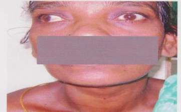
Both eyes Eyelid retraction and proptosis
Both eyes eyelid retraction and proptosis
Bibliography
[1]. Prabhakar BS, Bahn RS, Smith TJ. Current perspective on the pathogenesis of Graves’ disease and ophthalmology. Endocrinology
Review 2003;24:802-35
[2]. Am J Ophthalmol. 1996 Apr; 121(4) : 426-34. Chronolgy of Graves-ophthalmopathy in an incidence cohort.
[3]. Indian Journal of ophthalmology Vol.60 No.2 March 2012 Thyroid associatalorbotopathy.
[4]. Tellez M, Cooper J, Edmonds C. Graves ophthalmopathy in relation to cigarette smoking and ethnic origin.
ClinEndocrinol(Oxf)1992;36:291-4
[5]. Kendler DL, Lippa J, Rootman J. The initial clinical characteristics of Grave’s orbitopathy vary with age and sex.ArchOphthalmol.
1993; 111(2):197-201.
DOI: 10.9790/0853-1601045965 www.iosrjournals.org 65 | PageYou can also read