Giant Ovarian Cysts Treated by Single-Port Laparoscopic Surgery: A Case Series
←
→
Page content transcription
If your browser does not render page correctly, please read the page content below
Giant Ovarian Cysts Treated by Single-Port Laparoscopic Surgery: A
Case Series
Lili Jiang
Shengjing Hospital of China Medical University https://orcid.org/0000-0003-1628-7248
Kuiran Liu ( liukr@sj-hospital.org )
Shengjing Hospital of China Medical University
Research article
Keywords: Giant, Ovarian cyst, Single-port laparoscopic surgery
DOI: https://doi.org/10.21203/rs.3.rs-433441/v1
License: This work is licensed under a Creative Commons Attribution 4.0 International License. Read Full License
Page 1/8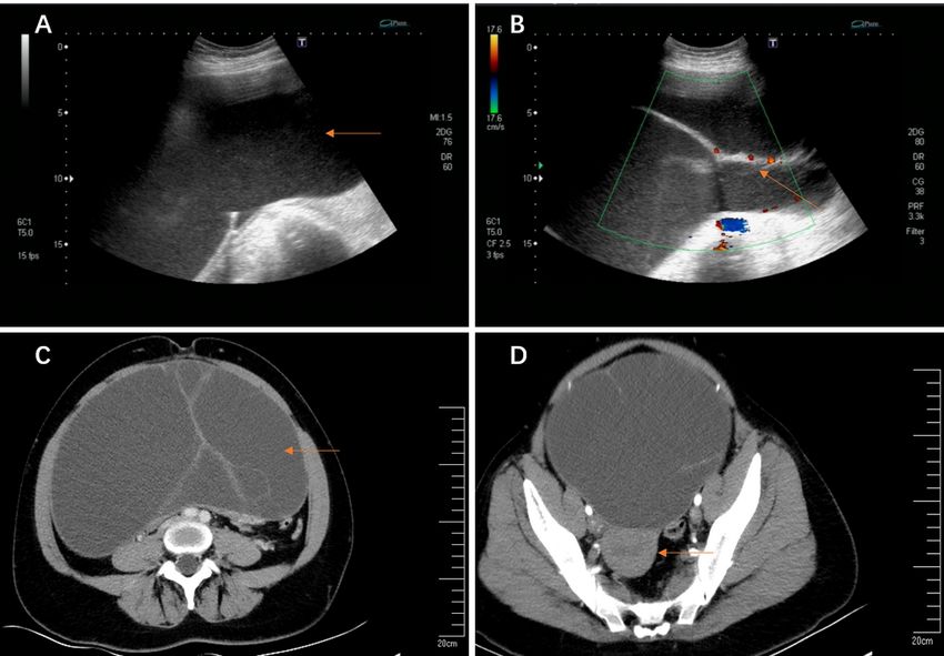
Abstract
Background:
Ovarian cysts are very common diseases of female reproductive system. Giant ovarian cysts refer to the tumors with diameters greater than
10 cm. In recent years, due to the development of clinical diagnosis, imaging modalities and the improvement of patients' cognition of the
diseases, the occurrence of giant ovarian cysts become rare. The purpose of this study was to show a new operation method of single-port
laparoscopy to treat giant ovarian cysts.

Methods:
We report a case series of 5 patients with giant ovarian cysts who underwent single-port laparoscopic surgery in gynecology department,
Shengjing Hospital of China Medical University between June 2020 and March 2021. The inclusion criteria were ovarian cysts at least 20
cm in diameters, and cases which the tumor might be malignant were excluded.

Results:
The patients' mean age was 26.2years. The most common clinical presentation was progressive abdominal distension. Median size of the
cysts at imaging was 39.2 cm (range 21–63 cm). All patients underwent single-port laparoscopic surgery, and none of them converted to
laparotomy. On nal pathological reports, two cysts were serous cystadenomas, and three were mucinous cystadenomas. All patients
recovered well and discharged on time.

Conclusion:
Giant ovarian cysts can be treated by single-port laparoscopic surgery. In addition to the well-known advantages of laparoscopic surgery
(e.g., small pelvic interference, fast postoperative recovery), it can also play the role of perfect cosmetic results, which has more advantages
for young women.

Background
Female pelvic cysts mostly come from the ovary with asymptomatic when they are small. The symptoms appears when they reach
enormous dimensions. Giant ovarian cysts (GOCs) are tumors larger than 10 cm in diameters or those cysts reaching above the
umbilicus[1]. Progressive abdominal distension, nonspeci c diffuse abdominal pain and organ compression (constipation, vomiting and
frequent urination) are the main clinical symptoms of ovarian cysts[2–4]. Most giant ovarian cysts are treated by surgery. Surgical
indications include a rapidly growing or symptomatic cyst, and when its malignant potential cannot be excluded[5]. In the past, exploratory
laparotomy was the most common surgical method, which had the advantage of minimizing the risk of intraperitoneal implantation caused
by cell over ow in case of unexpected malignant transformation of tumor. However, some giant ovarian cysts lled the abdominal cavity
and superior reaching the xiphoid process. The abdominal incision as long as tens of centimeters caused great trouble to patients,
especially young women. In recent years, minimally invasive surgery has been widely used in the eld of gynecology. Laparoscopy is the
choice for most benign ovarian cysts, but the size of the cysts may be a limiting factor. Giant ovarian cysts increase the complexity and
di culty of laparoscopic surgery. How to avoid the leakage of cyst uid has become a challenge[6]. We report a case of giant ovarian cyst
treated by single-port laparoscopy. This method tries to ensure the oncologic safety while treating the disease. The aim of this study is to
introduce a new, minimally invasive and effective surgical approach for the treatment of giant ovarian cysts.

Materials And Methods
Four female patients with giant ovarian cyst who underwent single-port laparoscopic surgery between June 2020 and March 2021 were
included from gynecology department, Shengjing Hospital of China Medical University. The study was approved by the China Medical
University Research Ethics Committee. The inclusion criteria: All patients were diagnosed as giant abdominal cysts by pelvic ultrasound,
MRI or CT-scan before operation(Fig. 1A-D). The patients had signed the informed consent. The umbilicus was normal. Exclusion criteria:
Conversion to open surgery or other surgical methods. Malignant transformation of cysts. Severe medical system diseases which could
not endure laparoscopic surgery. Four patients were con rmed by preoperative imaging (ultrasound, MRI or CT-scan) with giant abdominal
masses at least 20 cm, mainly cystic, without obvious solid components, showing no sign of malignancy. Blood tumor markers (CA125,

                                                                   Page 2/8
HE4, CA199 and CEA) were detected for each patient. The patients with complications were consulted in relevant departments to exclude
surgical contraindications. The operations were performed by experienced gynecologists. Data was collected with operative time, intra- and
post-operative complications, intracystic liquid volume, conversion to laparotomy and the length of postoperative stay. 30 days after
operation, the satisfaction of patients with abdominal scar was recorded.
Surgical procedure
The patients received standardized preoperative nursing preparation and general anesthesia. Single-port laparoscopic surgery was
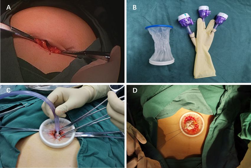
performed using the following techniques. After partial eversion of the umbilicus, a 2-3cm longitudinal incision was made at the
umbilicus(Figure 2A). The umbilical incision was lifted, the skin and subcutaneous tissue were incised layer by layer, and the peritoneum
was incised after con rming that there was no intestinal adhesion below the incision. The disposable incision protection sleeve (Lookmed,
Jiangsu, China) was placed in the incision, the inner ring was placed in the abdominal cavity, and the outer ring was left to the abdominal
wall to form a single-port laparoscopic approach platform Figure 2B . A giant cyst appeared under the incision and was liquid in the visual
eld. In order to prevent the adverse effects of sudden drop of abdominal pressure on patients, we used a syringe needle connected with a
suction device to suck out the liquid in the cyst slowly Figure 2C . If the cyst divided into several septums, we suck out the liquid in one
septum and then used the instruments to lift the wall of the cyst to prevent the leakage of the liquid in the cyst. We changed another septum
and continued to suck out the liquid to reduce the pressure of the cyst. When the liquid was sucked out completely, we used silk thread to
ligate the incision and returned the cyst to the abdominal cavity Figure 2D .
A sterile glove was connected with the outer ring. The thumb of the glove was cut, and 10mm trocar (Dike, Guangzhou, China) was placed
as the access of a scope and laparoscopic instruments. In order to prevent air leakage and loosening at the joint, No. 7 silk thread was used
to x and tie tightly, and the 5mm (Dike, Guangzhou, China) trocars were inserted into the other two ngers as the instrument port. This is a
self-made simple laparoscopic single-port (Fig. 3A). The advantage is that it can save the cost for the patients without affecting the
operation.
Carbon dioxide was injected at a pressure of 13 mm Hg and a rigid 30° 5-mm laparoscope was inserted (Karl Storz, tutlingen, Germany). 30°
laparoscope is a better choice because it provides a wide eld of vision. Then the standard laparoscopic surgery was performed. Giant
ovarian cysts were removed from the umbilicus using endopouch specimen retrieval bag (Wellead, Guangzhou, China) (Fig. 3B, C).
Results
The study consisted of 4 female patients and data are shown in Table 1. The mean age of the operated patients was 26.2 years (range, 19–
34 years). The most common symptom was progressive abdominal distension(patients 1,2,4 and 5),several of which were accompanied by
abdominal pain(patients 1,2 and 5). No obvious abdominal distension occurred in patient 3, mainly due to palpation of abdominal mass. All
patients were diagnosed by imaging, ultrasound, MRI or CT-scans. Median size of the cyst at imaging was 39.2 cm (range 21–63 cm), while
the maximum was 63.0 cm with the superiors reach the sword (patients 2). In particular, there were many comorbidities in patient 2.
Hypertension occurred 17 years ago. Now oral antihypertensive drugs are used to control blood pressure, and the blood pressure is
controlled at 130 / 80mmHg.In 2014, she suffered from cerebral thrombosis. The speci c location is unknown. She felt numb on the right
side of the body at the time of onset, which was improved after conservative treatment and now she is hemiplegic at the right limb. We
consulted the anesthesiology department, cardiology department and neurology department before operation to evaluate the safety of
operation and eliminate the operation contraindications. Based on the patient’s age and personal will, we decided to perform single-port
laparoscopic exploration after discussion.
Page 3/8Table 1
Patient’s data.
Patient Age Cyst Operative Fluid Intra-op. Post- Conversion Histology Post-op. Satisfactin
size(cm) time(min) volume blood op. to with
in loss(ml) complications
cyst(ml) stay laparotomy abdominal
(d) scar
1 23 32 100 7000 20 5 No Serous No Yes
cystadenoma
2 34 63 75 16000 20 5 No Mucinous No Yes
cystadenoma
3 19 21 37 3500 10 5 No Mucinous No Yes
cystadenoma
4 25 23 82 4000 50 4 No Serous No Yes
cystadenoma
5 30 57 132 13000 30 6 No Mucinous No Yes
cystadenoma
Mean 26.2 39.2 85.2 8700 26 5 - - - -
Four of the ve patients presented with a normal blood tumor markers. One patient presented with an elevated CA125 of 70.78 (normal
range 0–35mIU/ml) and CA-724 of 8.94(normal range 0–6.9 mIU/ml) (patient 3). In the postoperative reexamination, the blood tumor
markers returned to normal gradually. All patients underwent single-port laparoscopic surgery, no one converted to laparotomy.
Intraoperative suction of intracapsular uid range 3500-16000ml (Fig. 3D). Four patients underwent unilateral adnexectomy, and one
patient an ovarian cystectomy(Fig. 3E). We had a cosmetic suture of the single-port laparoscopic incision in patients’ navel (Fig. 3F). The
average operative time was 85.2min (range 37–132min). There was no extravasation of cyst uid and no decompression syndrome
happened due to gradual reduction of cyst pressure. Mean blood loss was 26ml (range 10–50ml). The average hospitalization time after
operation was 5days, such operative method did not increase the Post-operative stay. All patients recovered well, and no complications
related to the operation occurred. On nal pathological reports, two cysts were serous cystadenomas, and three were mucinous
cystadenomas. There was no borderline tumor or epithelial ovarian cancer in any of the ovarian cysts operated, but one case reported active
cell proliferation, which should be reexamined. All the patients were satis ed with the abdominal scar after 30 days after operation.
Discussion
Female pelvic cysts are very common gynecological diseases in women, most of which come from ovary. The clinical manifestations
appear when the cysts reach enormous dimensions. Giant ovarian cysts (GOCs) are tumors larger than 10 cm in diameters[1]. Due to
improved imaging techniques, giant abdominal cyst has become increasingly rare. The patients can present with rare complications such
as torsion, intestine obstruction, hydronephrosis in addition to causing non-speci c abdominal distension, pain, nausea and vomiting and
changes in defecation habits[7–10]. As the nonspeci c clinical manifestations of giant ovarian cysts, the differential diagnoses include the
giant cysts from other intra-abdominal organs (e.g. gastrointestinal, urological, or lymphatic)[11].
The treatment of ovarian cysts depends on the patient's age, the size of the cyst, and its histopathological feature. Excision of the intact
cysts for histology is the gold standard[12].Most giant ovarian cysts are benign and are treated by surgical excision generally either by
cystectomy or salpingo-oophorectomy [13]. It is utmost important to exclude any possibility of malignant tumor before operation[14].In the
past, resection of the cystic mass by exploratory laparotomy is the preferred management strategy[8]. But for laparotomy of benign giant
cysts, the huge incision caused trouble to the patients (especially young patients). A study shows that with the development of advanced
technology, it is feasible to use laparoscopic surgery to remove giant ovarian cysts on the basis of selecting suitable patients and
laparoscopic experts[15]. Recently, laparoscopic-assisted excision of these giant cystic masses has been reported in several literatures[6, 16,
17]. How to avoid the leakage of cyst uid has become a challenge in laparoscopic surgery for treating giant ovarian cysts.
In recent years, single-port laparoscopic surgery has become a hot spot as it uses the natural pores of the navel to hide the surgical incision
and has the characteristics of perfect cosmetic results and fast postoperative recovery. In our study,we used single-port laparoscope to
perform surgery on a slightly larger incision at the umbilicus, which exposed the visual eld better and avoided the exudation of liquid in the
giant cysts. In order to avoid the impact of sudden drop of intraperitoneal pressure on patients, we used the method previously described to
Page 4/8slowly reduce the uid in the giant cyst. Facts had proved that this method is effective, these patients did not appear related discomfort
symptoms. We use the wound protector-retractor to protect the incision and reduce the risk of cell spillage. The endopouch specimen
retrieval bag was used to take out the specimen after resection of the diseased tissue, which reduced the potential risks of the leakage of
cells and residual cystic uid. These measures ensured the safety of the operation. Although giant ovarian cysts are larger than 10 cm in
diameter, we still selected cysts larger than 20 cm in diameter for study, which are more rare in clinic. We analyzed the general information
and surgical outcomes of these patients and found that single-port laparoscopic surgery did not increase the adverse prognosis of patients.
On the contrary, minimally invasive surgery and perfect cosmetic results accelerated the recovery and satisfaction of patients.
Despite the advantages of single-port laparoscopic surgery, not all giant ovarian cysts are suitable for this type of surgery. We need to
evaluate the patient's condition before operation rigorously, and it is very important to exclude any possible malignant tumors before
operation. Single-port laparoscopic surgery is di cult to form an operation triangle as its limited operation space, relatively concentrated
instruments and mutual interference which places high demands on the surgeon. It is necessary for us to improve the safety of surgery
through more research.
Conclusion
In the treatment of giant ovarian cysts, it is safe and feasible to perform single-port laparoscopic surgery through screening of suitable
patients strictly. This operation method has the same advantages of traditional laparoscopy, it ensures the safety of operation as most as
possible and improves the cosmetic results perfectly, which are particularly important for young women.
Abbreviations
GOCs
Giant ovarian cysts
MRI
Magnetic Resonance Imaging
CT
Computerized Tomography
CA125
Carbohydrate antigen-125
HE4
Human Epididymis Protein 4
CA199
Carbohydrate antigen-199
CEA
Carcinoembryonic antigen
Declarations
ACKNOWLEDGMENTS
The authors thank the support of the funding “Scienti c research funding project of Liaoning Provincial Department of Science and
Technology (No.2020JH2/10300050)” to this article.
AUTHOR CONTRIBUTIONS
Jiang LL conducted a thorough literature review and was the major contributor in writing the manuscript; Liu KR was responsible for
reviewing and revising the article.
FUNDING
This research received the support from “Scienti c research funding project of Liaoning Provincial Department of Science and
Technology No.2020JH2/10300050 ”.
AVAILABILITY OF DATA AND MATERIALS
The datasets used and/or analyzed during the current study are available from the corresponding author upon reasonable request.
Page 5/8ETHICS APPROVAL AND CONSENT TO PARTICIPATE
The study was approved by the China Medical University Research Ethics Committee.
CONSENT FOR PUBLICATION
Each author agrees to the publication of the present study.
COMPETING INTERESTS
The authors declare that they have no competing interests.
References
1. de Lima SH, Dos Santos VM, Darós AC, Campos VP, Modesto FR. A 57-year-old Brazilian woman with a giant mucinous
cystadenocarcinoma of the ovary: a case report. Journal of medical case reports. 2014;8:82. doi:10.1186/1752-1947-8-82.
2. Cîrstoiu MM, Sajin M, Secară DC, Munteanu O, Cîrstoiu FC. Giant ovarian mucinous cystadenoma with borderline areas: a case report.
Romanian journal of morphology embryology = Revue roumaine de morphologie et embryologie. 2014;55(4):1443–7. PMID: 25611279.
3. Mehboob M, Naz S, Zubair M, Kasi MA. Giant ovarian cyst–an unusual nding. Journal of Ayub Medical College Abbottabad: JAMC.
2014;26(2):244–5. PMID: 25603687.
4. Abu Sulb A, Abu El Haija M, Muthukumar A. Incidental nding of a huge ovarian serous cystadenoma in an adolescent female with
menorrhagia. SAGE open medical case reports 2016, 4:2050313x16645755. doi:10.1177/2050313X16645755.
5. Leite C, Barbosa B, Santos N, Oliveira A, Casimiro C. Giant abdominal cyst in a young female patient: A case report. International journal
of surgery case reports 2020, 72:549–555. doi: 10.1016/j.ijscr.2020.06.085.
. Dubuisson J, Heersche S, Petignat P, Undurraga M. Laparoscopic Management of Giant Ovarian Cysts Using the Alexis Laparoscopic
System®: A Case Series. Frontiers in surgery 2020, 7:24. doi:10.3389/fsurg.2020.00024.
7. Bolukbas FF, Bolukbas C, Furuncuoglu Y, Tabandeh B, Saglam FY, Iyigun G, Orug T, Ozcan A, Yapicier O. Large abdominal cystic
masses: Report of seven cases. JPMA The Journal of the Pakistan Medical Association. 2016;66(2):226–8. PMID: 26819176.
. Yeika EV, E e DT, Tolefac PN, Fomengia JN. Giant ovarian cyst masquerading as a massive ascites: a case report. BMC Res Notes.
2017;10(1):749. doi:10.1186/s13104-017-3093-8.
9. Kim HY, Cho MK, Bae EH, Kim SW, Ma SK. Hydronephrosis caused by a giant ovarian cyst. Int Braz J Urol. 2016;42(4):848–9.
doi:10.1590/S1677-5538.IBJU.2015.0354.
10. Kim TJ, Yeh YT, Zobair K. Impending abdominal compartment syndrome from a giant ovarian cyst torsion. ANZ J Surg.
2019;89(4):E164–5. doi:10.1111/ans.14147. Epub 2017 Sep 18.
11. Vecchio R, Leanza V, Genovese F, Accardi M, Gelardi V, Intagliata E. Conservative laparoscopic treatment of a benign giant ovarian cyst
in a young woman. Journal of laparoendoscopic advanced surgical techniques Part A. 2009;19(5):647–8. doi:10.1089/lap.2009.0138.
12. American College of Obstetricians and Gynecologists’ Committee on Practice Bulletins—Gynecology. Practice Bulletin No. 174:
Evaluation and Management of Adnexal Masses. Obstetrics gynecology. 2016;128(5):e210–26. doi:10.1097/AOG.0000000000001768.
13. Katke RD. Giant mucinous cystadenocarcinoma of ovary: A case report and review of literature. Journal of mid-life health 2016,
7(1):41–44. doi: 10.4103/0976-7800.179167.
14. Ou CS, Liu YH, Zabriskie V, Rowbotham R. Alternate methods for laparoscopic management of adnexal masses greater than 10 cm in
diameter. Journal of laparoendoscopic advanced surgical techniques Part A. 2001;11(3):125–32. doi:10.1089/10926420152389251.
15. Alobaid A, Memon A, Alobaid S, Aldakhil L. Laparoscopic management of huge ovarian cysts. Obstetrics and gynecology international
2013, 2013:380854. doi: 10.1155/2013/380854. Epub 2013 May 8.
1 . Uyanikoglu H, Dusak A. A Huge Ovarian Dermoid Cyst: Successful Laparoscopic Total Excision. Journal of clinical and diagnostic
research: JCDR 2017, 11(8):Qd03-qd05. doi: 10.7860/JCDR/2017/29262.10436. Epub 2017 Aug 1.
17. Baradwan S, Sendy F, Sendy S. Complete Laparoscopic Extirpation of a Giant Ovarian Cyst in an Adolescent. Case reports in obstetrics
and gynecology 2017, 2017:7632989. doi: 10.1155/2017/7632989. Epub 2017 Aug 3.
Figures
Page 6/8Figure 1
A and B Transvaginal ultrasound imaging. A: A giant cyst in the abdominal and pelvic cavity (63.0cm x 44.0cm x 13.4 cm); B: The blood
ow signal detected at the separation; (C and D) Abdomen enhanced CT imaging. C: A giant cyst with septums; D: The uterus was pushed
to the back of the pelvis by a giant cyst.
Page 7/8Figure 2
(A) The 2-3cm longitudinal incision was made at the umbilicus. (B) Single-port laparoscopic approach connection instrument. (C) A syringe
needle connected with a suction device to suck out the liquid in the cyst. (D) Ligate the incision in order to avoid the leakage of cyst uid.
Figure 3
(A) The instruments enter the abdominal cavity through single-port laparoscopic access. B The excised tissue was put into endopouch
specimen retrieval bag under laparoscope.(C) The wall of a giant cyst removed through the navel.(D) Intraoperative suction of intracapsular
uid.(E) Unilateral salpingo-oophorectomy by laparoscope.(F) A cosmetic suture of the single-port laparoscopic incision in patients’ navel.
Page 8/8You can also read