Post Covid Fungal Infection: Histopathological and Microbiological Correlation
←
→
Page content transcription
If your browser does not render page correctly, please read the page content below
Dr. Priyanka Vaghasiya, Dr. Jignasa Bhalodia. Post Covid Fungal Infection: Histopathological and Microbiological
Correlation. IAIM, 2021; 8(8): 53-61.

Original Research Article

Post Covid Fungal Infection:
Histopathological and Microbiological
Correlation
Dr. Priyanka Vaghasiya1*, Dr. Jignasa Bhalodia2
1 rd
  3 year resident doctor, 2Professor and Head
Department of Pathology, GMERS Medical College, Sola, Ahmedabad, Gujarat
*
 Corresponding author email: pd.vaghasiya46@gmail.com

                     International Archives of Integrated Medicine, Vol. 8, Issue 8, August, 2021.
                                     Available online at http://iaimjournal.com/
                          ISSN: 2394-0026 (P)                          ISSN: 2394-0034 (O)
                           Received on: 15-08-2021                Accepted on: 25-08-2021
                   Source of support: Nil                       Conflict of interest: None declared.
                                   Article is under creative common license CC-BY
How to cite this article: Dr. Priyanka Vaghasiya, Dr. Jignasa Bhalodia. Post Covid Fungal Infection:
Histopathological and Microbiological Correlation. IAIM, 2021; 8(8): 53-61.

Abstract
Introduction: Mucormycosis is a life-threatening infection caused by saprophytic fungi belonging to
the genera Mucor, Rhizopus and Absidia which belong to the order Mucorales and class
Zygomycetes.Covid-19 is a life-threatening, infectious disease in which decreased CD4 and
CD8positive cell counts, indicating susceptibility to fungal co-infections. Extensive use of steroids in
Covid-19 management or associated with diabetes mallitus can also suppress immunity, allowing
opportunistic fungal infections to colonise.
Materials and methods: A Retrospective study of 50 patients with invasive fungal infection who
presented to the ENT department and who were either coronavirus-positive or had recovered from
coronavirus infection, were included in the study. Tissue samples from all suspected site were
received in formalin for histological examination and in without formalin were used for KOH smear
and culture.
Result: A total of 50 patients presented. Out of 50 patient 43 (86%) cases were found to be positive
based on direct microscopy, culture and histopathology. Among these 31 (62 %) cases were found to
be positive by direct microscopy - KOH, 40 (80%) by culture and 43(86 %) by histopathology.
Among 43 cases, 4 cases which was negative by KOH but positive by histopathology considering
histopathology as a gold standard. Mucormycosis was seen in 23 patients, candidiasis in 6 patient,
aspergillosis in 3 patient and mixed infection of mucormycosis with candidiasis in 6 patients, with
aspergillosis in 3 patients and with both candidiasis and aspergillosis in 3 patients.
Conclusion: Covid 19 associated with invasive mucormycosis sinusitis is dangerous. Uncontrolled
diabetes and over-zealous use of steroids are two of the main factors aggravating the illness. If
infected, early surgical intervention and intravenous anti-fungal treatment should be sought for

                                                                                                               Page 53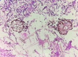
Dr. Priyanka Vaghasiya, Dr. Jignasa Bhalodia. Post Covid Fungal Infection: Histopathological and Microbiological
Correlation. IAIM, 2021; 8(8): 53-61.

management, as a good prognosis and less fulminant disease course can be achieved in cases of post-
coronavirus mucormycosis.

Key words
Covid 19, Diabetes, Mucormycosis, Steroid.

Introduction                                                  vein and cribriform plate along the perivascular
Mucormycosis infection of the sinuses is a form               channels can occur the intracranial spread [3, 5].
of life-threatening invasive fungal sinusitis that
typically       affects     immunocompromised                 Clinically, rhinocerebral mucormycosis can
individuals with an impaired neutrophilic                     present with atypical signs and symptoms similar
response.                                                     to complicated sinusitis, such as headache, nasal
                                                              blockade, crusting, proptosis, facial pain and
Patients can include those with uncontrolled                  oedema, ptosis, chemosis, black eschar over
diabetes mellitus, acquired immunodeficiency                  nasal cavity and even ophthalmoplegia, with
syndrome, iatrogenic immunosuppression and                    headache and fever and various neurological
hematological malignancies, and those who have                signs and symptoms if intracranial extension is
undergone organ transplantation [1].                          present [1, 2].

Systemic immune alterations of Covid-19                       Histological features include mycotic infiltration
infection itself may lead to secondary infections,            of blood vessels, vasculitis with thrombosis,
which are increasingly being recognized in view               tissue infarction, hemorrhage and acute
of their impact on morbidity and mortality [1, 2,             neutrophilic infiltrate [1, 3, 4].
3].
                                                              Furthermore, as Covid-19 is a life-threatening,
Rhinocerebral mucormycosis is a life-threatening              infectious disease, affected patients show an
infection caused by saprophytic fungi belonging               overexpression of inflammatory cytokines, and
to the genera Mucor, Rhizopus and Absidia. All                impaired cell-mediated immunity with decreased
of these belong to the order Mucorales and class              cluster of differentiation 4 and 8 positive T-
Zygomycetes [2, 3].                                           helper (CD4+ T and CD8+ T) cell counts,
                                                              indicating susceptibility to fungal co-infections
Inoculation by inhalation of fungal spores reach              [3, 4].
the nasal cavity, germination is favored by low
oxygen concentration, high glucose, acidic                    Critically ill patients, especially those admitted to
medium and high iron levels. They germinate                   intensive care units and those who required
into hyphae.                                                  mechanical ventilation, or who had a longer
                                                              duration of hospital stays, even as long as 50
Due to the metabolic hypoxic conditions, the                  days, were more likely to develop fungal co-
Polymorphonuclear cells are less effective at                 infections.
removing these hyphae, as it is often found in
patients with mucormycosis associated with                    Hence, it is important to be aware that Covid-19
Diabetes mellitus, thereby favoring the                       patients can develop further fungal infections
establishment of infection.                                   during the middle and latter stages of this
                                                              disease, especially severely ill individuals [1, 6].
Extension of the disease into the maxillary and
ethmoid sinus can lead to orbital involvement.                Here, we present our recent and still ongoing
Through the superior orbital fissure, ophthalmic              experience of 50 cases of mucormycosis of the

                                                                                                               Page 54
Dr. Priyanka Vaghasiya, Dr. Jignasa Bhalodia. Post Covid Fungal Infection: Histopathological and Microbiological
Correlation. IAIM, 2021; 8(8): 53-61.
sinuses seen over a time period of just three Histopathology: All histological tissue obtained
months, with these patients being, or having in formalin were fixed with 10% neutral
previously been, Covid-19 positive. formaldehyde for 24 h, routinely dehydrated and
embedded with paraffin, 4 μM sections were
Materials and methods serially cut on albumin coated slides and stained
A Retrospective study was undertaken at by Hematoxylin and Eosin (H&E) and Periodic
GMERS Medical College, Sola, Ahmedabad, Acid Schiff (PAS) stain.
India, over a period of three months, from May
to June 2021. KOH Microscopy and Culture: Tissue was
examined in 20% KOH. Culture was done on
Tissue samples from all suspected site were Sabouraud Dextrose Agar (SDA) with
received in formalin for histopathological chloramphenicol and incubated at 25⁰C and 37⁰C
examination and in without formalin were used respectively and were examined until 28 days.
for KOH smear and fungal culture.
Results
Once fungal hyphae demonstrated by KOH A total of 50 patients presented; 36 of these were
mount method, histological examination male and 14 were female with ages ranging from
performed. The various types of fungi were 30 to 74 years (mean = 67 years). The majority
confirmed by histopathological examination. of patients (88.0%) were aged over 40 years,
with those aged 30–60 years (6.0%) being most
affected (Table – 1).
Table – 1: Details of cases.
SR AG SEX DIAGNOSIS COVID DIABET STEROID KOH SITE
NO. E STATUS ON ES ADMINIS
ADMISSION TRATION
1 46 M MUCORMYCOS +VE Before 1 Yes Yes MUCORM Right nasal
IS month YCOSIS cavity
2 68 F MUCORMYCOS +VE Before 1 Yes Yes MUCORM Bilateral nasal
IS month YCOSIS cavity
3 60 F CHRONIC +VE Before 15 Yes Yes MUCORM Left nasal cavity,
SINUSITIS days YCOSIS left maxilla
4 75 M ASPERGILLOSI +VE Before 25 No Yes MUCORM Right maxilla
S days YCOSIS
5 53 F ASPERGILLOSI +VE Before 1 Yes Yes MUCORM Left sphenoid,
S month YCOSIS maxilla
6 42 F MUCORMYCOS +VE Before 16 Yes No MUCORM Left middle
IS days YCOSIS turbinate,
maxilla
7 30 F CHRONIC +VE Before 1 No Yes No growth All sinuses
SINUSITIS month
8 41 M CANDIDIASIS +VE Before 18 No Yes MUCORM Left and right
days YCOSIS maxilla
9 74 M MUCORMYCOS +VE Before 2 Yes Yes MUCORM Maxilla
IS month YCOSIS
10 45 M MUCORMYCOS +VE Before 6 Yes Yes MUCORM Middle turbinate
IS days YCOSIS
11 31 F ASPERGILLOSI +VE Before 12 No No No growth
S days
12 52 F MUCORMYCOS No H/O COVID Yes No No growth Left alveolus
IS
13 60 M MUCORMYCOS +VE Before 10 No Yes MUCORM Right and left
Page 55Dr. Priyanka Vaghasiya, Dr. Jignasa Bhalodia. Post Covid Fungal Infection: Histopathological and Microbiological
Correlation. IAIM, 2021; 8(8): 53-61.
IS days YCOSIS maxilla
14 67 M CHRONIC +VE Before 6 No Yes No growth All sinuses
SINUSITIS days
15 67 M CANDIDIASIS +VE Before 16 No Yes No growth Left maxilla
days
16 48 M MUCORMYCOS No H/O COVID Yes No MUCORM Right and left
IS+ YCOSIS maxilla, left
CANDIDIASIS ethmoid, left
alveolus, left
orbit, left frontal
17 42 M MUCORMYCOS +VE Before 26 No No MUCORM Left maxilla
IS days YCOSIS
18 53 F CHRONIC +VE Before 40 Yes Yes No growth Right maxilla
SINUSITIS days
19 54 M MUCORMYCOS No H/O COVID Yes No MUCORM Left maxilla, left
IS YCOSIS inferior turbinate
20 52 M MUCORMYCOS +VE Before 1 No Yes MUCORM Ethmoid, dental
IS month YCOSIS tissue
21 43 M MUCORMYCOS +VE Before 15 No Yes No growth Left inferior
IS + days turbinate
ASPERGILLOSI
S
22 32 M MUCORMYCOS +VE Before 45 No Yes No growth Right frontal
IS days
23 63 M MUCORMYCOS +VE Before 2.5 No Yes No growth Left alveolus
IS months
24 67 M MUCORMYCOS No H/O COVID Yes No MUCORM Left maxilla
IS YCOSIS
25 36 F CANDIDIASIS No H/O COVID No No MUCORM Right maxilla,
YCOSIS Right uncinate
26 55 M MUCORMYCOS No H/O COVID No No No growth Right middle
IS turbinate, Right
maxilla
27 60 M MUCORMYCOS +VE Before 2.5 No Yes MUCORM Left maxilla,
IS months YCOSIS Right uncinate
28 35 M CHRONIC +VE Before 2 Yes Yes MUCORM Left nasal cavity
SINUSITIS months YCOSIS
29 48 M ACUTE No H/O COVID Yes Yes No growth Left nasal cavity
INFLAMMATIO
N
30 59 M MIXED +VE Before 1 Yes No MUCORM Left nasal cavity
INFLAMMATIO month YCOSIS
N
31 54 M CANDIDIASIS +VE Before 12 No No No growth Right maxilla
days
32 50 M MUCORMYCOS No H/O COVID Yes No No growth Left middle
IS+ turbinate, left
CANDIDIASIS maxilla
33 34 M CANDIDIASIS +VE Before 10 No Yes MUCORM Right inferior
days YCOSIS turbinate, right
maxilla
34 58 F MUCORMYCOS +VE Before 6 No Yes No growth Left maxilla,
IS days Left ethmoid
35 56 M MUCORMYCOS +VE Before 16 No Yes No growth Left maxilla
IS+ASPERGILL days
US+
CANDIDIASIS
36 47 F MUCORMYCOS No H/O COVID Yes No MUCORM Right upper
IS YCOSIS alveolus
37 72 M MUCORMYCOS +VE Before 26 No No MUCORM Left & Right
IS days YCOSIS maxilla
38 58 M MUCORMYCOS +VE Before 40 Yes Yes No growth Alveolus
IS days
39 70 M MUCORMYCOS No H/O COVID Yes No MUCORM Right septum,
Page 56Dr. Priyanka Vaghasiya, Dr. Jignasa Bhalodia. Post Covid Fungal Infection: Histopathological and Microbiological
Correlation. IAIM, 2021; 8(8): 53-61.
IS YCOSIS right ethmoid
40 60 F MUCORMYCOS +VE Before 1 No Yes MUCORM Right maxilla
IS month YCOSIS
41 63 M MUCOR+ +VE Before 1 Yes Yes MUCORM Right ethmoid,
ASPERGILLOSI month YCOSIS maxilla
S+
CANDIDIASIS
42 45 M MUCORMYCOS +VE Before 1 Yes Yes MUCORM Right & left
IS + month YCOSIS + maxilla
CANDIDIASIS ASPERGIL
LOUS
43 65 M MUCORMYCOS +VE Before 3 Yes Yes No growth Left maxilla,
IS months alveolus
44 40 F CHRONIC +VE Before 15 No Yes No growth Right ethmoid
SINUSITIS days
45 48 M MUCORMYCOS +VE Before 1 No Yes MUCORM Maxilla
IS month YCOSIS
46 58 M MUCORMYCOS +VE Before 2 No Yes MUCORM Right & left
IS + months YCOSIS maxilla
CANDIDIASIS
47 62 M MUCORMYCOS +VE Before 1 No Yes ASPERGIL Left inferior
IS + months LOUS turbinate, Left
CADIDIASIS + ethmoid, left
ASPERGILLUS maxilla
48 39 F MUCORMYCOS +VE Before 2 Yes Yes No growth Right sphenoid
IS + months
CANDIDIASIS
49 53 M CANDIDIASIS +VE Before 1 No Yes No growth Left & right orbit
months
50 59 M MUCORMYCOS +VE Before 1 Yes No No growth Right maxilla
IS + month
CANDIDIASIS
Table - 2: Results of 50 clinically suspected cases of chronic fungal rhinosinusitis by different
methods.
Total cases=50 Direct microscopy=KOH Culture Histopathology
Positive 31 40 43
Negative 19 10 07
Table - 3: Incidence of sinus affected.
Sinus affected Case No.(%)
Maxillary 30
Ethmoid 10
Frontal 05
Sphenoid 03
Turbinate and uncinate process 17
Alveolus 07
Table – 4: Stages of presentation and status of co-morbidities.
Sr No. Stage Involvement Comorbid condition No. of
patients
1. Stage-1 Nose and PNS With diabetes mellitus 19
With Covid 19 infection
With no history of diabetes 03
mellitus and Covid 19
infection
Page 57Dr. Priyanka Vaghasiya, Dr. Jignasa Bhalodia. Post Covid Fungal Infection: Histopathological and Microbiological
Correlation. IAIM, 2021; 8(8): 53-61.
2. Stage-2 Nose and PNS with With diabetes mellitus 01
orbital extension With Covid 19 infection
With no history of diabetes
mellitus and Covid 19 00
infection
3. Stage-3 Nose and PNS with With diabetes mellitus 00
intracranial extension With Covid 19 infection
With no history of diabetes
mellitus and Covid 19 00
infection
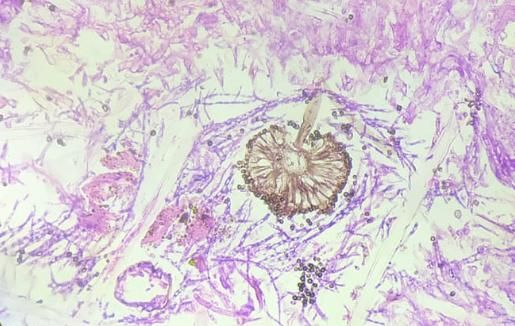
Figure - 1a and 1b: Hematoxylin and eosin staining showing angioinvasion by broad aseptate fungal
hyphae.
1a 1b
Figure - 2: Hematoxylin and eosin staining showing (2a)- thin septate fungal hyphae with fruititing
body of aspergillus fungus, (2b)- Broad aseptate fungal hyphae with fruititing body of mucormycosis.
2a 2b
Page 58Dr. Priyanka Vaghasiya, Dr. Jignasa Bhalodia. Post Covid Fungal Infection: Histopathological and Microbiological
Correlation. IAIM, 2021; 8(8): 53-61.
Four (04) of the patients were coronavirus- angle, fungal hyphae of aspergillus were thin,
positive at the time of presentation but had been septate with branching at acute angle.
infected for more than 14 days; the remaining 46 Submucosal infiltration was present. Both
had been infected earlier and had recovered. 34 hyphae were evident with Hematoxylin and
patients received steroids for management of Eosin stain and PAS stain. Mucor and aspergillus
their covid infection either previously or after sporangium with sporangiophore were evident on
diagnosis was confirmed upon admission (Table haematoxyline and eosin stain (Figure - 2a, b).
– 1). Angioinvasion was demonstrated by H & E stain
(Figure - 1a, b).
Out of 50 patient 43 (86%) cases were found to
be positive based on direct microscopy, culture Discussion
and histopathology. Among these 31 (62 %) The Covid-19 infection caused by the novel
cases were found to be positive by direct SARS-CoV-2 has been associated with a wide
microscopy-KOH, 40 (80%) by culture and range of disease patterns, ranging from a mild
43(86 %) by histopathology (Table - 1, 2), 4 case cough to life-threatening pneumonia [13]. A
which was negative by KOH but positive by myriad of manifestations and complications have
histopathology (Table – 1) considering been documented, and new ones are emerging
histopathology as a gold standard. and being reported on with each passing day as
we learn more about this novel Covid-19
30 patients had a primary disease infection pandemic Mucormycosis or zygomycosis, also
involving the maxillary group of sinus air cells. called phycomycosis, initially described in 1885
The turbinate and uncinate process was affected by Paltauf, is an uncommon and aggressive
in 17 cases, The ethmoid sinus was affected in 10 fungal infection that usually affects patients with
of 50 cases. Sphenoid and frontal and alveolus alteration of their immunological system [15]. It
involvement was less common (Table – 1). is a lethal fungal disease, with rhinocerebral
presentation being its most common form [16].
Mucormycosis was seen in 23 patients, Although it has a low incidence rate, varying
aspergillosis in 3 patient and mixed infection from 0.005 to 1.7 per million population, many
ofmucormycosis with candidiasis in 6 patients, cases have been seen recently, amounting to a
with aspergillosis in 3 patients and with both significant increase in its incidence in the wake
candidiasis and aspergillosis in 3 patients. of the ongoing coronavirus pandemic.
Nose and PNS were involved in 45 cases. In 5 Like SARS-CoV and Middle East respiratory
cases, there was Orbital involvement along with syndrome, SARS-CoV-2 is also responsible for
Nose and PNS while no cases had intracranial lower respiratory tract infection and can cause
extension from cases which we received for the acute respiratory distress syndrome.[17] Besides
histopathology processing (Table – 3). the diffuse alveolar damage with severe
inflammatory exudation, Covid-19 patients
The most common co-morbidity with covid 19 always have immunosuppression with a decrease
was the diabetes mellitus. Other co morbidity in in CD4+ T and CD8+ T cells.
our case was prolonged steroid use. In stage 1, 22
patients had DM and 34 patients were on During the SARS-CoV infection spread in 2003,
prolonged steroid use. In stage 2, 01 patients had the incidence of fungal infection was 14.8–27 per
DM with diabetic ketoacidosis (Table - 1, 4). cent, and it was the main cause of death for
severe acute respiratory syndrome patients,
On histopathology, the fungal hyphae of accounting for 25–73.7 per cent in all causes of
mucormycosis seen were broad, ribbon like, death.23–25 Studies have shown that SARS-CoV
irregular and aseptate with branching at right
Page 59Dr. Priyanka Vaghasiya, Dr. Jignasa Bhalodia. Post Covid Fungal Infection: Histopathological and Microbiological
Correlation. IAIM, 2021; 8(8): 53-61.
and SARS-CoV-2 belong to the same species, the disease suggesting early bone involvement
and have similar prevalence rates and biological can also occurs.
and clinical characteristics. Based on our
experience in 2003, it is important that Yohai, et al. reviewed 145 case reports of
physicians pay critical attention to the high ROCM, 60% of them had diabetes, and analysed
probability of increased incidence of fungal their ophthalmic and nonophthalmic signs and
infections in Covid-19 affected or recovered symptoms occurring at any time during the
patients, similar to the finding observed in course of disease. Similarly Ferry and Abedi
mucormycosis cases here. reported 16 cases of ROCM; 13 (81%) of them
had diabetes. We have compared our
Previously, few such incidental case reports have observations with these two available series
been published, but a firm association between where the majority of the patients had diabetes
Covid-19 and increased fungal infections can [3].
now be clearly seen. Mehta and Pandey reported
a single case of a 60-year-old male with rhino- Conclusion
orbital mucormycosis associated with Covid-19 We are learning more about the new and long-
in September 2020 [13]. Another such case term manifestations of the Covid-19 infection.
report was published by Werthman-Ehrenreich in Uncontrolled diabetes and over-zealous use of
the same month [14]. steroids in Covid 19 management can also
suppress immunity, allowing opportunistic
White et al. studied 135 adults with Covid-19 fungal infections to colonise which might
infection, and reported an incidence of 26.7 per aggravating the illness, and both of these must be
cent for invasive fungal infections [1]. Song, et properly checked.
al. studied the association between Covid-19 and
invasive fungal sinusitis in April 2020, and Its association with invasive mucormycosis
concluded that a large number of patients sinusitis is dangerous and must be given serious
affected by or recovered from Covid-19 are at consideration. Histopathological examination
increased risk of developing invasive fungal must be needed for diagnosing it properly.
diseases, and gave a management algorithm for
such cases [1]. In a recent review, 8 per cent of If infected, early surgical intervention and
coronavirus-positive or recovered patients had intravenous anti-fungal treatment should be
secondary bacterial or fungal infections during sought for management, as a good prognosis and
hospital admission, with widespread use of less fulminant disease course can be achieved in
broad-spectrum antibiotics and steroids [1]. cases of post-coronavirus mucormycosis.
Due to the angioinvasive nature of the disease,
References
skull base osteomyelitis and bone involvement is
usually not seen or seen only late in the disease 1. Sharma S, Grover M, Bhargava S,
[2]. Only few case reports of chronic Samdani S, Kataria T. Post coronavirus
mucormycosis involving bone are available [2]. disease mucormycosis: a deadly addition
However in our series 15 patients presenting with to the pandemic spectrum. J Laryngol
acute mucormycosis went on to develop chronic Otol., 2021; 1–6. https://doi.org/10.1017/
infection with bone involvement following the S0022215121000992
initial treatment. The involved bones showed 2. Jacob Therakathu, Shailesh Prabhu,
expansion, sclerosis, erosions and irregular lytic Aparna Irodi, Sniya Valsa Sudhakar,
destruction. Also many of our patients showed Vikas K. Yadav, V. Rupa. Imaging
destructive bony changes in the acute phase of features of rhinocerebral mucromycosis:
A study of 43 patients. The Egyptian
Page 60Dr. Priyanka Vaghasiya, Dr. Jignasa Bhalodia. Post Covid Fungal Infection: Histopathological and Microbiological
Correlation. IAIM, 2021; 8(8): 53-61.
Journal of Radiology and Nuclear findings and viral tropism in UK patients
Medicine, 2018; 49: 447–452. with severe fatal COVID-19: a post-
3. V. P. Singh, Chetan Bansal, Madhuri mortem study. Lancet Microbe, 2020; 1:
Kaintura. Sinonasal Mucormycosis: A to e245–53.
Z. Indian J Otolaryngol Head Neck 10. Ajay Kumar Singh, et al. Fungal
Surg., November 2019; 71(Suppl 3): Rhinosinusitis: Microbiological and
S1962–S1971. Histopathological Perspective, Journal of
4. A Bhansali, S Bhadada, A Sharma, V Clinical and Diagnostic Research, 2017
Suresh, A Gupta, P Singh, A Jul; Vol-11(7): DC10-DC12.
Chakarbarti, R J Dash. Presentation and 11. Yanling Feng,, Dong Zeng, Lvyin Hu,
outcome of rhino-orbital-cerebral Yuexiang Yang. Case report:
mucormycosis in patients with diabetes. histopathology and molecular pathology
Post Graduate Medical Journal, 2004; analysis on enteric tissue of a COVID-19
80: 670-674. patient. Diagnostic Pathology, 2021; 16:
5. Sharma S, Grover M, Bhargava S, 40.
Samdani S, Kataria T. Post coronavirus 12. Dora Y. Ho, Margaret Lin, Joanna
disease mucormycosis: a deadly addition Schaenman. Yield of diagnostic
to the pandemic spectrum. J Laryngol procedures for invasive fungal infections
Otol., 2021; 1–6. https://doi.org/10.1017/ in neutropenic febrile patients with chest
S0022215121000992 computed tomography abnormalities.
6. Aditya Moorthy, Rohith Gaikwad, Mycoses, 2011 January; 54(1): 59–70.
Shreya Krishna, Raghuraj Hegde, K. K. 13. Mehta S, Pandey A. Rhino-orbital
Tripathi, Preeti G. Kale, P. Subramanya mucormycosis associated with COVID-
Rao, Deepak Haldipur, Krishnamurthy 19. Cureus, 2020; 12: e10726.
Bonanthaya. SARS-CoV-2, 14. Werthman-Ehrenreich A. Mucormycosis
Uncontrolled Diabetes and with orbital compartment syndrome in a
Corticosteroids - An Unholy Trinity in patient with COVID-19. Am J Emerg
Invasive Fungal Infections of the Med., 2021; 42: 264 e5- 264 e8.
Maxillofacial Region? A Retrospective, 15. Paltauf A. Mycosis mucorina. Virchows
Multi-centric Analysis. J Maxillofac Oral Arch Pathol Anat Physiol Klin Med.,
Surg., 2021; 20(3): 1-8. 1885; 102: 543–64.
7. Katja Evert, Thomas Dienemann, 16. Arnáiz-García ME, Alonso-Peña D,
Christoph Brochhausen. Autopsy González-Vela Mdel C, García-Palomo
findings after long-term treatment of JD, Sanz-Giménez-Rico JR, Arnáiz-
COVID-19 patients with microbiological García AM. Cutaneous mucormycosis:
correlation. Virchows Arch., 2021; report of five cases and review of the
479(1): 97-108. literature. J Plast Reconstr Aesthet Surg.,
8. Stefania Caramaschi, Meghan E. Kapp, 2009; 62: e434–41.
Sara E. Miller. Histopathological 17. Wang Y, Wang Y, Chen Y, Qin Q.
findings and clinicopathologic Unique epidemiological and clinical
correlation in COVID-19: a systematic features of the emerging 2019 novel
review. Mod Pathol., 2021; 34(9): 1614- coronavirus pneumonia (COVID-19)
16633. implicate special control measures. J
9. Brian Hanley, Kikkeri N Naresh, Med Virol., 2020; 92: 568–76.
Candice Roufosse. Histopathological
Page 61You can also read