Case Report: Myopathy in Critically Ill COVID-19 Patients: A Consequence of Hyperinflammation? - Frontiers
←
→
Page content transcription
If your browser does not render page correctly, please read the page content below
CASE REPORT
                                                                                                                                               published: 29 January 2021
                                                                                                                                          doi: 10.3389/fneur.2021.625144

                                               Case Report: Myopathy in Critically Ill
                                               COVID-19 Patients: A Consequence
                                               of Hyperinflammation?
                                               Viviana Versace 1*, Luca Sebastianelli 1 , Davide Ferrazzoli 1 , Leopold Saltuari 1 ,
                                               Markus Kofler 2 , Wolfgang Löscher 3 and Antonino Uncini 4
                                               1
                                                Department of Neurorehabilitation, Hospital of Vipiteno (SABES-ASDAA), Vipiteno, Italy, 2 Department of Neurology, Hochzirl
                                               Hospital, Zirl, Austria, 3 Department of Neurology, Medical University Innsbruck, Innsbruck, Austria, 4 Department of
                                               Neuroscience, Imaging and Clinical Sciences, University “G. d’Annunzio”, Chieti, Italy

                                               Introduction: COVID-19-associated muscular complications may comprise myalgia,
                                               weakness, wasting, and rhabdomyolysis. Skeletal muscle damage in COVID-19 may
                                               be due to direct infection by the virus SARS-CoV-2 through interaction with the ACE2
                                               receptor, systemic hyper-inflammatory state with cytokine release and homeostatic
                           Edited by:
                 Jesús Porta-Etessam,          perturbation, an autoimmune process, or myotoxic drugs. Disclosing the cause of
    Hospital Clínico San Carlos, Spain         weakness in an individual patient is therefore difficult.
                        Reviewed by:
                 Raghav Govindarajan,
                                               Case Description: We report two patients, who survived typical COVID-19 pneumonia
  University of Missouri, United States        requiring intensive care treatment and who developed early on myalgia and severe
                      Marcus V. Pinto,
                                               proximal weakness in all four limbs. Laboratory exams revealed elevated serum creatine
            Mayo Clinic, United States
                                               kinase and markedly increased C-reactive protein and interleukin 6, concurring with a
                   *Correspondence:
                        Viviana Versace        systemic inflammatory response. On admission in neurorehabilitation (4 and 7 weeks after
              viviana.versace@sabes.it         COVID-19 onset, respectively), the patients presented with proximal flaccid tetraparesis
     orcid.org/0000-0003-4641-3532
                                               and limb-girdle muscle atrophy. Motor nerve conduction studies showed decreased
                    Specialty section:         amplitude and prolonged duration of compound muscle action potentials (CMAPs) with
          This article was submitted to        normal distal motor latencies and normal conduction velocities in median and ulnar
             Neuromuscular Diseases,
                                               nerves. Needle electromyography in proximal muscles revealed spontaneous activity in
                a section of the journal
                 Frontiers in Neurology        one and myopathic changes in both patients.
       Received: 02 November 2020              Discussion: Clinical, laboratory, and electrodiagnostic findings in these patients were
        Accepted: 13 January 2021
        Published: 29 January 2021
                                               unequivocally consistent with myopathy. Interestingly, increased distal CMAP duration
                              Citation:
                                               has been described in patients with critical illness myopathy (CIM) and reflects slow
            Versace V, Sebastianelli L,        muscle fiber conduction velocity due to membrane hypo-excitability, possibly induced
     Ferrazzoli D, Saltuari L, Kofler M,
                                               by inflammatory cytokines. By analogy with CIM, the pathogenesis of COVID-19-related
 Löscher W and Uncini A (2021) Case
      Report: Myopathy in Critically Ill       myopathy might also depend on hyperinflammation and metabolic pathways that may
  COVID-19 Patients: A Consequence             affect muscles in a pathophysiological continuum from hypo-excitability to necrosis.
                of Hyperinflammation?
            Front. Neurol. 12:625144.          Keywords: COVID-19, SARS-CoV-2, critical illness myopathy, compound muscle action potential duration,
     doi: 10.3389/fneur.2021.625144            interleukin 6

Frontiers in Neurology | www.frontiersin.org                                          1                                         January 2021 | Volume 12 | Article 625144
Versace et al. Case Report: COVID-19 and Myopathy
INTRODUCTION ventilation, the patients suffered mild dyspnea requiring oxygen
support (2 l/min), complained of myalgia and fatigue, and
Muscular complications in hospitalized coronavirus disease showed on examination severe proximal muscles weakness in
(COVID-19) patients may include myalgia, muscle weakness in both upper and lower limbs (Medical Research Council scale
and wasting, elevated serum creatine kinase (CK), and 2/5). Strength in distal muscle was normal. Deep tendon reflexes
rhabdomyolysis (1). Severe acute respiratory syndrome were hypoactive. No deep or superficial sensory disturbance
coronavirus 2 (SARS-CoV-2) binds to cells through the was noted. Cranial nerve examination was unremarkable; in
angiotensin-converting enzyme 2 (ACE2) receptor, which is particular, no bulbar muscles weakness was found.
expressed in skeletal muscle (2). However, SARS-CoV-2 particles, Laboratory examination revealed elevated creatine kinase
despite its broad organotropism beyond the respiratory tract, (CK) (peak-levels 4,002 and 6,732 U/l, respectively) which
have not been demonstrated in muscle samples so far (3). progressively normalized in the following 3 weeks, but no
Besides the possibility of direct skeletal muscle injury myoglobinuria nor acute renal failure signs. Due to the Covid-
by SARS-CoV-2, other conceivable causes of myopathies in 19-related emergency situation in Italian Intensive Care Units,
COVID-19 may comprise an autoimmune process, such as in no further neuroradiological or histopathological muscle studies
necrotizing autoimmune myositis, consequence of the systemic could be performed.
hyperinflammatory state, and myotoxicity by medication (e.g., On admission in the neurorehabilitation unit (4 and 7
hydroxychloroquine, anti-retroviral agents) (2, 4, 5). Moreover, weeks after onset of COVID-19, respectively), both patients
severely affected COVID-19 patients with systemic inflammatory presented with flaccid proximal tetraparesis and limb-girdle
response, prolonged intensive care treatment with ventilation, muscle atrophy. A timeline of the clinical course is presented in
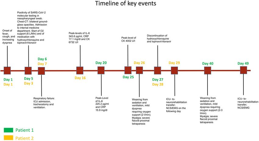
and immobilization are prone to develop critical illness Figure 1.
myopathy (CIM). Hence, explaining the exact cause of weakness Motor nerve conduction studies showed normal distal
in an individual patient may be difficult. latencies and normal conduction velocities. Distal compound
To date, CIM has been reported and at least muscle action potential (CMAP) amplitudes were decreased
electrodiagnostically confirmed in 20 patients with COVID-19 and CMAP durations were prolonged in median and ulnar
(6–9) (Table 1). Intensive care unit acquired muscle weakness nerves in both patients (Table 2, Figure 2). Sensory conduction
was clinically diagnosed in 72% of COVID-19 patients at velocities and sensory nerve action potential amplitudes were
awakening (11). Compared to patients without muscle weakness, normal. Needle EMG showed spontaneous activity (fibrillation
myopathic patients had longer ICU stays, prolonged duration of potentials) in patient 2 and a myopathic pattern with short
invasive mechanical ventilation, higher mean morning glycemia, duration motor unit action potentials, increased percentage
higher exposure to corticosteroids, sedatives, analgesics, and of polyphasic potentials, and early recruitment at voluntary
neuromuscular blocking agents (11). Half of critically ill COVID- effort in proximal muscles in both patients. Distal muscles
19 patients presented acute myopathy in a recent retrospective were unremarkable. Within 2 weeks from admission in
study (10). neurorehabilitation, serum CK returned to normal values (23 and
201 U/l, respectively).
CASE DESCRIPTION Clinical condition improved progressively in both patients,
who were discharged home after 6–8 weeks of rehabilitation,
Here, we report two patients who survived typical COVID-19 with a muscle strength of 3/5 in proximal upper limb and 4/5
pneumonia, confirmed by RT-PCR test on nasopharyngeal swab in proximal lower limb muscles, and normal walking capability.
and by chest computed tomography, which showed bilateral However, both complained of reduced endurance and increased
diffuse consolidations and ground-glass opacities. No personal fatigue during physical activity.
or family medical history of rhabdomyolisis or myoglobinuria
or any type of muscle pathology was known. Neither patient
had ever received statin therapy or other potentially myotoxic DISCUSSION
agents. In general, the patients did not suffer from any previous
relevant pathology. In the presented patients, clinical, laboratory and
Table 2 summarizes demographic, clinical, laboratory, and electrodiagnostic findings were consistent with a myopathy
electrophysiological data. except for increased distal CMAP duration that is usually
Because of respiratory failure, both patients required intensive considered a hallmark of acquired demyelination. However,
care treatment, including tracheostomy and ventilatory support prolonged duration of distal CMAPs that did not change between
for several weeks. Oral treatment with hydroxychloroquine distal and proximal stimulation (Figure 2), together with normal
200 mg twice a day and lopinavir/ritonavir 400/100 mg twice a distal motor latencies and conduction velocities, indicates that
day was administered for 3 weeks. No antibiotics, corticosteroids in these patients, temporal dispersion of distal CMAP is due
or analgesics were administered. High serum levels of C-reactive to slow muscle fibers conduction velocity. Indeed, prolonged
protein (CRP) and interleukin 6 (IL-6) were documented during distal CMAP duration, besides reduced CMAP amplitude, has
the acute phase (Table 2). previously been reported in patients with CIM, who presented,
After weaning from sedation (intravenous sufentanil/propofol as compared to healthy controls, with reduced mean muscle
together with rocuronium bromide as muscle relaxant) and fiber conduction velocity, which was inversely related to CMAP
Frontiers in Neurology | www.frontiersin.org 2 January 2021 | Volume 12 | Article 625144Versace et al. Case Report: COVID-19 and Myopathy
TABLE 1 | Studies on COVID-19-patients with intensive care unit acquired myopathy.
N of Age Sex ICU stay Medication Clinical NCS/EMG Muscle biopsy CK peak-level IL-6
patients mean mean feature peak-level
(range) (range)
[years] [days]
(9) 7 NA NA NA Antiretrovirals, Generalized Myopathy Three patients 181–3,228 N/A
neuromuscular muscular (scattered µmol/l
blockers, weakness necrotic and
corticosteroids, regenerative
antibiotics fibers, no
inflammatory
infiltrates)
(10) 5 N/A N/A N/A Antirheumatics, Generalized Myopathy ND 61–1,206 µg/l NA
antiretrovirals, muscular
corticosteroids, weakness
antibiotics
(8) 6 61 1F 6-14 until Antirheumatics, Acute flaccid Myopathy; ND 55–1,274 UI/L 18.4–5,402.2
(51-72) NCS/EMG antiretrovirals, quadriplegia reduced CMAP ng/ml
corticosteroids, amplitude with
antibiotics, markedly
anticoagulants prolonged
duration
(6) 1 68 M 65 Antibiotics Severe Myopathy and ND NA NA
symmetrical bilateral peroneal
proximal and compression
distal neuropathy.
weakness
and diffuse
muscle
wasting
(7) 1 62 F 30 Antirheumatics, Symmetrical Myopathy ND Normal NA
antiretrovirals, muscle
antibiotics, weakness
neuromuscular predominant
blockers, in lower limbs
antifungal drugs, and proximal
corticosteroids. muscles.
ICU, intensive care unit; NCS/EMG, nerve conduction studies/electromyography; CK, creatine kinase; IL-6, interleukine 6; F, female; M, male; CMAP, compound muscle action potential;
ND not done; NA not available.
duration (13, 14). Moreover, in an in vitro model, sera from with slow muscle fiber conduction velocity in regenerating
patients with CIM applied to single muscle fibers induced muscle fibers.
depolarization of the resting membrane potential, reduced the Interestingly also six reported COVID-10 patients with
action potential rise time, and increased inward sodium current acute quadriplegic myopathy (8), showed markedly prolonged
peak amplitude (15). Evidence from human studies and animal CMAP durations without evidence of acute myonecrosis (CK
models indicates that in CIM associated with sepsis (the so- were slightly elevated in half patients and decreased in few
called “SIM,” sepsis-induced myopathy), systemic inflammatory days) and, with exception of one patient who died due
response, and cytokine release induce a depolarizing shift of the to sepsis, showed rapid improvement of weakness (14–20
muscle cell membrane potential, sodium channel inactivation, days). This can concur with the proposed mechanism of
slowing of muscle fiber conduction velocity until total membrane muscular impairment in COVID-19, ranging from membrane
inexcitability, increase of membrane permeability for Ca2+ , and excitability dysfunction (which reflects in reduced amplitude and
eventually Ca2+ -dependent muscle necrosis by proteasome increased duration of CMAPs) with possible prompt recovery
activation (16). to myonecrosis, CK elevation, consequent muscle atrophy, and
We hypothesize that in the reported patients, by analogy poorer outcome.
with SIM, myopathy was caused by the COVID-associated Serum IL-6 elevation is common in critically ill patients (16),
hyper-inflammatory state, as demonstrated by high initial serum but it also plays a central role in the COVID-19 inflammation
levels of CRP and IL-6. Prolonged distal CMAP durations can cascade already at an early stage, preceding need for intensive
be explained by muscle membrane hypo-excitability combined care, and it correlates with disease severity (17).
Frontiers in Neurology | www.frontiersin.org 3 January 2021 | Volume 12 | Article 625144Frontiers in Neurology | www.frontiersin.org
Versace et al.
TABLE 2 | Demographic, clinical, laboratory, and electrodiagnostic data.
Patient Age Sex ICU stay Clinical features Laboratory findings (peak levels) NCS/EMG: Sensory NCS Motor NCS EMG
time since
disease
onset
CK CRP IL-6 D-dimer WBC Lymphocyte SNAP sNCV CMAP CMAP DML mNCV Proximal
amplitude amplitude duration muscles*
[weeks] [U/l] [mg/l] [pg/ml] [mg/l] [× 103 /µl] [× 103 /µl] [weeks] [µV] [m/s] [mV] [ms] [ms] [m/s]
40–220Versace et al. Case Report: COVID-19 and Myopathy FIGURE 1 | Timeline of key events related to COVID-19 and myopathy in patient 1 (green labels) and patient 2 (yellow labels). FIGURE 2 | Motor nerve conduction studies of patient 1 (A,B) compared to a healthy control subject (C,D). Amplitude and duration of the negative phase of compound muscle action potentials (CMAPs) were measured at a sensitivity of 0.5 mV with a 2 Hz low frequency filter. The cut-off values for distal CMAP duration are according to reported normal values + 2 SD (12). (A) median nerve: distal CMAP amplitude is reduced (4.2 mV), distal motor latency (DML) is normal (3.9 ms), distal CMAP duration is increased (9.3 ms, 127% of upper limit of normal = 7.3 ms), conduction velocity (CV) is 48 m/s. CMAP amplitude and duration did not change between proximal and distal stimulation. Note the broadening and smooth contour of the negative phase of the distal CMAP and the reduction of the ensuing positive phase compared to panel (C) (CMAP duration = 5.7 ms); (B) ulnar nerve: distal CMAP amplitude is slightly reduced (5.7 mV), DML is normal (2.9 ms), distal CMAP duration is increased (13.7 ms, 183% upper limit of normal = 7.5 ms), CV is 55 m/s. CMAP amplitude and duration did not change with proximal stimulation. Note the very prolonged negative phase of the distal CMAP with a long tail and the absence of the ensuing positive phase compared to panel (D) (CMAP duration = 6.3 ms). Frontiers in Neurology | www.frontiersin.org 5 January 2021 | Volume 12 | Article 625144
Versace et al.                                                                                                                     Case Report: COVID-19 and Myopathy

   In conclusion, the same pathogenetic mechanism                                      ETHICS STATEMENT
that causes interstitial pneumonia and damage to
extrapulmonary tissues and organs in COVID-19, i.e., the                               Ethical review and approval was not required for the study
inflammatory cytokine storm together with coagulopathy                                 on human participants in accordance with the local legislation
and macrophage activation, could contribute, in patients                               and institutional requirements. Written informed consent for
requiring prolonged critical care, to skeletal muscle                                  participation was not required for this study in accordance
damage (17).                                                                           with the national legislation and the institutional requirements.
   Further studies are necessary to elucidate the                                      Written informed consent was obtained from the patients for the
pathogenesis of COVID-19-associated myopathy and                                       submission of their data and for the publication of any potentially
to differentiate among direct infection, autoimmune                                    identifiable images or data included in this article.
process, and CIM due to hyperinflammation; in particular,
muscle biopsy with specific investigations would be of                                 AUTHOR CONTRIBUTIONS
crucial importance.
                                                                                       Material preparation, data collection, and analysis were
                                                                                       performed by VV, LSe, and DF. The first draft of the manuscript
DATA AVAILABILITY STATEMENT                                                            was written by VV and MK. AU contributed substantially to
                                                                                       the interpretation of the results, provided critical feedback, and
The original contributions presented in the study are included                         revised the manuscript. All authors contributed in review and
in the article/supplementary material, further inquiries can be                        editing of the manuscript and approved its final version. All
directed to the corresponding author/s.                                                authors contributed to the study conception and design.

REFERENCES                                                                                   Intensive Care Med. (2020) 46:2083–5. doi: 10.1007/s00134-020-
                                                                                             06244-7
 1. Paliwal VK, Garg RK, Gupta A, Tejan N. Neuromuscular presentations                 12.   Mitsuma S, Van den Bergh P, Rajabally YA, Van Parijs V, Martin-Lamb
    in patients with COVID-19. Neurol Sci. (2020) 41:3039–56.                                D, Sonoo M, et al. Tokyo Metropolitan Neuromuscular Electrodiagnosis
    doi: 10.1007/s10072-020-04708-8                                                          Study, Effects of low frequency filtering on distal compound muscle
 2. Ferrandi PJ, Alway SE, Mohamed JS. The interaction between SARS-                         action potential duration for diagnosis of CIDP: a Japanese-European
    CoV-2 and ACE2 may have consequences for skeletal muscle viral                           multicenter prospective study. Clin Neurophysiol. (2015) 126:1805–10.
    susceptibility and myopathies. J Appl Physiol (1985). (2020) 129:864–7.                  doi: 10.1016/j.clinph.2014.11.027
    doi: 10.1152/japplphysiol.00321.2020                                               13.   Kramer CL, Boon AJ, Harper CM, Goodman BP. Compound muscle action
 3. Puelles VG, Lütgehetmann M, Lindenmeyer MT, Sperhake JP, Wong MN,                        potential duration in critical illness neuromyopathy. Muscle Nerve. (2018)
    Allweiss L, et al. Multiorgan and renal tropism of SARS-CoV-2. N Engl J Med.             57:395–400. doi: 10.1002/mus.25732
    (2020) 383:590–2. doi: 10.1056/NEJMc2011400                                        14.   Allen DC, Arunachalam R, Mills KR. Critical illness myopathy: further
 4. Guidon AC, Amato AA. COVID-19 and neuromuscular disorders. Neurology.                    evidence from muscle-fiber excitability studies of an acquired channelopathy.
    (2020) 94:959–69. doi: 10.1212/WNL.0000000000009566                                      Muscle Nerve. (2008) 37:14–22. doi: 10.1002/mus.20884
 5. Dalakas MC. Guillain-Barré syndrome: The first documented COVID-19-                15.   Friedrich O, Hund E, Weber C, Hacke W, Fink RH. Critical illness
    triggered autoimmune neurologic disease: more to come with myositis                      myopathy serum fractions affect membrane excitability and intracellular
    in the offing. Neurol Neuroimmunol Neuroinflamm. (2020) 7:e781.                          calcium release in mammalian skeletal muscle. J Neurol. (2004) 251:53–65.
    doi: 10.1212/NXI.0000000000000781                                                        doi: 10.1007/s00415-004-0272-z
 6. Tankisi H, Tankisi A, Harbo T, Markvardsen LK, Andersen H, Pedersen                16.   Friedrich O, Reid MB, Van den Berghe G, Vanhorebeek I, Hermans G, Rich
    TH. Critical illness myopathy as a consequence of Covid-19 infection. Clin               MM, et al. The sick and the weak: neuropathies/myopathies in the critically
    Neurophysiol. (2020) 131:1931–2. doi: 10.1016/j.clinph.2020.06.003                       ill. Physiol Rev. (2015) 95:1025–9. doi: 10.1152/physrev.00028.2014
 7. Bagnato S, Boccagni C, Marino G, Prestandrea C, D’Agostino T, Rubino F.            17.   McGonagle D, Sharif K, O’Regan A, Bridgewood C. The role of cytokines
    Critical illness myopathy after COVID-19. Int J Infect Dis. (2020) 99:276–8.             including interleukin-6 in COVID-19 induced pneumonia and macrophage
    doi: 10.1016/j.ijid.2020.07.072                                                          activation syndrome-like disease. Autoimmun Rev. (2020) 19:102537.
 8. Madia F, Merico B, Primiano G, Cutuli SL, De Pascale G, Servidei S. Acute                doi: 10.1016/j.autrev.2020.102537
    myopathic quadriplegia in patients with COVID-19 in the intensive care unit.
    Neurology. (2020) 95:492–4. doi: 10.1212/WNL.0000000000010280                      Conflict of Interest: The authors declare that the research was conducted in the
 9. Cabañes-Martínez L, Villadóniga M, González-Rodríguez L, Araque L,                 absence of any commercial or financial relationships that could be construed as a
    Díaz-Cid A, Ruz-Caracuel I, et al. Neuromuscular involvement in                    potential conflict of interest.
    COVID-19 critically ill patients. Clin Neurophysiol. (2020) 131:2809–16.
    doi: 10.1016/j.clinph.2020.09.017                                                  Copyright © 2021 Versace, Sebastianelli, Ferrazzoli, Saltuari, Kofler, Löscher and
10. Abenza-Abildúa MJ, Ramírez-Prieto MT, Moreno-Zabaleta R, Arenas-                   Uncini. This is an open-access article distributed under the terms of the Creative
    Valls N, Salvador-Maya MA, Algarra-Lucas C, et al. Neurological                    Commons Attribution License (CC BY). The use, distribution or reproduction in
    complications in critical patients with COVID-19. Neurologia. (2020)               other forums is permitted, provided the original author(s) and the copyright owner(s)
    35:621–7. doi: 10.1016/j.nrleng.2020.07.012                                        are credited and that the original publication in this journal is cited, in accordance
11. Van Aerde N, Van den Berghe G, Wilmer A, Gosselink R, Hermans G.                   with accepted academic practice. No use, distribution or reproduction is permitted
    Intensive care unit acquired muscle weakness in COVID-19 patients.                 which does not comply with these terms.

Frontiers in Neurology | www.frontiersin.org                                       6                                            January 2021 | Volume 12 | Article 625144
You can also read