Pan-Facial Fractures: A Retrospective Study and Review of Literature - Scientific Research Publishing
←
→
Page content transcription
If your browser does not render page correctly, please read the page content below
Open Journal of Stomatology, 2018, 8, 110-119
                                                                                               http://www.scirp.org/journal/ojst
                                                                                                         ISSN Online: 2160-8717
                                                                                                           ISSN Print: 2160-8709

Pan-Facial Fractures: A Retrospective Study
and Review of Literature

Abdeljalil Abouchadi, Hind Taoufik, Oussama Nacir, Adil Arrob

Department of Maxillo Facial, Stomatology and Plastic Surgery, Avicenne Military Teaching Hospital, Marrakech, Morocco

How to cite this paper: Abouchadi, A.,        Abstract
Taoufik, H., Nacir, O. and Arrob, A. (2018)
Pan-Facial Fractures: A Retrospective Study   Purpose: The aim of this retrospective study was to analyze the characteristics
and Review of Literature. Open Journal of     of panfacial fractures and evaluate treatment results at the Maxillofacial, Sto-
Stomatology, 8, 110-119.                      matology and plastic surgery department at the AVICENNE military hospital
https://doi.org/10.4236/ojst.2018.84010
                                              over a period of 5 years. Patients and Methods: Forty eight patients with
Received: February 27, 2018                   panfacial fractures were treated in Maxillofacial, stomatology and plastic sur-
Accepted: April 15, 2018                      gery department of the AVICENNE Military Teaching Hospital between 2012
Published: April 18, 2018                     and 2017. The criteria for inclusion in the study were patients who had frac-
Copyright © 2018 by authors and
                                              tures of at least three of the four axial segments of the facial skeleton: frontal,
Scientific Research Publishing Inc.           upper midface, lower midface, and mandible. Results: 48 patients with panfa-
This work is licensed under the Creative      cial fractures had a total of 116 subtypes of facial bone fractures. A total of se-
Commons Attribution International             venteen (14.6%) LeFort II fractures in 16 (33.4%) patients were recorded, fif-
License (CC BY 4.0).
http://creativecommons.org/licenses/by/4.0/
                                              teen LeFort I fractures were recorded in 3 (6.2%) cases; seven (6%) LeFort III
                Open Access                   fractures were recorded in 5 (10.4%) cases, thirteen (11.2%) fractures of the
                                              NOF complex were recorded in 6 (12.5%) patients; sixteen (33.4%) patients
                                              had thirty eight (32.7%) fractures involving the mandible. Ten (8.6%) NOM
                                              (naso-orbito-maxilla) complex fractures occurred in 9 (18.7%) cases. 5 (10.4%)
                                              patients had a total of five (4.3%) CNEMFO (naso-ethmoido-maxillo-fronto-
                                              orbital) complex fractures. Our case series included five Comminuted pre-
                                              maxillary fractures and six Intermaxillary disjunctions. All 48 cases had facial
                                              deformities and thirty six had malocclusions. The treatment plan to reduce
                                              and fix the facial bone fractures was sequenced “Bottom up, Outside in”.
                                              Postoperative complications were reported, there were 5 cases whose maloc-
                                              clusions, 4 cases of zygomatic non-union or partial defects, 13 had enoph-
                                              thalmos and hypoglobus. Seven had scars from the trauma, 2 had lower eyelid
                                              ectropion, and 2 had temporal muscle atrophy. Conclusion: Panfacial frac-
                                              tures seem to be complex and difficult to treat, but with an organized and
                                              flexible approach, appropriate reduction of fractures is accomplishable, yet
                                              post-surgical complications mainly caused by soft tissue problems, including
                                              lacerations and asymmetries, can’t be easily avoided.

DOI: 10.4236/ojst.2018.84010       Apr. 18, 2018                 110                                 Open Journal of Stomatology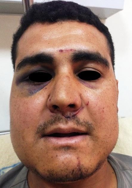
A. Abouchadi et al.

                                 Keywords
                                 Panfacial, Trauma, Fracture, Treatment, Complications

                               1. Introduction
                               Panfacial fractures are defined as those that simultaneously involve the upper,
                               mid and lower face [1] [2]. There is no clear definition and classification for
                               panfacial fractures in the literature. Panfacial fractures, as defined by Follmar et
                               al. are fracture patterns that involve at least three of the four axial segments of
                               the facial skeleton: frontal, upper midface, lower midface, and mandible [3]
                               (Figure 1).
                                  Panfacial fractures are due to road traffic accidents, interpersonal violence,
                               sports-related accidents, industrial accidents, and gunshot wounds. The me-
                               chanism of injury helps identify the energy of impact as well as the probable ex-
                               tent of injury [4].
                                  Panfacial trauma is commonly associated with Multisystem injury; thus, treat-
                               ment is often multidisciplinary. When the patient is stabilized, early and total res-
                               toration of facial form and function should be the goal. The management of pan-
                               facial trauma went from a conservative, delayed, multiple-staged surgery to early,
                               aggressive, and one-stage process. High resolution computed tomography (CT),
                               sufficient surgical exposure, proper anatomic reduction, rigid fixation, primary
                               bone grafting, and soft tissue suspension are the basics for optimum results [4] [5].
                                  The goal of management of panfacial fractures is the restoration of the func-
                               tion and aesthetic three-dimensional facial contours, the earliest possible, while
                               minimizing the patient pain at the lowest possible cost to the victim and society.
                               However, the ideal sequencing of a complex panfacial trauma remains the great-
                               est challenge to every maxillo facial surgeon [6]. Fracture dislocation and the
                               degree of comminution are decisive guidelines in the choice of the surgical pro-
                               cedures [7] [8]. In published literature, two classic approaches have been de-
                               scribed for the management of panfacial trauma; namely “bottom up and inside
                               out” or “top down and outside in”. The preferred sequence starts with mandibu-
                               lar reconstruction, including fractures of the temporo-mandibular joints. In the
                               next step, the fronto-facial and zygomatico-orbital compartments are recon-
                               structed; these are key for subsequent midfacial reconstruction [8] [9].
                                   This paper analyzes the characteristics of panfacial fractures and evaluates
                               treatment results of a series of cases treated at AVICENNE teaching military
                               hospital over a period of 5 years. The outcome is then discussed in the light of
                               preexisting literature.

                               2. Patients and Methods
                               The study group consisted of 48 patients who had panfacial fractures and treated
                               between January 2012 and July 2017 in the Maxillofacial Trauma Center at

DOI: 10.4236/ojst.2018.84010                        111                                 Open Journal of Stomatology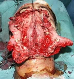
A. Abouchadi et al.

                                                   Figure 1. Four axial segments of facial skele-
                                                   ton [1]: Upper face: Frontal bone and supe-
                                                   rior orbital rim, Midface: Inferior orbital rim,
                                                   nasal bone and zygomatic arch + Maxilla,
                                                   Lower face: Mandible.

                               AVICENNE military hospital. The criteria for inclusion in the study were pa-
                               tients who had fractures of at least three of the four axial segments of the facial
                               skeleton: frontal, upper midface, lower midface, and mandible. All of our patient
                               had a CT (Computed Tomography) scan with 3D reconstruction for precise
                               identification of the fractures. The 48 patients with panfacial features had a total
                               of 116 subtypes of facial bone fractures. Road traffic accidents were the predo-
                               minant etiology (71%) (Table 1).
                                  A retrospective chart review was carried out. Demographic information, de-
                               tails of treatment, and results of follow-up were tabulated for descriptive anal-
                               ysis. The protocol used in their management was also retrospectively analyzed.
                               The study was approved by an ethics committee and patients’ consent was
                               taken.

                               3. Results
                               The 48 patients with panfacial fractures had a total of 78 subtypes of facial
                               bone fractures. Road traffic accidents were the predominant etiology (71%).
                               There were 43 male and 5 female (Sex Ratio ≈ 9/1), aged between 13 and 58
                               years with a main age of 34 years,30 of them were military patients, 18 were ci-
                               vilians.
                                 All patients (100%) had midface fractures in different combinations (Table 2)
                               (Figure 2).
                                 A total of 17 (14.6%) LeFort II fractures in 16 (33.4%) patients were recorded;
                               10 patients had bilateral and 6 had unilateral fractures. Fifteen (13%) LeFort I
                               fractures were recorded in 3 (6.2%) cases; 2 patients had bilateral fractures and 1
                               had unilateral fracture. Seven (6%) LeFort III fractures were recorded in 5
                               (10.4%) cases; 3 patients had bilateral and 2 had unilateral fractures. Thirteen

DOI: 10.4236/ojst.2018.84010                        112                                    Open Journal of Stomatology
A. Abouchadi et al.
(11.2%) fractures of the NOF (naso-orbito-frontal) complex were recorded in 6
(12.5%) patients. Ten (8.6%) NOM (naso-orbito-maxilla) complex fractures oc-
curred in 9 (18.7%) cases. 5 (10.4%) patients had a total of five (4.3%) CNEMFO
(naso-ethmoido-maxillo-fronto-orbital) complex fractures.
Sixteen (33.4%) patients had thirty eight (32.7%) fractures involving the
mandible (Figure 2). 11parasymphysis fractures were recorded in6 patients; 4
patients had bilateral and 2 had unilateral fractures. A total of 9 condylar frac-
tures were recorded in 4 patients; 2 patients each had unilateral and bilateral
fractures. 16 angle fractures were recorded; 3 patients had bilateral fracture.
All 48 cases had facial deformities (Figure 3) and some had functional dis-
orders, in our study thirty six (75%) had malocclusions. Twenty six cases had
limitation of mouth opening (54.17%) to under 30 mm, among which 9 had TMJ
(Temporomandibular Joint) ankylosis with a range of mouth opening of less
than 15 mm. Thirteen cases (27%) had enophthalmos or hypoglobus and three
Table 1. Circumstances of the trauma for our case series.
Patients
Circumstances of the trauma:
Number Percentage (%)
Road traffic accidents 34 cases 71%
Falls:
- 1 accidental fall of the 5th floor
3 cases 6.25%
- 1 fall of the 2nd floor in an epileptic context
- 1syncope
Brawls or aggressions 8 cases 16.7%
Ballistic trauma due to a suicide attempt (Figure 4) 1 case 2%
Trauma by hoofbeats 2 cases 4.17%
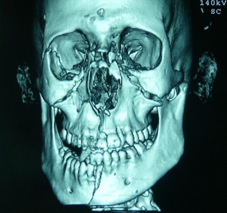
Figure 2. Panfacial fracture scannography
showing mandibular symphisal fracture, bi-
lateral zygomatic fracture with a right lateral
orbital wall fracture, mediomaxillar fracture
and communited nasal bone fracture.
DOI: 10.4236/ojst.2018.84010 113 Open Journal of Stomatology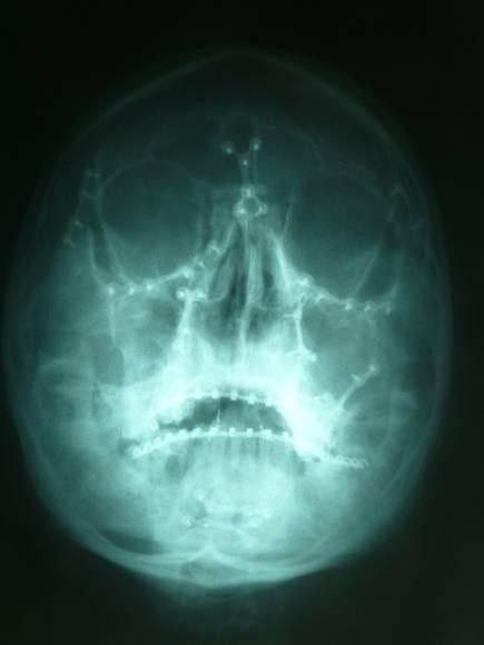
A. Abouchadi et al.

                               Table 2. Different fractures were seen in our caseseries.

                                                                      Fractures                    Patients
                                         Type of fracture               Number    Percentage (%)      Number    Percentage (%)

                                           CNEMFO                          5           4.3                5          10.4

                                                         Unilateral                                       1
                                                                           15          13
                                     LeFort I            Bilateral                                        2          6.2

                                                         Unilateral                                       6
                                                                           17          14.6
                                    LeFort II            Bilateral                                       10          33.4

                                                         Unilateral                                       2
                                                                           7            6
                                    LeFort III           Bilateral                                        3          10.4

                                                         Unilateral
                                      NOM                                  10          8.6                9          18.7
                                                         Bilateral

                                         NOF Complex                       13          11.2               6          12.5

                                    Comminuted premaxillary                5           4.3                2          4.2

                                   Intermaxillary disjunctions             6           5.2                1           2

                                                                           38
                                                                            2
                                           Mandible                         9
                                       Symphysis Condyle                    1
                                       Unilateral Bilateral                 4
                                         Parasymphysis                     11          32.7              16          33.4
                                       Unilateral Bilateral                 3
                                             Angle                          4
                                       Unilateral Bilateral                16
                                                                            4
                                                                            6

                                                 Total                    116          100               48          100

                                                Figure 3. Panfacial fracture appearance: swelling, periorbital
                                                ecchymosis, flattening of the malar prominence, enophtalmos.

                               (6.25%) had 1 globe removed before treatment of their maxillofacial injuries
                               (Table 3).
                                 Postoperative complications were not easily avoided; most of our patients

DOI: 10.4236/ojst.2018.84010                              114                                      Open Journal of Stomatology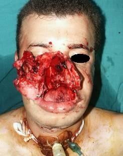
A. Abouchadi et al.

                                                             (a)                         (b)

                                                (c)                         (d)                        (e)

                                  Figure 4. A 31 year old patient presenting a ballistic injury due to a suicide at-
                                  tempt causing an open panfacial trauma. (a) Scannographic image showing 3
                                  staged facial fractures; (b) Pre-operative evaluation, open fractures of the nasal
                                  bone and maxilla with soft tissuesuffering; (c) Per operative procedure starting
                                  “bottom, up”; (d) Immediate post-operative result; (e) Long term post-operative
                                  evaluation: satisfying mouth opening and good occlusion.

                                           Figure 5. Post-operative osteosynthesis control on a Blondeau
                                           radiography.

                               suffered from esthetic and functional problems in the 3 and 6 months follow up
                               (Table 4). Esthetic problems were at the lead of all complications: Soft tissue
                               problems (23%) and asymmetry (16.7%). Patients also suffered from functional
                               issues, 5 had malocclusion (10.4%), 5 had mandibular hypomotility (10.4%) and
                               3 had enophtalmos (6.25%). Infections were seen in 3 of our patients (6.25%)
                               and were treated by antibiotics. Non-union occurred in 4 cases (8.34%).
                                 No intra-operative complications took place for any patient. Postoperatively,

DOI: 10.4236/ojst.2018.84010                          115                                  Open Journal of Stomatology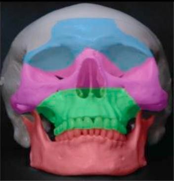
A. Abouchadi et al.

                               Table 3. Clinical findings in 48 panfacial fracture cases.

                                                 Symptoms                         Patients (n)            Percentage %

                                              Facial deformity                        48                      100

                                                Malocclusion                          36                      75

                                         Enophthalmos/hypoglobus                      13                      27

                                             One globe removed                            3                   6.25

                                          Mouth opening limitation                    26                     54.17

                               Table 4. Postoperative complications.

                                            Complications                         Number                           %

                                             Malocclusion                             5                         10.4

                                              Non-union                               4                         8.34

                                               Infection                              3                         6.25

                                            Enophthalmos                              3                         6.25

                                             Asymmetry                                8                         16.7

                                       Mandibular hypomotility                        5                         10.4

                                         Soft tissue problems                        11                         23

                               control radiographs noted adequate reduction (Figure 5) and after resolution of
                               the edema, mouth opening and mandibular movements were fairly adequate.

                               4. Discussion
                               There is no accepted definition of panfacial fracture in the literature. Some au-
                               thors define it as fracture patterns involving both midface and mandible. Others
                               think it must involve the upper, middle, and lower face that means the NOE
                               complex; zygomatic complex, Le Fort midfacial area, and the mandible are all
                               simultaneously fractured. In addition to the possible facial deformities, maloc-
                               clusion and limited facial movement, panfacial trauma can impact the patient’s
                               psychological state [10].
                                 Panfacial fractures are often associated with soft tissue injuries and loss of
                               bone structures that can lead to severe post-traumatic deformities and disabili-
                               ties. Planning the treatment of panfacial fracture is a challenging process. The
                               timing of operative management remains controversial. Multisystem injury is
                               commonly associated; therefore the treatment is often multidisciplinary. Frac-
                               ture dislocation and the degree of comminution are decisive guidelines in the
                               choice of the surgical procedures. Early management of fractures facilitates re-
                               duction and avoids the damage of soft tissues.
                                  In our study, the average time of maxillofacial correction was 9 days since the
                               majority of our patients had associated systemic or neurologic injuries; time was
                               also needed for the edema to be resolved. All of our patients received oral corti-
                               costeroids for a quicker edema reduction; antibiotics were also given to prevent

DOI: 10.4236/ojst.2018.84010                           116                                       Open Journal of Stomatology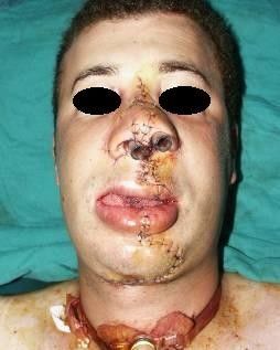
A. Abouchadi et al.

                               infection. Open fractures are considered to be an emergency, which makes the
                               treatment planning of panfacial fracture very challenging.
                                  A CT scanning with coronal cut was achieved from the cranium to the sub-
                               mental region to all of our patients for a good diagnosis. Three-dimensional fa-
                               cial models are also very helpful in treating delayed panfacial fractures. They
                               give the surgeon an appreciation of the spatial relationships of the displaced ske-
                               letal components and help plan treatment.
                                 In the OR, nasotracheal intubation using fibroscopy was used in the majority
                               of our cases, it is a reasonable way for establishing and maintaining an airway
                               when treating panfacial fractures with an intact nasal bone, in other cases oro-
                               tracheal intubation was used in patients with teeth loss; the probe fixated in the
                               area of the lost teeth. When IMF (Inter Maxillary Fixation) was needed, it was
                               done post operatively after extubation. Tracheotomy was necessary in one case
                               of ballistic injury where the nasal bone was completely destructed (Figure 4).
                               Ramanujamet et al. believe that submental intubation is a safer, effective and
                               time efficient method for securing the airway [11].
                                 The treatment was based on an open reduction with titanium mini plate os-
                               teosynthesis. Multiple surgical approaches were used in accordance with the
                               fractured bones. Most cases needed IMF, liquid alimentation of variant dura-
                               tions (30 to 45 days) and reeducation. A bone graft taken from the iliac crest was
                               necessary for bone reconstruction in a number of cases where bone defect was
                               important to stabilize the bone structure and reestablish the buttresses.
                                 Some authors recommend that surgical correction of facial fractures be per-
                               formed immediately after completion of cranial repairs. They advocate the re-
                               duction and fixation of complex injuries within 48 h when initial edema has re-
                               solved and a thorough clinical and radiological exam has been completed. For
                               patients who are medically unstable because of associated neurologic or systemic
                               injuries, facial fracture repair may have to be delayed beyond a reasonable time.
                               A delay of 2 weeks for definitive repair increases the difficulty in obtaining ade-
                               quate reduction of fracture dislocations. Carr and Mathog believe bone healing
                               beyond 3 weeks is in a “grey stage”—the edges of the fragment begin to absorb
                               and remodel, which makes it very difficult to obtain anatomic reduction. This
                               can lead to bone malunion, delayed union, nonunion, and bone defect. Quick
                               management is also critical within 10 days because soft-tissue stiffening and in-
                               terfragmentary healing make delayed corrections very difficult [12].
                                 A review of published literature on panfacial fractures revealed that two ap-
                               proaches of management have enjoyed universal acceptance; “top-to-bottom”
                               and “bottom-to-top” [13] [14]. Marciani et al. [9] recommend first restoring the
                               occlusion and alveolar ridge continuity and alignment and then repairing the
                               mandibular body and angle fractures. Next, the vertical height of the mandibular
                               condyles and ramus is established. They advocate exposing all mid-face and up-
                               per face fractures to allow good visualization of the fracture segments. Then, the
                               transverse width of the face is restored by using the zygomatic arches as a guide.

DOI: 10.4236/ojst.2018.84010                        117                                Open Journal of Stomatology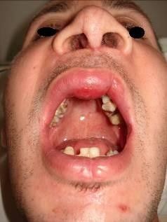
A. Abouchadi et al.

                               The vertical height of the face is restored by aligning and fixing all facial vertical
                               buttresses. The continuity of the orbital and sinus floors and walls is reestab-
                               lished and finally the nasoethmoidal fractures are aligned and fixed.
                                  At our center, our approach to reduce and fix the facial bone fractures is
                               “Bottom up, Outside in”. Facial bone fractures were reduced first and then fixed
                               in a sequential manner. The mandible was reconstructed to establish a stable
                               base. Next, the maxilla was guided into occlusion using the intact mandible as
                               reference and IMF was done. After simultaneously visualizing all fracture sites,
                               the midface fractures were reduced using the “Outside in” principle. The ZMC
                               unit was fixed first and if indicated, the zygomatic arch was lifted into position.
                               Finally the nasal bone was reduced.
                                  Overall, some patients had postoperative functional problems: visual and
                               oculomotor disorders, sensory disturbances of the orbital nerve (V2) territory,
                               morphologic deformities, limitation of oral opening, articular and dental dis-
                               orders. Hence, reeducation is a weighty treatment chapter for optimal results.
                               Moreover, aesthetic sequelae is a painful experience that every panfacial trauma
                               victim goes through; consequently psychological support should also be pro-
                               vided.
                                  On the whole, satisfaction rate of 41/48 patients, most of our patients wished
                               to be operated for removal of osteosynthesis material and/or for rhinoplasty.

                               5. Conclusion
                               Management of panfacial trauma allows proper restoration of facial form and
                               function. Panfacial fractures seem to be complex and difficult to treat, but with
                               an organized and flexible approach, appropriate reduction of fractures is accom-
                               plishable, yet post-surgical complications can’t be easily avoided.

                               References
                               [1]   Yang, R., Zhang, C., Liu, Y., Li, Z. and Li, Z. (2012) Why Should We Start from
                                     Mandibular Fractures in the Treatment of Panfacial Fractures? Journal of Oral and
                                     Maxillofacial Surgery, 70, 1386-1392. https://doi.org/10.1016/j.joms.2011.11.006
                               [2]   Wenig, B.L. (1991) Management of Panfacial Fractures. Otolaryngologic Clinics of
                                     North America, 24, 93-101.
                               [3]   Follmar, K.E., DeBruijn, M., Baccarani, A., Bruno, A.D., Mukundan, S., Erdmann,
                                     D., et al. (2007) Concomitant Injuries in Patients with Panfacial Fractures. Journal
                                     of Trauma and Acute Care Surgery, 63, 831-835.
                                     https://doi.org/10.1097/TA.0b013e3181492f41
                               [4]   Louis, P. (2004) Management of Panfacial Fractures. In: Miloro, M., Ed., Peterson’s
                                     Principles of Oral and Maxillofacial Surgery, 2nd Edition, BCDecker Inc., Hamilton.
                               [5]   David, D.J. (1999) Maxillofacial Trauma: Principles of Management, Priorities and
                                     Basic Techniques. Trauma, 1, 215-226.
                                     https://doi.org/10.1177/146040869900100305
                               [6]   Fritz, M.A. and Koltai, .PJ. (2002) Sequencing and Organization of the Repair of
                                     Panfacial Fractures. Operative Techniques in Otolaryngology-Head and Neck Sur-
                                     gery, 13, 261-264. https://doi.org/10.1016/S1043-1810(02)80056-2

DOI: 10.4236/ojst.2018.84010                          118                                  Open Journal of Stomatology
A. Abouchadi et al.
[7] He, D., Zhang, Y. and Ellis, E. (2007) Panfacial Fractures: Analysis of 33 Cases
Treated Late. Journal of Oral and Maxillofacial Surgery, 65, 2459-2465.
https://doi.org/10.1016/j.joms.2007.06.625
[8] Hardt, N. and Kuttenberger, J. (2010) Surgical Strategy for Complex Craniofacial
Fractures. Craniofacial Trauma. Springer-Verlag, Berlin, 205-238.
https://doi.org/10.1007/978-3-540-33041-7_12
[9] Marciani, R. (2009) Integrating the Care and Treatment of the Complex Facial-
trauma Patient. In: Fonseca, R., Marciani, R., Eds., Oral and Maxillofacial Surgery,
2nd Edition, Saunders/Elsevier, 395-411.
[10] Tang, W., Feng, F., Long, J., Lin, Y., Wang, H., Liu, L., et al. (2009) Sequential Sur-
gical Treatment for Panfacial Fractures and Significance of Biological Osteosynthe-
sis. Dental Traumatology, 25, 171-175.
https://doi.org/10.1111/j.1600-9657.2008.00739.x
[11] Ramanujam, L., Sehgal, S., Krishnappa, R. and Prasad, K. (2013) Panfacial Frac-
tures—A Retrospective Analysis at MS Ramaiah Group of Hospitals, Bangalore.
Journal of Oral and Maxillofacial Surgery, Medicine, and Pathology, 25, 333-340.
https://doi.org/10.1016/j.ajoms.2013.02.006
[12] Carr, R.M. and Mathog, R.H. (1997) Early and Delayed Repair of Orbitozygomatic
Complex Fractures. Journal of Oral and Maxillofacial Surgery, 55, 253-258.
https://doi.org/10.1016/S0278-2391(97)90537-1
[13] Powers, D.B., Will, M.J., Bourgeois, S.L. and Hatt, H.D. (2005) Maxillofacial Trau-
ma Treatment Protocol. Oral & Maxillofacial Surgery Clinics of North America, 17,
341-355. https://doi.org/10.1016/j.coms.2005.05.003
[14] Bagheri, S.C., Khan, H.A., Jahangirnia, A., Rad, S.S. and Mortazavi, H. (2012) An
Analysis of 101 Primary Cosmetic Rhinoplasties. Journal of Oral and Maxillofacial
Surgery, 70, 902-909. https://doi.org/10.1016/j.joms.2011.02.075
DOI: 10.4236/ojst.2018.84010 119 Open Journal of StomatologyYou can also read