Loxoscelism: Cutaneous and Hematologic Manifestations
←
→
Page content transcription
If your browser does not render page correctly, please read the page content below
Hindawi
Advances in Hematology
Volume 2019, Article ID 4091278, 6 pages
https://doi.org/10.1155/2019/4091278

Research Article
Loxoscelism: Cutaneous and Hematologic Manifestations

          Ngan Nguyen1 and Manjari Pandey                         2,3

          1
            Department of Internal Medicine, University of Tennessee Health Science Center, 956 Court Ave., Suite H314, Memphis,
            TN 38163, USA
          2
            Department of Hematology and Oncology, West Cancer Clinic, 7945 Wolf River Blvd, Germantown, TN 38138, USA
          3
            Department of Hematology and Oncology, University of Tennessee Health Science Center, Memphis, TN 38163, USA

          Correspondence should be addressed to Manjari Pandey; mpandey@westclinic.com

          Received 30 November 2018; Accepted 18 February 2019; Published 20 March 2019

          Academic Editor: Lawrence Rice

          Copyright © 2019 Ngan Nguyen and Manjari Pandey. This is an open access article distributed under the Creative Commons
          Attribution License, which permits unrestricted use, distribution, and reproduction in any medium, provided the original work is
          properly cited.

          Background. Brown recluse spider (BRS) envenomation can lead to significant morbidity through severe local reaction and systemic
          illness including acute hemolytic anemia, rhabdomyolysis, disseminated intravascular coagulopathy (DIC), and even death. We aim
          to describe the clinical features and the roles of antibiotics and steroids in the treatment of loxoscelism. Methods. We retrospectively
          identified nine patients (pts) at our institution who were admitted with moderate to severe loxoscelism. A chart review was
          performed to highlight important clinical features and effect of interventions. Results. Nine pts (age 18 to 53) presented with fever (6),
          rash (9), pain/swelling (4), and jaundice (2). Of these, 6 pts had antecedent spider bites documented. Five pts were discharged from
          Emergency Room (ER) with oral antibiotics for “cellulitis” and were readmitted with severe systemic symptoms, with almost half
          (45%) of the pts being admitted to the intensive care unit. The most common admission diagnosis was sepsis secondary to cellulitis
          (6). Four pts developed worsening dermonecrosis, and 3 received prompt incision and drainage (I&D) with debridement. Hemolytic
          anemia developed around day 5 after spider bite (average); the lowest mean hemoglobin level was 5.8g/dL, with average drop of 3.1
          g/dL. Direct antiglobulin test (DAT) (for both complement and surface immunoglobulin) was positive in 4 out of 9 patients. Four
          pts received glucocorticoid therapy for their hemolytic anemia. The use of steroid and intravenous immunoglobulin (IV Ig) did not
          seem to show a difference in the time of recovery although those who received steroids required less blood transfusion (2.1 units less).
          All pts had a complete recovery within two weeks. Conclusion. Treatment of systemic loxoscelism involves aggressive supportive care
          including appropriate wound management, blood transfusions, intravenous fluid replacement, and appropriate antibiotic coverage.
          It is unclear at this time if glucocorticoids or IVIg has any beneficial impact on the treatment of severe loxoscelism.

1. Introduction                                                                 Recluse spider venom contains various enzymes which
                                                                            are enriched in phospholipase D, sphingomyelinase, astacin-
Spiders of the genus Loxosceles have a common name                          like metalloproteases, and Inhibitor Cystine Knot peptides
“brown spiders.” Among these, the brown recluse spiders,                    [2]. Among these, phospholipase D is unique to Loxosceles
or Loxosceles reclusa, have gained notoriety in the medical                 and has clinical significance. It exerts its effects by activating
literature. Their bites can cause clinical manifestations like              complement and inducing neutrophil chemotaxis and apop-
skin necrosis and occasionally severe systemic manifestations               tosis of keratinocytes. It is known to cause the local effect
such as acute hemolytic anemia, rhabdomyolysis, and DIC.                    of dermonecrosis and systemic manifestations including
The brown recluse spider is commonly found in homes in                      hemolysis, thrombocytopenia, and renal failure [3].
endemic areas, in the United States; this includes parts of                     There have been no available commercial tests to detect
South, West, and Central Midwestern United States [1]. They                 spider venom [4]. A presumptive diagnosis of spider bites is
prefer isolated spaces such as closets, attics, or basement.                made based on the history and physical exam findings, while
Most of the recluse bites occur only when they feel disturbed               a definitive diagnosis can be considered only if the spider was
or endangered.                                                              observed biting and recovered. Otherwise the diagnosis of a
2                                                                                                              Advances in Hematology

       Table 1: Admission data and patient characteristics.

            Admission data and patient characteristics
                                            Incidence Rate; % (n)
Age in years (median, range)                       30,18-53
                              Gender
Male                                               11.1% (1)
Female                                            89.9% (8)
Antecedent spider bite documented
Yes                                               67.7% (6)
No                                                33.3% (3)
                           Bite location                                                                (a)
Distal upper extremity                            33.3% (3)
Proximal upper extremity                           11.1% (1)
Proximal lower extremity                          55.6% (5)
                          Admission type
General Med-Surg                                  55.6% (5)
ICU                                               45.4% (4)
Hospital stay length in days (mean ± SD)           7.3 ± 2.8
                  Chief complaints at admission
Fever                                             66.7% (6)
Rash                                              100% (9)
Pain and swelling                                 44.4% (4)
                                                                                                        (b)
Jaundice                                          22.2% (2)

Loxosceles spider bite should be considered after ruling out
other causes. As a result of usually not being able to appre-
hend a spider at the time of the bite, the diagnosis can often
be missed by clinicians and mislabeled as a skin infection
or cellulitis. For patients with systemic findings positive for
fever, myalgia, nausea, and/or vomiting, laboratory studies
are warranted to look for hemolytic anemia, coagulopathy,
and acute kidney injury. To date, there is no definitive
intervention or guideline treatment for loxoscelism besides                                             (c)
supportive care [5]. In this paper, we retrospectively identified     Figure 1: Recluse spider bite with skin necrosis. (a), (b): area of
nine patients with moderate to severe loxoscelism treated at          ecchymosis (a) and black eschar (b) after recluse spider bite. (c) Black
our institution. We will focus on both local and systemic             eschar surrounded by miliary rash as a result of recluse spider bite
manifestations of the disease to highlight the need for high          local reaction.
index of suspicion and review the role of commonly used
treatments: antibiotics, steroids, and IVIg for loxoscelism.
                                                                      were admitted to the intensive care unit (ICU), while five
2. Case Reports                                                       were treated in the regular medicine floor. Patients #2 and 3
                                                                      developed septic shock and required vasopressor in the ICU.
2.1. Initial Presentations. Nine patients whose ages ranged           The average hospital stay length was 7.3 days.
from 18 to 53 presented with cellulitis. The most common
initial presentations were fever (6), rash (9), pain and swelling     2.2. General Findings. All patients initially presented with
(4), and jaundice (2) (Table 1). Interestingly, 8 out of 9 patients   an indurated erythematous rash and received wound care
were female. Six patients could recall an antecedent spider           with daily dressing changes during their hospital stay. In
bite and identify the exact location, while the other three           four patients, the rash progressed to a local black eschar
had no recollection of a bite. All of them presented with a           with surrounding desquamation over an average of 5 days
rash on their distal upper extremities (3), proximal upper            (Figure 1). Three patients with skin necrosis received prompt
extremity (1), or proximal lower extremities (5) (Table 1).           open incision and drainage and debridement. The average
Five patients were discharged from the ER with local care             time to recovery after prompt debridement was 3 days.
and antibiotics, only to be readmitted within a week for              Patient 8 was the only one with skin necrosis who did not
worsening systemic symptoms. The most common admission                receive debridement in her first hospital stay. Thirty days after
diagnosis was sepsis secondary to cellulitis (6). Four patients       discharge, her wound in the left inner thigh had progressed
Advances in Hematology 3
Table 2: Clinical findings and associated treatments.
Clinical findings Percentage (n) of patients
Skin necrosis 44.4% (4)
Sepsis 66.7% (6)
Fever 66.7% (6)
Tachycardia 77.8% (7)
Hypotension 66.7% (6)
Hemolytic anemia 100% (9)
Acute kidney injury 33.3% (3)
Transaminitis 55.6% (5)
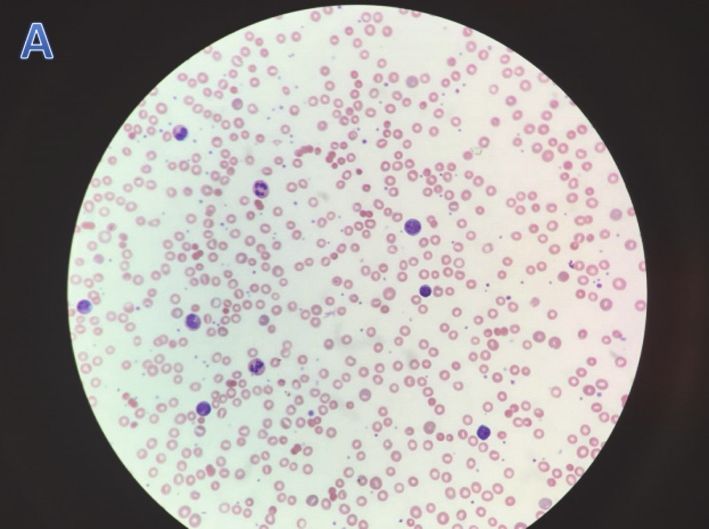
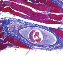
Figure 2: Peripheral blood smears in patients with hemolytic
anemia: peripheral smear showed microspherocytes and some
increased bands.
3. Discussion
Brown recluse spider bites often occur indoors in uninhabited
into abscess. She had to be readmitted for wound debride- places such as in basement or closets. The classical derma-
ment with wound vacuum-assisted closure. The most com- tologic findings caused by brown recluse spider bites are an
mon bacteria grew in surgical wound culture were Staphylo- initial inflammatory reaction at the bite sites followed by local
coccus epidermidis (2 out of 4), Pseudomonas (1), and E. coli eschar. Patients who present with a skin lesion centered by
(1). two puncture marks and surrounding ecchymosis should be
Six patients at presentation had fever, tachycardia, and evaluated for possible spider bite [6]. Of note, 8 out of 9
leukocytosis and were diagnosed with sepsis secondary to patients in this case series were female. The most common
cellulitis based on systemic inflammatory response syndrome bite location is the proximal lower extremity, then distal
(SIRS) criteria. Patients 2, 3, 4, and 8 had remarkable peak upper extremity. No bite was observed on trunk, face, or
white blood cell count of 50.9, 65, 44.8, and 50.2, respectively. hands. Location of bite did not seem to affect severity of
In patients 2 and 7, their sepsis did not improve in spite of local reaction or systemic illness. Cellulitis and skin necrosis
broad spectrum antibiotics and aggressive fluid resuscitation are the most common findings of local loxoscelism. In this
and only subsided after surgical debridement was done. In review, 4 out of 9 patients (44.4%) developed skin necrosis.
this case series, antibiotic use included vancomycin (55.5%), Sphingomyelinase is believed to be the main agent causing
clindamycin (44.4%), zosyn (22.2%), cefepime (22.2%), doxy- skin necrosis as it activates complements and induces apop-
cycline (22.2%), cephalexin (11.1%), and meropenem (11.1%). tosis of keratinocytes [7]. Appropriate wound care, prompt
Hemolytic anemia occurred in all 9 patients with the I&D, and debridement are necessary to permit appropriate
lowest mean hemoglobin level of 5.8 g/dL. On average, healing. Of note, patient 7 who developed a black tender
patients experienced hemolysis beginning day 5 post-bite. eschar continued to spike fevers on three consecutive days
Four of them developed hemolysis earlier, on days 2, 3, in the ICU despite being on broad spectrum antibiotics.
and 4, while five had hemolysis later, on days 5, 6, and Her fever only subsided after debridement was performed.
7. The DAT test was positive for both surface complement Similarly, patient 8 had a 5x2cm black eschar on her inner
component C3 and IgG in 4 out of 9 patients; none of our thigh at admission, and debridement was deferred. Thirty
pts had IgG or C3 alone positivity. Peripheral smears in days after discharge, patient was readmitted due to worsening
these patients often showed the presence of microspheres eschar (now 11x3cm) with abscess formation. She required
with some increased bands (Figure 2). None of nine patients extensive debridement with wound VAC. These findings
had palpable hepatosplenomegaly or lymphadenopathy doc- suggest that delays in debridement in patients with moderate
umented on physical exam. Computed tomography (CT) size of dermonecrosis can lead to progressive skin lesions and
scans of the abdomen and pelvis were not performed. Patients delay wound healing. Average time to recovery after prompt
received an average of 3.1 units of pRBC (packed Red Blood debridement in patients with skin necrosis was 3 days. It is
Cell) transfusions. Four patients received glucocorticoid also good to note that most necrotic skin lesions are very
therapy for their hemolytic anemia. In addition to steroid, tender in these patients; thus appropriate pain control is
patient 8 received IV Ig therapy. indicated.
AKI was present in 3 out of 9 patients (33.3%), and mild Surgical wound cultures showed both Gram positive
transaminitis was found in 5 patients (55.6%) (Table 2). Both bacteria such as Staph and Strep and Gram negative rods
AKI and transaminitis resolved at the time of discharge. such as Pseudomonas and E. coli; therefore, antibiotics should
There seemed to be no difference in the time to recovery have coverage of both Gram positive cocci and Gram negative
in patients receiving additional steroid or steroid and IV Ig rods. None of our patients developed bacteremia, DIC, or
versus those who did not; average time to recovery was within death as a result of systemic loxoscelism.
7 days in all patients. However, those who received steroid Loxoscelism can also cause significant leukocytosis even
were found to require less blood transfusion: 2 vs 3.4 units of in the absence of infection; 3 out 9 patients had peak
pRBCs. WBC above 40,000/mL (Table 3). While the mechanism of
4

                                                          Table 3: Laboratory and clinical course of patients with systemic loxoscelism.
                                   Patient 1          Patient 2          Patient 3          Patient 4          Patient 5          Patient 6     Patient 7    Patient 8   Patient 9
Initial WBC count (x103 /mL)         5.6                40.7                17.1              44.8                14                11.4            13          9.3         9.3
Peak WBC count (x103 /mL)            15.9               50.9                65                44.8               17.3               29.4           16          50.2        13.4
Lowest platelet (x103 /mL)           136                365                 152                179               136                124            117         144         320
Initial Hgb (g/dL)                   10.1                5.3                3.5               11.7                4.7                12            8.8          9.3        14.2
Lowest Hgb (g/dL)                     6.3                5.3                3.5                8.2                4.7                4.4           6.0          7.6         4.8
Onset of hemolysis relative
                                       5                  3                  4                  5                  3                 7             6             1          6
to bite (days)
Hgb at discharge (g/dL)               7.5                8.2               8.8                9.1                11.1                6.9           7.8           12          9.1
Peak Reticulocyte count (%)           3.9               10.8               0.6               3.93                1.6                 2.6           2.7          10.5        13.2
Haptoglobin (mg/dL)
Advances in Hematology                                                                                                             5

venom-induced leukocytosis is not completely understood,            sepsis resolved. While only 3 out of 9 patients had their CPK
leukocytosis is believed to be partly from the effect of            checked (found to be within the normal limit), we suggest
phospholipase D in inducing neutrophil chemotaxis [2].              checking CPK levels for all the patients because rhabdomy-
Further, a high index of suspicion needs to be maintained           olysis is a known complication of systemic loxoscelism.
for development of systemic loxoscelism. Five of the nine
patients in our series were discharged from ER with no              4. Conclusion
planned follow-up. Equally important is to consider that
patients with systemic findings such as fever, myalgia, and         Diagnosis of systemic loxoscelism is often delayed; improved
jaundice warrant laboratory studies to assess for hemolytic         patient education and close follow-up for systemic manifes-
anemia, rhabdomyolysis, and kidney injury. Acute intravas-          tations are warranted. Most patients develop the clinical syn-
cular hemolysis has been described as the main process of sys-      drome within 7-10 days of bite; common symptoms include
temic loxoscelism [8]. The pathogenesis of venom-induced            fever, rash, and jaundice and require further laboratory eval-
hemolysis is not completely understood, but it is believed          uation to look for hemolytic anemia, AKI, rhabdomyolysis,
to be from direct sphingomyelinase venom toxin-induced-             or DIC. The favorable clinical outcomes in our case-series
hemolysis and complement mediated immune destruction                suggest that treatment of systemic loxoscelism is mainly
[2]. In our review, hemolysis was present in all 9 patients,        supportive. Appropriate wound care with prompt surgical
accompanied by markers of hemolysis such as elevated LDH,           debridement if skin necrosis develops facilitates rapid wound
hyperbilirubinemia, and low haptoglobin levels (
6                                                                          Advances in Hematology

Acknowledgments
This paper is funded by West Cancer Clinic and University of
Tennessee Health Science Center Memphis.

References
 [1] R. S. Vetter and D. K. Barger, “An infestation of 2,055 brown
     recluse spiders (Araneae: Sicariidae) and no envenomations in
     a Kansas home: implications for bite diagnoses in nonendemic
     areas,” Journal of Medical Entomology, vol. 39, no. 6, pp. 948–951,
     2002.
 [2] L. H. Gremski, D. Trevisan-Silva, V. P. Ferrer et al., “Recent
     advances in the understanding of brown spider venoms: from
     the biology of spiders to the molecular mechanisms of toxins,”
     Toxicon: Official Journal of the International Society on Toxinol-
     ogy, vol. 83, pp. 91–120, 2014.
 [3] D. V. Tambourgi, D. Paixão-Cavalcante, R. M. Gonçalves De
     Andrade et al., “Loxosceles sphingomyelinase induces com-
     plement-dependent dermonecrosis, neutrophil infiltration, and
     endogenous gelatinase expression,” Journal of Investigative Der-
     matology, vol. 124, no. 4, pp. 725–731, 2005.
 [4] W. V. Stoecker, J. A. Green, and H. F. Gomez, “Diagnosis of
     loxoscelism in a child confirmed with an enzyme-linked im-
     munosorbent assay and noninvasive tissue sampling,” Journal of
     the American Academy of Dermatology, vol. 55, no. 5, pp. 888–
     890, 2006.
 [5] C. J. Hogan, K. C. Barbaro, and K. Winkel, “Loxoscelism: old
     obstacles, new directions,” Annals of Emergency Medicine, vol.
     44, no. 6, pp. 608–624, 2004.
 [6] G. K. Isbister and H. W. Fan, “Spider bite,” The Lancet, vol. 378,
     no. 9808, pp. 2039–2047, 2011.
 [7] M. D. F. Fernandes Pedrosa, I. D. L. M. Junqueira de Azevedo,
     R. M. Gonçalves-de-Andrade et al., “Molecular cloning and
     expression of a functional dermonecrotic and haemolytic factor
     from loxosceles laeta venom,” Biochemical and Biophysical
     Research Communications, vol. 298, no. 5, pp. 638–645, 2002.
 [8] S. Anwar, R. Torosyan, C. Ginsberg, H. Liapis, and A. R. Morri-
     son, “Clinicopathological course of acute kidney injury follow-
     ing brown recluse (Loxoscles reclusa) envenomation,” Clinical
     Kidney Journal, vol. 6, no. 6, pp. 609–612, 2013.
 [9] E. A. Gehrie, H. Nian, and P. P. Young, “Brown recluse spider
     bite mediated hemolysis: clinical features, a possible role for
     complement inhibitor therapy, and reduced rbc surface gly-
     cophorin a as a potential biomarker of venom exposure,” PLoS
     ONE, vol. 8, no. 9, Article ID e76558, 2013.
[10] D. R. Lane and J. S. Youse, “Coombs-positive hemolytic anemia
     secondary to brown recluse spider bite: a review of the literature
     and discussion of treatment,” Cutis; Cutaneous Medicine for the
     Practitioner, vol. 74, no. 6, pp. 341–347, 2004.
[11] A. Nag, J. Datta, A. Das et al., “Acute kidney injury and der-
     monecrosis after Loxosceles reclusa envenomation,” Indian
     Journal of Nephrology, vol. 24, no. 4, pp. 246–248, 2014.
MEDIATORS         of

                                                                                   INFLAMMATION

The Scientific                                 Gastroenterology                                                              Journal of
World Journal
Hindawi Publishing Corporation
                                               Research and Practice
                                               Hindawi
                                                                                   Hindawi
                                                                                                                             Diabetes Research
                                                                                                                             Hindawi
                                                                                                                                                              Disease Markers
                                                                                                                                                              Hindawi
                                                                                   www.hindawi.com        Volume 2018
http://www.hindawi.com
www.hindawi.com                  Volume 2018
                                        2013   www.hindawi.com   Volume 2018                                                 www.hindawi.com    Volume 2018   www.hindawi.com            Volume 2018

Journal of                                                                                                                                                    International Journal of
Immunology Research
Hindawi
                                                                                                                                                              Endocrinology
                                                                                                                                                              Hindawi
www.hindawi.com                  Volume 2018                                                                                                                  www.hindawi.com            Volume 2018

                                                                 Submit your manuscripts at
                                                                    www.hindawi.com

                                                                                                                                                              BioMed
PPAR Research
Hindawi
                                                                                                                                                              Research International
                                                                                                                                                              Hindawi
www.hindawi.com                  Volume 2018                                                                                                                  www.hindawi.com            Volume 2018

                                                                                                                             Journal of
                                                                                                                             Obesity

                                                                               Evidence-Based
Journal of                                     Stem Cells                      Complementary and                                                              Journal of
Ophthalmology
Hindawi
                                               International
                                               Hindawi
                                                                               Alternative Medicine
                                                                               Hindawi                                       Hindawi
                                                                                                                                                              Oncology
                                                                                                                                                              Hindawi
www.hindawi.com                  Volume 2018   www.hindawi.com   Volume 2018   www.hindawi.com                 Volume 2018   www.hindawi.com    Volume 2018   www.hindawi.com            Volume 2013

                                                                               Parkinson’s
                                                                               Disease

Computational and
Mathematical Methods
in Medicine
                                               Behavioural
                                               Neurology
                                                                                                                             AIDS
                                                                                                                             Research and Treatment
                                                                                                                                                              Oxidative Medicine and
                                                                                                                                                              Cellular Longevity
Hindawi                                        Hindawi                         Hindawi                                       Hindawi                          Hindawi
www.hindawi.com                  Volume 2018   www.hindawi.com   Volume 2018   www.hindawi.com                 Volume 2018   www.hindawi.com    Volume 2018   www.hindawi.com            Volume 2018

You can also read