Case Report Percutaneous Aspiration of a Mobile Infected Thrombus from the Right Ventricular Outflow Tract Using the AngioVac System
←
→
Page content transcription
If your browser does not render page correctly, please read the page content below
Hindawi
Case Reports in Cardiology
Volume 2019, Article ID 6279019, 4 pages
https://doi.org/10.1155/2019/6279019

Case Report
Percutaneous Aspiration of a Mobile Infected Thrombus from the
Right Ventricular Outflow Tract Using the AngioVac System

          Justin Ugwu ,1 Umbreen Hussein,2 Sampson Alliu,3 and Ravi Gurujal4
          1
            Department of Hospital Medicine, Miami Valley Hospital, Dayton, Ohio, USA
          2
            Cardiovascular Disease Fellowship, Wright State University, Dayton, Ohio, USA
          3
            Department of Cardiovascular Medicine, Maimonides Medical Center, Brooklyn, New York, USA
          4
            Department of Cardiology, Miami Valley Hospital, Dayton, Ohio, USA

          Correspondence should be addressed to Justin Ugwu; ugwujustin@yahoo.com

          Received 6 December 2018; Accepted 1 April 2019; Published 17 April 2019

          Academic Editor: Man-Hong Jim

          Copyright © 2019 Justin Ugwu et al. This is an open access article distributed under the Creative Commons Attribution License,
          which permits unrestricted use, distribution, and reproduction in any medium, provided the original work is properly cited.

          The AngioVac system was invented in 2012 and was originally designed for the removal of thrombi from the venous system. It has
          been successfully used in the management of iliocaval and right endocardial thrombi but is reportedly less effective in the
          management of pulmonary emboli (PE). Since its advent, there has been interest in its application towards other medical
          situations. One of the most revolutionary uses thus far has been for percutaneous debridement of valvular and cardiac
          electronic device-associated vegetations. In most instances, the AngioVac device has been used to obviate the need for
          surgery in high-risk patients. Here, we describe a novel use of this device in the successful retrieval of a large, mobile,
          infected thrombus from the right ventricular outflow tract in a high surgical-risk patient.

1. Introduction                                                       setting of a non-ST elevation myocardial infarction,
                                                                      hypertension, hyperlipidemia, diabetes mellitus type 2,
An isolated right ventricular (RV) outflow mass is a rare              chronic kidney disease stage III, and prior bariatric surgery.
finding in clinical practice. The differential diagnosis of a           He presented to the emergency department with complaints
mass appearing six days after a negative transesophageal              of fatigue and progressive drainage from a left foot wound
echocardiogram (TEE) in this location includes thrombus,              with associated pain. The patient was hypotensive (blood
embolus, tumor embolus, and endocardial vegetation. Fre-              pressure 89/53 mmHg) and bradycardic (43 beats/minute)
quently, initial management options are conservative, includ-         with complete heart block noted on telemetry monitoring
ing anticoagulation for thrombus/embolus and antibiotics              and confirmed on 12-lead electrocardiogram. Initial labora-
for vegetations. When surgical intervention is indicated,             tory tests were significant for leukocytosis (white blood cell
thrombectomy, embolectomy, or vegetectomy is usually                  count 32.1 K/mm3), anemia (hemoglobin 9.5 g/dl), and
invasive and associated with increased morbidity and mortal-          elevated BUN (51 mg/dl) and creatinine (3.6 mg/dl) from
ity. As described in several case reports, the AngioVac               baseline. A temporary transvenous pacemaker was placed
catheter has proven to be a suitable alternative to surgery in        emergently in the cardiac catheterization laboratory. The
these scenarios [1–3].                                                patient was subsequently hospitalized for the management
                                                                      of septic shock, wet gangrene of the left second toe, and
2. Case Description                                                   complete heart block. Empiric antibiotic therapy was initi-
                                                                      ated with vancomycin and piperacillin/tazobactam, and he
Our patient is a 64-year-old African American male with               underwent urgent toe amputation followed by left ankle
a past medical history significant for coronary artery dis-            disarticulation. Postoperatively, the patient developed respi-
ease status post percutaneous coronary intervention in the            ratory failure requiring intubation. Initial blood cultures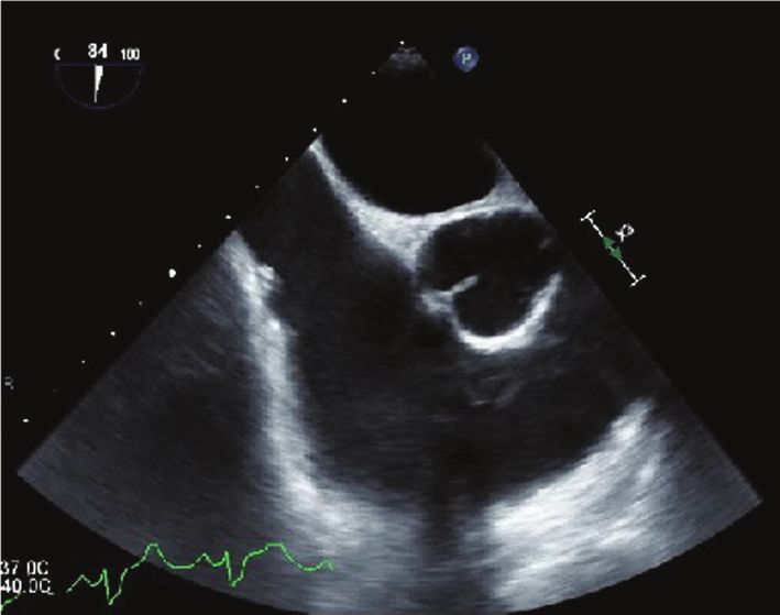
2 Case Reports in Cardiology
grew MRSA. Given persistent hemodynamic instability
despite presumed infectious source control, echocardiogra-
phy was performed to rule out infective endocarditis. Trans-
thoracic echocardiogram (TTE) on hospital day 1 and TEE
on day 3 were, however, noncontributory (Figure 1). Bac-
teremia was persistent through the second week of hospi-
talization on multiple sets of blood cultures, and antibiotic
regimen was changed to ceftaroline. Due to the persistent
bacteremia, a whole-body 18-florodeoxyglucose positron
emission tomography/computed tomography (18F-
FDGPET/CT) scan was then obtained on hospital day 8
which revealed multiple PET-avid lung nodules and cavitary
lesions concerning for septic pulmonary emboli (SPE). TEE
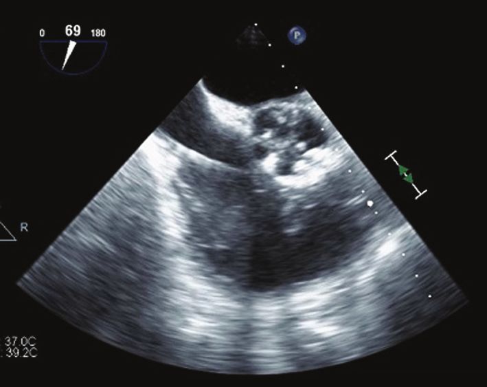
Figure 1: Midesophageal short axis view obtained on
was then repeated on hospital day 9 revealing a highly mobile
transesophageal echocardiogram performed on hospital day 3 with
echodensity in the right ventricular outflow tract (RVOT)
a focus on the right atrium, tricuspid valve, and RVOT. There was
measuring 3.4 cm by 1.6 cm (Figure 2) without valve leaflet no clear, identifiable source of infection on this initial evaluation.
or chordae involvement. The patient was evaluated for surgi-
cal vegetectomy; however, his surgical risk was deemed pro-
hibitive in the acute setting, and thus conservative
management was pursued with antibiotic therapy alone.
Infection control was not successfully achieved. From a Heart
Team approach, the decision was made to attempt an
AngioVac evacuation of the RV mass on hospital day 12.
Venous access was obtained at both femoral venous sites
with 6-French sheaths, and a right internal jugular access was
obtained for the replacement of a temporary pacemaker. The
left 6-French sheath was changed out with a 17-French can-
nula after serial dilatation while the right sheath was replaced
with a number 26 DrySeal Sheath. Using a Swan-Ganz cath-
eter, a Swan wire was positioned into the right pulmonary
artery (PA). Using an MPA catheter, a Lunderquist wire
was positioned in the right PA. The AngioVac return cannula
was then attached with a wet-to-wet seal to the perfusion
pump. The return cannula was loaded over the Lunderquist Figure 2: Focused midesophageal view of the RVOT on repeat
wire after removing air from the cannula. Under TEE guid- TEE performed on hospital day 9. Note the large echodense
ance, the return cannula was advanced into the right atrium. structure measuring 3 4 cm × 1 6 cm that is independent of the
When the return cannula could not be advanced further into tricuspid valve apparatus and is highly mobile with a narrow
the RV outflow tract, it was pulled back and the Lunderquist peduncle. The temporary pacemaker lead is also independent of
wire was repositioned into the left PA using the MPA cathe- this structure.
ter. It was then possible to advance the aspiration catheter
back and forth, and this resulted in the return of a large
amount of material visually consistent with clots. Activated The recommended initial management of right endocar-
clotting time was maintained above 300 seconds through- dial infection is antibiotics [4]. Surgical intervention is
out the procedure with heparin. Postprocedure TEE considered in specific situations. In our patient, persistent
showed up to 90% resolution of the mass (Figure 3). Aspi- bacteremia for more than 7 days despite adequate antibi-
rated blood sampled from the PA was positive for MRSA. otic therapy was an indication for surgery. As is often
Antibiotic therapy was continued, and subsequent blood cul- the case in endocardial infections, our patient was too
tures were sterile. Resolution of complete heart block was unstable clinically to undergo surgery. Recent reports have
verified on continued telemetry monitoring, and the tempo- suggested that AngioVac aspiration could be a minimally
rary pacemaker was removed. The patient was eventually invasive alternative to surgery and the decision was made
discharged in stable condition on hospital day 27. to pursue this option.
The AngioVac venous drainage system, invented in 2012,
3. Discussion was developed for the removal of fresh, soft thrombi or
emboli from the vascular system. It was intended to be used
In the case presented here, persistent MRSA bacteremia as an alternative to anticoagulation, catheter-directed throm-
and SPE on 18F-FDG PET/CT heightened the suspicion bolysis, mechanical thrombectomy, pharmacomechanical
for endocardial infection. The RV outflow mass seen on thrombectomy, or open surgical embolectomy [5, 6]. In a
repeat TEE appeared within 6 days of initial negative TEE single-center analysis of 16 procedures, AngioVac aspiration
and was suspected to be an infected thrombus or vegetation. achieved the removal of more than 70% of thrombi/masses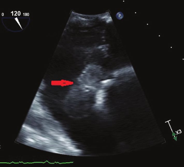
Case Reports in Cardiology                                                                                                          3

                                                                   the use of the AngioVac device if evacuation is desired.
                                                                   Use of this new device may be a safer alternative to sur-
                                                                   gical vegetectomy, particularly when the perioperative risk
                                                                   is prohibitive.

                                                                   Conflicts of Interest
                                                                   The authors declare that they have no conflicts of interest.

                                                                   References
                                                                    [1] N. Patel, T. Azemi, F. Zaeem et al., “Vacuum assisted vege-
                                                                        tation extraction for the management of large lead vegeta-
Figure 3: Midesophageal RVOT-focused view on intraoperative             tions,” Journal of Cardiac Surgery, vol. 28, no. 3, pp. 321–
TEE immediately postaspiration using the AngioVac system                324, 2013.
(catheter can be visualized in this image.) Although residual       [2] A. K. Thiagaraj, M. Malviya, W. W. Htun et al., “A novel
vegetation is visible, there is approximately 90% resolution            approach in the management of right-sided endocarditis:
postaspiration.                                                         percutaneous vegectomy using the AngioVac cannula,” Future
                                                                        Cardiology, vol. 13, no. 3, pp. 211–217, 2017.
                                                                    [3] B. Nickel, T. McClure, and J. Moriarty, “A novel technique for
with 67% and 100% success rate for the right atrial and                 endovascular removal of large volume right atrial tumor
iliocaval sites, respectively [7]. Similarly, Worku et al. ana-         thrombus,” CardioVascular and Interventional Radiology,
lyzed case reports involving 56 procedures which showed                 vol. 38, no. 4, pp. 1021–1024, 2015.
the removal of 100% of material in 87% and 82% of iliocaval         [4] G. Habib, P. Lancellotti, M. J. Antunes et al., “2015 ESC
and intracardiac thrombus, respectively. On the other hand,             guidelines for the management of infective endocarditis:
the success rate for the removal of PE was remarkably low               The Task Force for the Management of Infective Endocardi-
                                                                        tis of the European Society of Cardiology (ESC). Endorsed
at 12.5% [8]. Pasha et al. had alternatively reported successful
                                                                        by: European Association for Cardio-Thoracic Surgery
management of an acute massive PE in a hemodynamically
                                                                        (EACTS), the European Association of Nuclear Medicine
unstable patient using the AngioVac system [9]. Other novel             (EANM),” European Heart Journal, vol. 36, no. 44,
applications of the AngioVac catheter have been explored.               pp. 3075–3128, 2015.
Abubakar et al. published a detailed compilation of case            [5] Angiodynamics, “AngioVac Cannula and Circuit,” http://
reports including 65 procedures, noting an increasing pop-              www.angiovac.com/wp-content/uploads/2017/10/AngioVac-
ularity of AngioVac aspiration for vegetation debulking                 Product-Brochure-ANGB-257-US-Rev-02.pdf.
from right endocardial locations including the right atrium,        [6] G. Behrens and H. Bjarnason, “Venous thromboembolic
tricuspid valve, pulmonic valve, and intracardiac device                disease: the use of the aspiration thrombectomy device Angio-
leads. In nearly all cases, successful vegetectomy led to infec-        Vac,” Seminars in Interventional Radiology, vol. 32, no. 4,
tion control and clinical improvement [10]. In some cases,              pp. 374–378, 2015.
the AngioVac device has been used as a bridge to surgery            [7] J. M. Moriarty, R. al-Hakim, A. Bansal, and J. K. Park,
[11, 12]. The most serious reported complication associated             “Removal of caval and right atrial thrombi and masses using
with the device is access site hematoma [8, 13].                        the AngioVac device: initial operative experience,” Journal of
     With the finding of a large RV outflow mass initially                Vascular and Interventional Radiology, vol. 27, no. 10,
suspected to be an infected thrombus or vegetation, we                  pp. 1584–1591, 2016.
concluded that this was the source of persistent bacteremia         [8] B. Worku, A. Salemi, M. D. D’Ayala, R. F. Tranbaugh, L. N.
and SPE. We decided that urgent thrombectomy or vegetect-               Girardi, and I. M. Gulkarov, “The AngioVac device: under-
omy was the most viable option for infection control and for            standing the failures on the road to success,” Innovations:
                                                                        Technology and Techniques in Cardiothoracic and Vascular
preventing further potentially catastrophic embolization into
                                                                        Surgery, vol. 11, no. 6, pp. 430–433, 2016.
the pulmonary vasculature. Since our patient was at prohibi-
                                                                    [9] A. K. Pasha, M. D. Elder, D. Khurram, B. A. Snyder, and
tive perioperative risk, we elected to attempt retrieval by
                                                                        M. R. Movahed, “Successful management of acute massive
AngioVac aspiration. Following successful extraction of                 pulmonary embolism using Angiovac suction catheter tech-
90% of material visibly consistent with thrombus, we were               nique in a hemodynamically unstable patient,” Cardiovascu-
able to achieve rapid control of the infection. To the best of          lar Revascularization Medicine, vol. 15, no. 4, pp. 240–243,
our knowledge, this is the first report detailing successful             2014.
AngioVac aspiration of a large infected thrombus from the          [10] H. Abubakar, A. Rashed, A. Subahi, A. S. Yassin, M. Shokr,
RV outflow tract without serious complications.                          and M. Elder, “AngioVac system used for vegetation
                                                                        debulking in a patient with tricuspid valve endocarditis: a
                                                                        case report and review of the literature,” Case Reports in
4. Conclusion                                                           Cardiology, vol. 2017, Article ID 1923505, 7 pages, 2017.
                                                                   [11] A. A. Divekar, T. Scholz, and J. D. Fernandez, “Novel per-
When a right endocardial mass of any etiology is encoun-                cutaneous transcatheter intervention for refractory active
tered in clinical practice, consideration should be given to            endocarditis as a bridge to surgery-angiovac aspiration
4                                                                     Case Reports in Cardiology

     system,” Catheterization and Cardiovascular Interventions,
     vol. 81, no. 6, pp. 1008–1012, 2013.
[12] G. Makdisi, T. Casciani, T. C. Wozniak, D. W. Roe, and Z. A.
     Hashmi, “A successful percutaneous mechanical vegetation
     debulking used as a bridge to surgery in acute tricuspid valve
     endocarditis,” Journal of Thoracic Disease, vol. 8, no. 1,
     pp. E137–E139, 2016.
[13] J. Salsamendi, M. Doshi, S. Bhatia et al., “Single center
     experience with the AngioVac aspiration system,” Cardio-
     Vascular and Interventional Radiology, vol. 38, no. 4,
     pp. 998–1004, 2015.
MEDIATORS         of

                                                                                   INFLAMMATION

The Scientific                                 Gastroenterology                                                              Journal of
World Journal
Hindawi Publishing Corporation
                                               Research and Practice
                                               Hindawi
                                                                                   Hindawi
                                                                                                                             Diabetes Research
                                                                                                                             Hindawi
                                                                                                                                                              Disease Markers
                                                                                                                                                              Hindawi
                                                                                   www.hindawi.com        Volume 2018
http://www.hindawi.com
www.hindawi.com                  Volume 2018
                                        2013   www.hindawi.com   Volume 2018                                                 www.hindawi.com    Volume 2018   www.hindawi.com            Volume 2018

Journal of                                                                                                                                                    International Journal of
Immunology Research
Hindawi
                                                                                                                                                              Endocrinology
                                                                                                                                                              Hindawi
www.hindawi.com                  Volume 2018                                                                                                                  www.hindawi.com            Volume 2018

                                                                 Submit your manuscripts at
                                                                    www.hindawi.com

                                                                                                                                                              BioMed
PPAR Research
Hindawi
                                                                                                                                                              Research International
                                                                                                                                                              Hindawi
www.hindawi.com                  Volume 2018                                                                                                                  www.hindawi.com            Volume 2018

                                                                                                                             Journal of
                                                                                                                             Obesity

                                                                               Evidence-Based
Journal of                                     Stem Cells                      Complementary and                                                              Journal of
Ophthalmology
Hindawi
                                               International
                                               Hindawi
                                                                               Alternative Medicine
                                                                               Hindawi                                       Hindawi
                                                                                                                                                              Oncology
                                                                                                                                                              Hindawi
www.hindawi.com                  Volume 2018   www.hindawi.com   Volume 2018   www.hindawi.com                 Volume 2018   www.hindawi.com    Volume 2018   www.hindawi.com            Volume 2013

                                                                               Parkinson’s
                                                                               Disease

Computational and
Mathematical Methods
in Medicine
                                               Behavioural
                                               Neurology
                                                                                                                             AIDS
                                                                                                                             Research and Treatment
                                                                                                                                                              Oxidative Medicine and
                                                                                                                                                              Cellular Longevity
Hindawi                                        Hindawi                         Hindawi                                       Hindawi                          Hindawi
www.hindawi.com                  Volume 2018   www.hindawi.com   Volume 2018   www.hindawi.com                 Volume 2018   www.hindawi.com    Volume 2018   www.hindawi.com            Volume 2018


You can also read