Computational fluid dynamics (CFD) simulation analysis on retinal gas cover rates using computational eye models - Nature
←
→
Page content transcription
If your browser does not render page correctly, please read the page content below
www.nature.com/scientificreports
OPEN Computational fluid dynamics
(CFD) simulation analysis
on retinal gas cover rates using
computational eye models
Makoto Gozawa*, Yoshihiro Takamura, Tomoe Aoki, Kentaro Iwasaki & Masaru Inatani
We investigated the change in the retinal gas cover rates due to intraocular gas volume and
positions using computational eye models and demonstrated the appropriate position after pars
plana vitrectomy (PPV) with gas tamponade for rhegmatogenous retinal detachments (RRDs).
Computational fluid dynamic (CFD) software was used to calculate the retinal wall wettability of a
computational pseudophakic eye models using fluid analysis. The model utilized different gas volumes
from 10 to 90%, in increments of 10% to the vitreous cavity in the supine, sitting, lateral, prone
with closed eyes, and prone positions. Then, the gas cover rates of the retina were measured in each
quadrant. When breaks are limited to the inferior retina anterior to the equator or multiple breaks
are observed in two or more quadrants anterior to the equator, supine position maintained 100% gas
cover rates in all breaks for the longest duration compared with other positions. When breaks are
limited to either superior, nasal, or temporal retina, sitting, lower temporal, and lower nasal position
were maintained at 100% gas cover rates for the longest duration, respectively. Our results may
contribute to better surgical outcomes of RRDs and a reduction in the duration of the postoperative
prone position.
In recent years, pars plana vitrectomy (PPV) with gas tamponade has become the standard treatment for rhegma-
togenous retinal detachments (RRDs)1–3. The principle of gas tamponade is that the gas closes the retinal beaks
and prevents the vitreous fluid from entering through breaks into the subretinal s pace4,5. Therefore, a proper
postoperative position should be maintained after PPV with gas tamponade so that the gas can adequately cover
the retinal breaks until the retina is well attached.
The postoperative position after PPV with gas tamponade, particularly for inferior breaks, remains contro-
versial. At present, a strict prone position is r ecommended6–11 to prevent postoperative complications, such as
pupillary block, intraocular lens (IOL) dislocation/iris capture, and IOL/cornea touch. However, some patients
may experience difficulty in maintaining a strict prone position for medical or physical conditions12–14. In addi-
tion, Martinez-Castillo et al. reported that PPV alone, with no prone position in the postoperative period,
achieves a high reattachment rate without severe postoperative complications in the management of pseudopha-
kic RRDs due to inferior retinal breaks15. We have previously demonstrated using magnetic resonance imaging
that intraocular gas may cover the peripheral retina better in the supine position than in the prone position16.
However, sitting and lateral positions are also used postoperatively. In addition, intraocular gas decreases with
time after surgery, and the intraocular gas makes the contact angle to the retina that affects retinal gas coverage5.
Therefore, determining how much area the gas covers in the target retina after PPV with gas tamponade is
difficult, and no previous reports have examined changes in retinal gas coverage based on the intraocular gas
volume and positions.
Computational fluid dynamic (CFD) analysis is suitable to observe the distribution of gas and fluid consider-
ing the physical properties, such as contact angles and surface tensions. Angunawela et al. reported on fluid shear
stresses using CFD software on the retinal wall in a model eye after vitrectomy and gas tamponade in relation
to eye and head movements17. However, no previous report has examined changes in retinal gas coverage based
on the intraocular gas volume and positions using fluid analysis. Therefore, this study aimed to investigate how
the retinal gas coverage changes due to the intraocular gas volume and positions using fluid analysis as well as
Department of Ophthalmology, Faculty of Medical Sciences, University of Fukui, 23‑3 Shimoaizuki, Matsuoka, Eiheiji,
Yoshida, Fukui 910‑1193, Japan. *email: makoto.gozawa@gmail.com
Scientific Reports | (2021) 11:4937 | https://doi.org/10.1038/s41598-021-84574-2 1
Vol.:(0123456789)
www.nature.com/scientificreports/
Figure 1. The 24-mm eye model of a pseudophakic eye and resting positions. (A) Supine; (B) sitting, (C) prone
with closed eyes, (D) prone, (E) lower temporal, and (F) lower nasal positions.
to demonstrate the appropriate postoperative positions after PPV with gas tamponade for RRDs based on the
gas volume and the location of breaks.
Methods
Computational eye model. A computer model of a pseudophakic eye was designed using three-dimen-
sional (3D) modeling software (3D Builder; Microsoft Corporation, WA, USA). The model eye was simplified
to a sphere, with the anterior 2.5 mm portion of the sphere removed to best reflect the vitreous cavity of a pseu-
dophakic eye18, and separated at 1:30, 4:30, 7:30, and 10:30 into four quadrants (Fig. 1). The majority of human
eyes with RRDs have an axial length of > 24 mm, therefore, we used 24-mm, 25-mm, 26-mm, and 31-mm diam-
eter sphere for analysis. The maximum vertical and horizontal linear dimensions of the eyeball have been shown
to be linearly related to axial length19. Hence, when changing the axial length of the eye, the simplified model
with the anterior 2.5 mm part removed was maintained, and its overall diameter was proportionately increased
or decreased. We then defined the position of the ora serrata of the superior, nasal, temporal, and inferior retina
as per the anatomical differences as 7.4 mm, 5.5 mm, 6.9 mm, and 6.7 mm posterior to the simulated limbus,
respectively. Next, we defined the equator, according to the vortex vein ampullae, as 6.5 mm posterior to the ora
serrata in each retinal a rea20. Therefore, the anterior retina to the equator, where retinal tears are usually f ound21,
was separated into four parts: superior, nasal, inferior, and temporal (Fig. 1).
Direction of the eye model. Figure 1A–F shows resting positions of the 24 mm eye model, which repre-
sents the postoperative body positions. We have previously reported that even if patients superficially maintain
a strict prone position with eyes closed, the mean supraduction angle (°) of the eyeball to the perpendicular line
is positive (16.1°)16. Therefore, six resting positions of the eyeball examined were as follows:
1. Supine: The direction of the eyeball is 90° upward from the horizontal line
2. Sitting: The direction of the eyeball is parallel to the horizontal line
3. Prone with closed eyes: The supraduction angle of the eyeball to the perpendicular line is 16.1°
4. Prone: The direction of the eyeball is 90° downward from the horizontal line
5. Lower temporal: The eyeball was rotated 90° from the sitting position, such that the temporal retina was on
the horizon side
6. Lower nasal: The eyeball was rotated 90° from the sitting position, such that the nasal retina was on the
horizon side
Calculating the wettability of the surface. CFD software (PHOENICS; Concentration Heat and
Momentum Ltd, London) was used to calculate the wettability of the surface of the eye model using cut cell
method, with different gas volumes from 10 to 90%, in increments of 10% to the vitreous cavity. The cut cell
method is developed particularly for improving the calculation accuracy of diagonally arranged objects. In this
method, the intersection of the outline and mesh of the arranged object are investigated, and the area and vol-
ume in which the fluid flows are calculated. When friction occurs, the friction force is given by vector-decom-
posing the intersection coordinates in the direction of the solid inclination. The water properties were as fol-
lows: temperature, 37 °C; density, 998.23 kg/m3; kinematic viscosity, 1.006E−6 m
2/s; and volume expansion rate,
Scientific Reports | (2021) 11:4937 | https://doi.org/10.1038/s41598-021-84574-2 2
Vol:.(1234567890)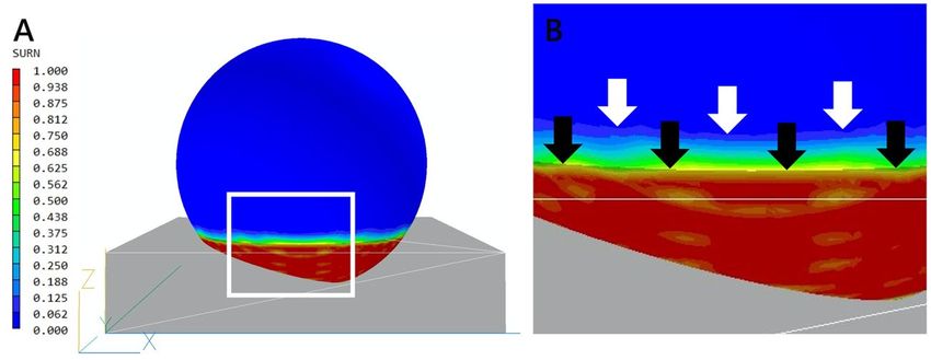
www.nature.com/scientificreports/
Figure 2. Process of measuring the gas cover rate of the inferior retina in the prone position with closed eyes
with 90% gas to the vitreous cavity. (A) The surface contour of the SURN variable and wireframe of the eye
model are displayed simultaneously. (B) The retinal area covered by the gas where the SURN is 0.0, and (C) the
total retinal area was measured.
1.18E−4 1/K. We used air properties as the tamponade gas and they were as follows: temperature, 37 °C; density,
1.1892 kg/m3; kinematic viscosity, 1.544E−05 m 2/s; and volume expansion rate, 3.41E−3 1/K. The air–water–
retina interface was characterized by a surface tension of 0.072 N/m and a contact angle of 38.8°5. A pressure
relief point was set in the gas, and the surface wettability of the eye model was calculated as the SURN variable,
which represents the volume fraction of the water on the eye model and was shown diagrammatically as surface
contour.
Measurement of gas cover rates of the retina. Figure 2 shows the process of measuring the gas cover
rate. The surface contour of the SURN variable and the wireframe of the eye model are displayed simultane-
ously, and the gas covered retinal area where the SURN was 0.0 was measured using 3D modeling software
(Blender 2.80). The gas cover rate was calculated as (the gas covered area) / (the total area of the retina at each
quadrant) × 100% in the supine, sitting, lower nasal, lower temporal, prone with closed eyes, and prone positions.
Results
Gas cover rate in each retinal area. Superior retina. Table 1 show the gas cover rates of the superior
retina of 24-, 25-, 26-, and 31-mm eye model, respectively. In the sitting position, the gas cover rate was 100%
until the gas volume was reduced to 10% in all models. In the supine position, the gas cover rate was 100% until
the gas volume was reduced to 70% in all models. In the prone with closed eyes position, the gas cover rate was
maintained at 100% until the gas volume was reduced to 80% in all models. In the lateral position, the gas cover
rate was maintained at 100% until the gas volume was reduced to 90% in all models. In the prone position, the
gas cover rate was maintained at 100% until the gas volume was reduced to 90% in 24-mm model and 80% in
25-, 26- and 31-mm models, respectively.
Nasal retina. Table 2 show the gas cover rates of the nasal retina of 24-, 25-, 26-, and 31-mm eye model, respec-
tively. In the lower temporal position, the gas cover rate was 100% until the gas volume was reduced to 10% in
all models. In the supine position, the gas cover rate was 100% until the gas volume was reduced to 60% in all
models. In the sitting and prone positions, the gas cover rate was maintained at 100% until the gas volume was
reduced to 90% in all models. In the prone with closed eyes position, the gas cover rate was < 100%, despite the
gas volume being 90%. In the lower nasal position, the gas cover rate was already 0%, despite the gas volume
being 90% in all models.
Temporal retina. Tables 3 show the gas cover rates of the temporal retina of 24-, 25-, 26-, and 31-mm eye
model, respectively. In the lower nasal position, the gas cover rate was 100% until the gas volume was reduced to
10% in all models. In the supine position, the gas cover rate was 100% until the gas volume was reduced to 70%.
In the sitting and prone positions, the gas cover rate was maintained at 100% until the gas volume was reduced
to 90% in all models. In the prone with closed eyes position, the gas cover rate was < 100%, despite the gas vol-
ume being 90% in all models. In the lower temporal position, the gas cover rate was already 0%, despite the gas
volume being 90% in all models.
Inferior retina. Tables 4 show the gas cover rates of the inferior retina of 24-, 25-, 26-, and 31-mm eye model,
respectively. In the supine position, the gas cover rate was 100% until the gas volume was reduced to 70% in
all models. In the prone and lateral positions, the gas cover rate was 100% until the gas volume was reduced to
90% in all models. In the prone with closed eyes position, the gas cover rate was already 75% in 24-mm model
and < 60% in 25-, 26-, and 31-mm models, respectively, although the gas volume was 90%. In the sitting position,
the gas cover rate was already 0%, despite the gas volume being 90% in all models.
Discussion
To the best of our knowledge, this is the first study to demonstrate using fluid analysis to show how the retinal
gas cover rate changes due to the intraocular gas volume and positions. Our results demonstrate that if retinal
breaks were located anterior to the equator, the prone position may not provide an adequate gas coverage for
Scientific Reports | (2021) 11:4937 | https://doi.org/10.1038/s41598-021-84574-2 3
Vol.:(0123456789)www.nature.com/scientificreports/
Gas volume to the vitreous cavity (%)
100 90 80 70 60 50 40 30 20 10 0
24-mm
Supine 100 100 100 100 77 50 24 0 0 0 0
Sitting 100 100 100 100 100 100 100 100 100 100 0
Lateral 100 100 94 66 52 45 34 26 12 4 0
Prone 100 100 89 60 36 11 0 0 0 0 0
Prone with closed eyes 100 100 100 80 67 55 27 10 0 0 0
25-mm
Supine 100 100 100 100 76 66 26 0 0 0 0
Sitting 100 100 100 100 100 100 100 100 100 100 0
Lateral 100 100 83 67 56 44 31 19 8 0 0
Prone 100 100 100 71 38 13 0 0 0 0 0
Prone with closed eyes 100 100 100 94 60 37 15 3 0 0 0
26-mm
Supine 100 100 100 100 80 55 30 0 0 0 0
Sitting 100 100 100 100 100 100 100 100 100 100 0
Lateral 100 100 76 65 51 42 30 18 8 0 0
Prone 100 100 100 71 40 9 0 0 0 0 0
Prone with closed eyes 100 100 100 90 60 38 17 5 0 0 0
31-mm
Supine 100 100 100 100 74 62 27 0 0 0 0
Sitting 100 100 100 100 100 100 100 100 100 100 0
Lateral 100 100 73 62 48 39 29 18 9 0 0
Prone 100 100 100 65 37 8 0 0 0 0 0
Prone with closed eyes 100 100 100 89 59 33 20 6 0 0 0
Table 1. Gas cover rates of the superior retina of 24-, 25-, 26- and 31-mm eye models.
the retina. In contrast, if breaks are limited to the inferior retina anterior to the equator or if multiple breaks are
located in two or more quadrants anterior to the equator, the supine position maintains a 100% gas cover rate
in all retinal breaks for longer than other positions. Although the potential postoperative complications caused
by the supine position require careful attention, our result may contribute to better surgical outcomes of RRDs,
a reduction in the duration of the postoperative prone position and may reduce the patients’ discomfort after
PPV with gas tamponade for RRDs.
The contact angle between a given tamponade gas and the retinal surface depends on the interactions of three
phases: gas, retina, and fluid. It has been reported that the mean contact angle measured for air bubble against the
retina in the fluid was 38.8°5. In the current study, fluid analysis, considering the contact angle, and the surface
tension allowed the gas cover rates of the retina to be calculated more accurately. The existence of a contact angle
and meniscus leads to a reduction in the gas cover rate for a given volume of gas (Fig. 3).
Yoon et al. reported that the adhesive force was transiently reduced after laser photocoagulation of the retina,
but increased beyond normal, and remained twice that of normal between 3 days and 4 weeks22. These findings
indicate that all retinal breaks should be closed with the intraocular gas after PPV with gas tamponade until the
strength of the adhesion by photocoagulation sufficiently prevents retinal redetachment. In the current study, all
gas cover rates in each quadrant in prone position were < 100% when the gas volume was 80%. Furthermore, we
have previously reported that if patients were superficially maintained in a strict prone position, the supraduction
angle (°) of the eyeball to the perpendicular line was positive (16.1°; range, 6.3–29.9)16. In the current study, in
the prone with closed eyes position, the gas cover rates of the nasal and temporal retinas were < 100%, despite
the intraocular gas volume being 90%. Surprisingly, the gas cover rate of the inferior retina in the prone with
closed eyes position was < 80%, despite the intraocular gas volume being 90%. Martinez-Castillo et al. reported
that only 14.4% cases presented with a vitreous cavity that was filled ≥ 90% by air or gas at the 1 and 3 days
postoperative days after PPV with air or gas tamponade for R RDs15. Therefore, if the location of the breaks are
limited to either superior, nasal, or temporal, the most appropriate position is sitting, lower temporal, and lower
nasal, respectively. However, in the supine position, the gas cover rates of the superior, nasal, and temporal retina
were maintained at 100% until the intraocular gas volume was 70%, 60%, and 70% in all models, respectively.
Therefore, the supine position may be appropriate for better gas coverage for the retina as the second choice of
the position in cases where the breaks are localized at either the superior, nasal, or temporal retina. In addition,
if the breaks are limited to the inferior retina anterior to the equator or if multiple breaks are in two or more
quadrants anterior to the equator, the supine position may provide 100% gas cover rate in all retinal breaks for
longer than other positions.
Scientific Reports | (2021) 11:4937 | https://doi.org/10.1038/s41598-021-84574-2 4
Vol:.(1234567890)www.nature.com/scientificreports/
Gas volume to the vitreous cavity (%)
100 90 80 70 60 50 40 30 20 10 0
24-mm
Supine 100 100 100 100 100 78 53 29 0 0 0
Sitting 100 100 92 68 51 43 33 26 14 4 0
Lower nasal 100 0 0 0 0 0 0 0 0 0 0
Lower temporal 100 100 100 100 100 100 100 100 100 100 0
Prone 100 100 67 33 9 0 0 0 0 0 0
Prone with closed eyes 100 91 59 11 0 0 0 0 0 0 0
25-mm
Supine 100 100 100 100 100 89 52 25 0 0 0
Sitting 100 100 78 65 54 44 30 17 8 0 0
Lower nasal 100 0 0 0 0 0 0 0 0 0 0
Lower temporal 100 100 100 100 100 100 100 100 100 100 0
Prone 100 100 72 47 14 0 0 0 0 0 0
Prone with closed eyes 100 95 62 37 0 0 0 0 0 0 0
26-mm
Supine 100 100 100 100 100 80 54 30 0 0 0
Sitting 100 100 73 62 49 39 27 16 7 0 0
Lower nasal 100 0 0 0 0 0 0 0 0 0 0
Lower temporal 100 100 100 100 100 100 100 100 100 100 0
Prone 100 100 70 47 18 0 0 0 0 0 0
Prone with closed eyes 100 92 61 34 0 0 0 0 0 0 0
31-mm
Supine 100 100 100 100 100 90 52 24 0 0 0
Sitting 100 100 73 62 49 41 31 18 8 0 0
Lower nasal 100 0 0 0 0 0 0 0 0 0 0
Lower temporal 100 100 100 100 100 100 100 100 100 100 0
Prone 100 100 68 46 15 0 0 0 0 0 0
Prone with closed eyes 100 90 63 38 0 0 0 0 0 0 0
Table 2. Gas cover rates of the nasal retina of 24-, 25-, 26- and 31-mm eye models.
The supine position may cause some postoperative complications, such as pupillary block, anterior chamber
shallowing, and IOL dislocation/iris capture. Furthermore, Shiragami et al. demonstrated that the immediate
prone or facedown position after PPV with gas tamponade may prevent the retina from postoperative retinal
translocation if the retinal detachment was large or if macular detachment was p resent6. In contrast, the post-
operative facedown position reported for only 2 h prevents retinal translocation after PPV with gas tamponade
for RRDs23. In addition, Otsuka et al. reported no significant difference in postoperative complications between
patients with RRDs in a prone position and those in a prone position on the day of surgery followed by the supine
position24. Therefore, considering these reports as well as our results, patients with RRDs should take a strict
prone or facedown position immediately following PPV with gas tamponade and maintain their position on
the day of surgery. This should be followed by maintaining the appropriate position, including supine position
as above on the basis of the location of breaks and the intraocular gas volume until the gas volume decreases to
70% in the vitreous cavity.
This study has some limitations. High myopia has been reported to have an irregular shape25; however, because
this study involves simulation using a 3D eye model, irregular-shaped eyes could not be analyzed. This study did
not consider eyeball movement, such as rotation, adduction, and abduction, as the simulation is of a stationary
3D eye model. Moreover, we considered that only one bubble was observed in the vitreous cavity and did not
consider the case of multiple bubbles. Often a computer model needs a physical model for verification, which
has not been performed in this study. Gas properties used in this study were of air, but clinically, different types
and concentrations of gases (SF6, C2F6, and C3F8) other than air are used to treat RRD eyes. This study only
considered the location of retinal breaks, not the extent of retinal detachment.
In conclusion, we demonstrated that the supine position may provide 100% gas cover rate for all retinal breaks
longer than other positions when the breaks are limited to the inferior retina or multiple breaks are in two or
more quadrants anterior to the equator. Although complications occurring due to the supine position should
be considered, our result may contribute to better surgical outcomes of RRDs and reduced duration of a strict
prone position that leads to reduced patient discomfort after PPV with gas tamponade for RRDs.
Scientific Reports | (2021) 11:4937 | https://doi.org/10.1038/s41598-021-84574-2 5
Vol.:(0123456789)www.nature.com/scientificreports/
Gas volume to the vitreous cavity (%)
100 90 80 70 60 50 40 30 20 10 0
24-mm
Supine 100 100 100 100 87 62 38 11 0 0 0
Sitting 100 100 91 67 51 44 33 26 12 4 0
Lower nasal 100 100 100 100 100 100 100 100 100 100 0
Lower temporal 100 0 0 0 0 0 0 0 0 0 0
Prone 100 100 86 47 25 0 0 0 0 0 0
Prone with closed eyes 100 95 78 38 13 6 0 0 0 0 0
25-mm
Supine 100 100 100 100 87 73 37 11 0 0 0
Sitting 100 100 79 65 54 42 30 19 8 0 0
Lower nasal 100 100 100 100 100 100 100 100 100 100 0
Lower temporal 100 0 0 0 0 0 0 0 0 0 0
Prone 100 100 78 60 22 0 0 0 0 0 0
Prone with closed eyes 100 93 77 59 18 4 0 0 0 0 0
26-mm
Supine 100 100 100 100 87 65 39 13 0 0 0
Sitting 100 100 74 62 49 39 29 19 7 0 0
Lower nasal 100 100 100 100 100 100 100 100 100 100 0
Lower temporal 100 0 0 0 0 0 0 0 0 0 0
Prone 100 100 71 59 24 0 0 0 0 0 0
Prone with closed eyes 100 86 72 54 20 3 0 0 0 0 0
31-mm
Supine 100 100 100 100 86 74 37 15 0 0 0
Sitting 100 100 75 64 50 41 29 17 8 0 0
Lower nasal 100 100 100 100 100 100 100 100 100 100 0
Lower temporal 100 0 0 0 0 0 0 0 0 0 0
Prone 100 100 78 58 20 0 0 0 0 0 0
Prone with closed eyes 100 89 77 58 22 4 0 0 0 0 0
Table 3. Gas cover rates of the temporal retina of 24-, 25-, 26- and 31-mm eye models.
Scientific Reports | (2021) 11:4937 | https://doi.org/10.1038/s41598-021-84574-2 6
Vol:.(1234567890)www.nature.com/scientificreports/
Gas volume to the vitreous cavity (%)
100 90 80 70 60 50 40 30 20 10 0
24-mm
Supine 100 100 100 100 88 62 39 13 0 0 0
Sitting 100 0 0 0 0 0 0 0 0 0 0
Lateral 100 100 94 67 52 44 33 26 12 4 0
Prone 100 100 86 47 24 0 0 0 0 0 0
Prone with closed eyes 100 75 38 0 0 0 0 0 0 0 0
25-mm
Supine 100 100 100 100 88 74 39 13 0 0 0
Sitting 100 0 0 0 0 0 0 0 0 0 0
Lateral 100 100 79 65 53 43 30 17 8 0 0
Prone 100 100 77 59 22 0 0 0 0 0 0
Prone with closed eyes 100 54 21 0 0 0 0 0 0 0 0
26-mm
Supine 100 100 100 100 88 66 42 15 0 0 0
Sitting 100 0 0 0 0 0 0 0 0 0 0
Lateral 100 100 74 62 50 39 27 16 7 0 0
Prone 100 100 70 58 24 0 0 0 0 0 0
Prone with closed eyes 100 55 21 0 0 0 0 0 0 0 0
31-mm
Supine 100 100 100 100 88 75 38 16 0 0 0
Sitting 100 0 0 0 0 0 0 0 0 0 0
Lateral 100 100 75 64 49 41 29 18 8 0 0
Prone 100 100 78 56 20 0 0 0 0 0 0
Prone with closed eyes 100 57 21 0 0 0 0 0 0 0 0
Table 4. Gas cover rates of the inferior retina of 24-, 25-, 26- and 31-mm eye models.
Figure 3. Surface contour in the prone with closed eyes position, with 90% gas to the vitreous cavity. (A) From
the side view. The gray translucent blockage represents the water object set to have a 10% volume to the vitreous
cavity before fluid analysis. The surface contour represents after analysis. (B) Enlarged image of (A). The black
arrows indicate the initial height of the water before analysis, and the white arrows indicate the line where the
SURN is 0.0, which indicates complete coverage by the gas after analysis. The existence of surface tension and
contact angle cause the line of the SURN = 0.0 to rise above the initial water level, which leads to a reduction in
the gas cover rate of the retina.
Data availability
The datasets generated during and/or analyzed during this study are not publicly available but are available from
the corresponding author on reasonable request.
Received: 27 July 2020; Accepted: 15 February 2021
References
1. Wong, C. W. et al. Trends and factors related to outcomes for primary rhegmatogenous retinal detachment surgery in a large asian
tertiary eye center. Retina 34, 684–692 (2014).
Scientific Reports | (2021) 11:4937 | https://doi.org/10.1038/s41598-021-84574-2 7
Vol.:(0123456789)www.nature.com/scientificreports/
2. Falkner-Radler, C. I. et al. Trends in primary retinal detachment surgery: results of a Bicenter study. Retina 31, 928–936 (2011).
3. Ho, J.-D., Liou, S.-W., Tsai, C.-Y., Tsai, R.J.-F. & Lin, H.-C. Trends and outcomes of treatment for primary rhegmatogenous retinal
detachment: a 9-year nationwide population-based study. Eye (London) 23, 669–675 (2009).
4. Williams, R. W. D. The influence of explants on the physical efficiency of tamponade agents. Graefe’s Arch. Clin. Exp. Ophthalmol.
237, 870–874 (1999).
5. Fawcett, I. M., Williams, R. L. & Wong, D. Contact angles of substances used for internal tamponade in retinal detachment surgery.
Graefes Arch. Clin. Exp. Ophthalmol. 232, 438–444 (1994).
6. Shiragami, C. et al. Unintentional displacement of the retina after standard vitrectomy for rhegmatogenous retinal detachment.
Ophthalmology 117, 86-92.e1 (2010).
7. Bartz-Schmidt, K. U., Kirchhof, B. & Heimann, K. Primary vitrectomy for pseudophakic retinal detachment. Br. J. Ophthalmol.
80, 346–349 (1996).
8. Campo, R. V. et al. Pars plana vitrectomy without scleral buckle for pseudophakic retinal detachments. Ophthalmology 106,
1811–1815 (1999) (discussion 1816).
9. Speicher, M. A., Fu, A. D., Martin, J. P. & von Fricken, M. A. Primary vitrectomy alone for repair of retinal detachments following
cataract surgery. Retina 20, 459–464 (2000).
10. Tanner, V., Minihan, M. & Williamson, T. H. Management of inferior retinal breaks during pars plana vitrectomy for retinal
detachment. Br. J. Ophthalmol. 85, 480–482 (2001).
11. Heimann, H. et al. Primary vitrectomy without scleral buckling for rhegmatogenous retinal detachment. Graefes Arch. Clin. Exp.
Ophthalmol. 234, 561–568 (1996).
12. Salam, A., Harrington, P., Raj, A. & Babar, A. Bilateral Ulnar nerve palsies: an unusual complication of posturing after macular
hole surgery. Eye (London) 18, 95–97 (2004).
13. Vincent, J. M., Peyman, G. A. & Ratnakaram, R. Bilateral ulnar decubitus as a complication of macular hole surgery. Ophthalmic
Surg. Lasers Imaging 34, 485–486 (2003).
14. Holekamp, N. M. et al. Ulnar neuropathy as a complication of macular hole surgery. Arch. Ophthalmol. (Chicago, Ill. 1960) 117,
1607–1610 (1999).
15. Martínez-Castillo, V. J., García-Arumí, J. & Boixadera, A. Pars plana vitrectomy alone for the management of pseudophakic
rhegmatogenous retinal detachment with only inferior breaks. Ophthalmology 123, 1563–1569 (2016).
16. Gozawa, M. et al. Evaluation of intraocular gas using magnetic resonance imaging after pars plana vitrectomy with gas tamponade
for rhegmatogenous retinal detachment. Sci. Rep. 10, 1521. https://doi.org/10.1038/s41598-020-58508-3 (2020).
17. Angunawela, R. I., Azarbadegan, A., Aylward, G. W. & Eames, I. Intraocular fluid dynamics and retinal shear stress after vitrectomy
and gas tamponade. Investig. Ophthalmol. Vis. Sci. 52, 7046–7051 (2011).
18. Shunmugam, M., Shunmugam, S., Williamson, T. H. & Laidlaw, D. A. Air-gas exchange reevaluated: clinically important results
of a computer simulation. Investig. Ophthalmol. Vis. Sci. 52, 8262–8265 (2011).
19. Song, H. T., Kim, Y. J., Lee, S. J. & Moon, Y. S. Relations between age, weight, refractive error and eye shape by computerized
tomography in children. Korean J. Ophthalmol. 21, 163–168 (2007).
20. Prokopich, C. L., Hrynchak, P. & Elliott, D. B. Ocular health assessment. In Clinical Procedures in Primary Eye Care (ed. Elliott, D.
B.) (Elsevier Ltd, Amsterdam, 2007). https://doi.org/10.1016/B978-0-7506-8896-3.50010-9.
21. Ishikawa, K. et al. Preoperative estimation of distance between retinal break and limbus with wide-field fundus imaging: potential
clinical utility for conventional scleral buckling. PLoS ONE 14, e0212284. https://doi.org/10.1371/journal.pone.0212284 (2019).
22. Yoon, Y. H. & Marmor, M. F. Rapid enhancement of retinal adhesion by laser photocoagulation. Ophthalmology 95, 1385–1388
(1988).
23. Dell’Omo, R. et al. Short-time prone posturing is well-tolerated and reduces the rate of unintentional retinal displacement in elderly
patients operated on for retinal detachment. BMC Surg. https://doi.org/10.1186/1471-2482-13-S2-S55 (2013).
24. Otsuka, K., Imai, H., Miki, A. & Nakamura, M. Impact of postoperative positioning on the outcome of pars plana vitrectomy
with gas tamponade for primary rhegmatogenous retinal detachment: comparison between supine and prone positioning. Acta
Ophthalmol. 96, e189–e194 (2018).
25. Ohno-Matsui, K. Proposed classification of posterior staphylomas based on analyses of eye shape by three-dimensional magnetic
resonance imaging and wide-field fundus imaging. Ophthalmology 121, 1798–1809 (2014).
Author contributions
M.G. had full access to all study data and takes responsibility for the data integrity and accuracy of data analysis.
M.G., Y.T., and T.A. designed the study concept. The acquisition, analysis, and interpretation of data were per-
formed by M.G., K.I., and M.I.. M.G. drafted the manuscript. All authors reviewed and approved the manuscript.
Competing interests
The authors declare no competing interests.
Additional information
Correspondence and requests for materials should be addressed to M.G.
Reprints and permissions information is available at www.nature.com/reprints.
Publisher’s note Springer Nature remains neutral with regard to jurisdictional claims in published maps and
institutional affiliations.
Open Access This article is licensed under a Creative Commons Attribution 4.0 International
License, which permits use, sharing, adaptation, distribution and reproduction in any medium or
format, as long as you give appropriate credit to the original author(s) and the source, provide a link to the
Creative Commons licence, and indicate if changes were made. The images or other third party material in this
article are included in the article’s Creative Commons licence, unless indicated otherwise in a credit line to the
material. If material is not included in the article’s Creative Commons licence and your intended use is not
permitted by statutory regulation or exceeds the permitted use, you will need to obtain permission directly from
the copyright holder. To view a copy of this licence, visit http://creativecommons.org/licenses/by/4.0/.
© The Author(s) 2021
Scientific Reports | (2021) 11:4937 | https://doi.org/10.1038/s41598-021-84574-2 8
Vol:.(1234567890)You can also read