Case Report Metastatic Seminoma with Positive Staining of Cytokeratin and MOC31: A Diagnostic Pitfall
←
→
Page content transcription
If your browser does not render page correctly, please read the page content below
Hindawi
Case Reports in Pathology
Volume 2021, Article ID 9992978, 5 pages
https://doi.org/10.1155/2021/9992978

Case Report
Metastatic Seminoma with Positive Staining of Cytokeratin and
MOC31: A Diagnostic Pitfall

          Jiaming Fan,1 Ren Yuan,2 David Stefanelli,3 and Gang Wang                             4

          1
            Tianjin Medical University, Tianjin, China
          2
            Department of Radiology, British Columbia Cancer Vancouver Centre, Vancouver, BC, Canada
          3
            Department of Pathology, Royal Inland Hospital, Kamloops, BC, Canada
          4
            Department of Pathology, British Columbia Cancer Vancouver Centre, Vancouver, BC, Canada

          Correspondence should be addressed to Gang Wang; gang.wang1@bccancer.bc.ca

          Received 2 April 2021; Revised 29 May 2021; Accepted 11 June 2021; Published 30 June 2021

          Academic Editor: Evelina Miele

          Copyright © 2021 Jiaming Fan et al. This is an open access article distributed under the Creative Commons Attribution License,
          which permits unrestricted use, distribution, and reproduction in any medium, provided the original work is properly cited.

          Retroperitoneal metastasis of seminoma often occurs in the higher stage through lymph nodes. Generally, seminoma expresses
          specific germ cell markers while being negative for carcinoma markers. We present a unique case of cytokeratin positive
          seminoma initially presented as retroperitoneal metastasis. The diagnosis was made based on the histological features and
          immunohistochemical stains. Testicular ultrasound confirmed the primary tumor in the patient’s left testicle. Pathologists
          should always be aware of germ cell tumors when encountering a metastasis of an unknown primary.

1. Introduction                                                       2. Case Presentation
Testicular germ cell tumors are composed of seminomas                 A 52-year-old man complained about severe lower abdomi-
and nonseminomas. Seminoma accounts for more than                     nal pain for days but without any digestive symptoms such
half of the diagnosis of germ cell tumors [1]. Most semi-             as nausea, vomiting, diarrhea, or constipation, as well as
nomas are localized to the testis, but some tend to present           any thoracic discomforts. The abdominal CT showed a 12:1
with lymph node metastases, mainly to the retroperito-                 × 10:8 × 8:6 cm mass encasing the aorta circumferentially
neum [2]. Although in some cases, the morphology of ret-              in the retroperitoneum (Figure 1). Containing considerable
roperitoneal metastasis shows a little difference with the             internal necrosis, the 9:5 × 7:5 × 10:5 cm left-side mass dis-
tumor in the primary site, and the metastatic and primary             placed the left kidney and renal pelvis, which caused mild left
tumors share similar germ cell features such as positivity            hydronephrosis. In comparison, the 3:5 × 5:5 × 4:5 cm right-
of tumor markers OCT3/4, CD117, and PLAP [3]. There                   side one squeezed the inferior vena cava slightly. Then, a core
are few reports of other epithelial or vascular markers               biopsy was taken from the sizeable left retroperitoneal mass.
being positive in seminoma [4]. Epithelial markers like                   Microscopic examination revealed that the lesion was
pan-cytokeratin, MOC31 often implicate that tumors have               comprised of infiltrative malignant cells mixed with lympho-
epithelial origin or epithelial differentiation tendency, so           cytes (Figure 2(a)). The infiltrative tumor cells displayed a
they have been applied to diagnose carcinomas. Here, we               sheet-like growth pattern with medium to large size, pale to
report a unique case of metastatic seminoma in the retro-             clear cytoplasm, and polyclonal nuclei. Some of them were
peritoneum with positive staining of cytokeratin and                  undergoing mitosis (Figure 2(b)). The immunostains showed
MOC31.                                                                that the tumor cells were negative for lymphoid and organ-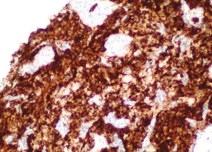
2 Case Reports in Pathology
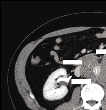
G: 94.5 mm ⁎
H: 75.1 mm
I: 35.2 mm
I
H C: 45.5 mm
J: 55.5 mm
A: 105.3 mm
(a) (b)
Figure 1: (a) Conglomerating bilateral paraaortic nodal mass circumferentially encases without narrowing the aorta. (b) The large left-side
mass measures 9:5 × 7:5 × 10:5 cm containing a large internal necrosis. It is inseparable from the left gonadal vessels (arrow), superiorly
extends to the renal vein level (arrowhead), and laterally it displaced the kidney and renal pelvis (∗ ) causing mild hydronephrosis. The 3:5
× 5:5 × 4:5 cm right-side mass indents the inferior vena cava.
specific markers, including CD3, CD20, CD45, CD30, S100, analysis reports that the classic seminoma is fleshy, solid, lob-
GATA3, calretinin, PAX8, TTF-1, CK7, CK20, CDX2, synap- ular, tan to pale yellow or gray-white [3, 6]. At the histologi-
tophysin, chromogranin, and NKX3.1. The malignant tumor cal examination, seminoma shows a nest-like or sheet-like
showed positive for pan-cytokeratin (Figure 2(c)) and growth mode with large polygonal nuclei, pale to clear to
MOC31 (Figure 2(d)). In comparison, the cells were diffusely eosinophilic cytoplasm, distinct cell membrane, and inter-
and strongly positive for OCT-3/4 (Figure 2(e)) and CD117 vening thin fibrous septa. The septa may be composed of
(Figure 2(f)) which are the markers of germ cells. Ki-67 lymphocytes [3]. However, the histological morphology is a
stained about 70% to 80% of the tumor cell nuclei. The over- little unusual in some metastasis cases [7], so the diagnosis
all immunostaining findings were consistent with metastatic of seminoma should include the morphological assessment
seminoma, classic type. and the immunohistochemical evaluation to separate other
Since the retroperitoneal biopsy suggested a metastatic tumors [5]. Seminoma is usually positive for the staining of
seminoma, a further image study was done. Testicular ultra- germ cell markers, including OCT3/4, CD117, PLAP,
sound revealed a 2:4 × 3:7 × 1:6 cm heterogeneous hypoe- SOX17, SALL4, and D2-40 [3, 6, 8]. Some testicular tumor
choic mass in the left testicle (Figures 3(a) and 3(b)). The markers (LDH, β-HCG, α-FP) can also be detected in classic
Color Doppler showed asymmetrical enlargement of the left seminomas [9], even in some cases of retroperitoneal metas-
testicle containing a hypervascular tissue mass compared to tasis [6]. Noticeably, seminoma typically has a negative
the right testicle (Figures 3(c) and 3(d)). This testicular mass immunoreactivity to other organs/lineage-specific markers
was considered the primary lesion of the classic seminoma. [10]. However, here, we report a case of classic seminoma
The patient’s serum tumor markers (LDH, hCG, AFP) were showing positive for cytokeratin and MOC31.
all in the normal range. Commonly, cytokeratin can be widely detected in the
After completing curative intent chemotherapy with four normal epithelium by AE1/AE3, the pan-cytokeratin
cycles of cisplatin etoposide, the patient underwent left radi- monoclonal antibody [7, 11]. Cytokeratin is a significant
cal orchiectomy. The pathology sections demonstrated a component of intermediate filaments in epithelial cells,
nodular scar confined to the testis but with the involvement and they almost account for 80% of the total protein con-
of rete testis. The background testicular parenchyma showed tent of stratified epithelia [12, 13]. Hence, cytokeratin
significant atrophy of the seminiferous tubules. There was no expression’s positive results in a tumor are always consid-
residual germ cell tumor or germ cell neoplasia in situ ered a symbol of epithelial origin. Similarly, MOC-31 is
identified. another common epithelial marker frequently applied in
clinical practice [14, 15]. As a kind of cluster that can rec-
3. Discussion ognize a transmembrane glycoprotein of cells [16], MOC-
31 could react with normal epithelia and adenocarcinomas
Seminoma tends to have retroperitoneal or mediastinal but not with mesotheliomas [17, 18], which is helpful in
metastasis through lymph nodes [5]. The gross pathologic differential diagnosis. Hence, in the present case, these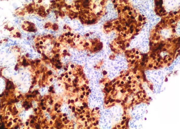
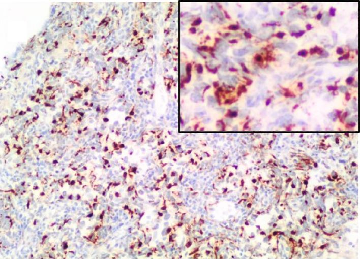
Case Reports in Pathology 3
(a) (b)
(c) (d)
(e) (f)
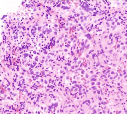
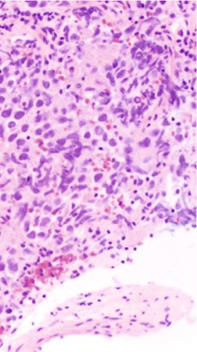
Figure 2: (a) Low power view infiltrative tumor cells mixed with lymphocytes. (b) High power view showing the infiltrating tumor cells with a
sheet-like growth pattern with medium to large size, pale to clear cytoplasm, and polyclonal nuclei. (c) The tumor cells were positive for pan-
cytokeratin. (d) The tumor cells are positive for MOC31. (e) The tumor cells were diffusely and strongly positive for Oct-3/4. (f) The tumor
cells were diffusely and strongly positive for CD117.
two markers’ positivity may lead to a misdiagnosis of met- sic seminoma while negative in embryonal carcinomas, while
astatic carcinoma. CD30 should be positive in embryonal carcinomas but nega-
Once the germ cell origin of this metastasis was estab- tive in seminomas [4, 7, 19, 21, 22].
lished, we should exclude the nonseminomatous component, Interestingly, the staining results of two markers in the
especially embryonal carcinoma. Indeed, we noticed that case we present may suggest epithelial differentiation in
some tumor cells showed increased nuclear pleomorphism seminoma. The incidences of classic seminoma being posi-
and cell crowding, making it hard to distinguish from embry- tive for any epithelial marker were between 39% and 48%,
onal carcinoma in the morphology [19]. On the other hand, usually weak and focal [22]. Kommoss et al. reported
both seminoma and embryonal carcinoma show similar pat- HEA125 (a different clone of EpCAM from MOC31) immu-
terns of immunohistochemical OCT3/4 expression [20]. noreactivity in 3/12 testicular seminomas, some of which are
While embryonal carcinoma is usually strongly positive for diffuse and strong [23]. Cheville et al. revealed that semi-
cytokeratin, once classic seminoma shows positivity for cyto- noma could express cytokeratins of stratified epithelia [21].
keratin markers like in the current case, it would be difficult Tickoo et al. demonstrated that expressing cytokeratin might
to distinguish between them. In this dilemma, other germ cell have been “seminomas with atypia,” a subset of seminomas
markers would be useful. CD117 should be expressed in clas- presenting at a higher clinical stage [4]. However, another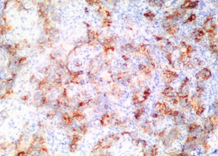
4 Case Reports in Pathology
(a) (b)
(c) (d)
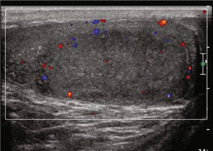
Figure 3: (a, b) US demonstrate a 2:4 × 1:6 × 3:7 cm left intratesticular heterogenous hypoechoic mass ((a) sagittal view; (b) transverse view).
(c) Color Doppler transverse view demonstrates asymmetrical enlargement of the left testicle (right side) containing a heterogeneous mass,
compare to the right testicle (right side). (d) Mild internal vascularity of the mass in the left testicle.
study showed no differences in patient age, stage, tumor size, References
or outcome between CK-positive and CK-negative semino-
mas [21]. Currently, there is no convincing evidence imply- [1] C. Durer, I. Y. Comba, S. Durer, N. Torres Luna, P. Jignesh,
ing that seminoma with epithelial differentiation has more and A. Carilli, “Seminoma metastasized to the prostate: a case
aggressive behavior. The staining patterns of cytokeratin in report and literature review,” Urology Case Reports, vol. 29,
seminoma vary among the reported cases, ranging from a p. 101096, 2020.
dot-like pattern, cytoplasmic, to membranous staining, [2] C. A. Dieker, B. R. Davis, and L. E. de Las Casas, “Retroperito-
depending on different labs and antibodies. The current case neal metastatic germ cell tumor presenting as a psoas abscess: a
diagnostic pitfall,” The American Journal of the Medical Sci-
showed globular and dot-like staining for pan-CK and mod-
ences, vol. 346, no. 1, pp. 70–72, 2013.
erate cytoplasmic staining for MOC31. More cases need to be
studied to investigate whether there is any particular staining [3] J. Marko, D. J. Wolfman, A. L. Aubin, and I. A. Sesterhenn,
“Testicular seminoma and its mimics: from the radiologic
pattern(s) of cytokeratin associated with classic seminoma.
pathology archives,” Radiographics, vol. 37, no. 4, pp. 1085–
In summary, pathologists should be aware that some 1098, 2017.
classic seminoma may express epithelial markers, including
[4] S. K. Tickoo, B. Hutchinson, J. Bacik et al., “Testicular semi-
pan-cytokeratin and MOC31. When dealing with metastasis
noma: a clinicopathologic and immunohistochemical study
with unknown primary, epithelial markers’ positivity should of 105 cases with special reference to seminomas with atypical
not exclude the possibility of germ cell tumors, including features,” International Journal of Surgical Pathology, vol. 10,
classic seminoma. no. 1, pp. 23–32, 2002.
[5] H. J. Schmoll, K. Jordan, R. Huddart et al., “Testicular semi-
noma: ESMO clinical recommendations for diagnosis, treat-
Data Availability ment and follow-up,” Annals of Oncology, vol. 20 Suppl 4,
pp. 83–88, 2009.
The data used to support the findings of this study are
included within the article. [6] C. V. Gingu, M. Mihai, C. Baston et al., “Primary retroperito-
neal seminoma - embryology, histopathology and treatment
particularities,” Romanian Journal of Morphology and Embry-
Conflicts of Interest ology, vol. 57, no. 3, pp. 1045–1050, 2016.
[7] M. T. Sung, G. T. MacLennan, and L. Cheng, “Retroperitoneal
The authors declare that they have no conflicts of interest. seminoma in limited biopsies: morphologic criteria and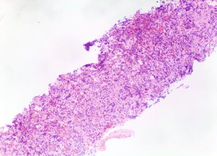
Case Reports in Pathology                                                                                                                      5

       immunohistochemical findings in 30 cases,” The American                   The American Journal of Surgical Pathology, vol. 28, no. 7,
       Journal of Surgical Pathology, vol. 30, no. 6, pp. 766–773, 2006.        pp. 935–940, 2004.
 [8]   L. H. J. Looijenga, T. H. Van der Kwast, D. Grignon et al.,         [21] J. C. Cheville, S. Rao, K. A. Iczkowski, C. M. Lohse, and V. S.
       “Report from the International Society of Urological Pathol-             Pankratz, “Cytokeratin expression in seminoma of the human
       ogy (ISUP) consultation conference on molecular pathology                testis,” American Journal of Clinical Pathology, vol. 113, no. 4,
       of urogenital cancers: IV: current and future utilization of             pp. 583–588, 2000.
       molecular-genetic tests for testicular germ cell tumors,” The       [22] M. T. Sung, G. T. Maclennan, A. Lopez-Beltran, S. Zhang,
       American Journal of Surgical Pathology, vol. 44, no. 7,                  R. Montironi, and L. Cheng, “Primary mediastinal seminoma:
       pp. e66–e79, 2020.                                                       a comprehensive assessment integrated with histology, immu-
 [9]   R. Yuan, C. Zhou, V. Meneghetti, J. M. Lavoie,                           nohistochemistry, and fluorescence in situ hybridization for
       C. Kollmannsberger, and G. Wang, “Seminoma presenting as                 chromosome 12p abnormalities in 23 cases,” The American
       a solitary metastasis in gastric mucosa with regressed testicular        Journal of Surgical Pathology, vol. 32, no. 1, pp. 146–155, 2008.
       mass,” Urology Case Reports, vol. 29, p. 101083, 2020.              [23] F. Kommoss, E. Oliva, F. Bittinger et al., “Inhibin-α, CD99,
[10]   B. Delahunt, J. N. Eble, D. King, P. B. Bethwaite, J. N. Nacey,          HEA125, PLAP, and chromogranin immunoreactivity in tes-
       and A. Thornton, “Immunohistochemical evidence for meso-                 ticular neoplasms and the androgen insensitivity syndrome,”
       thelial origin of paratesticular adenomatoid tumour,” Histopa-           Pathology, vol. 31, no. 9, pp. 1055–1061, 2000.
       thology, vol. 36, no. 2, pp. 109–115, 2000.
[11]   G. E. Orchard, K. Wojcik, F. Shams et al., “Pan-cytokeratin
       markers for rapid frozen section immunocytochemistry from
       head and facial Mohs cases of basal cell carcinoma: a compar-
       ison and evaluation to determine the marker of choice,” British
       Journal of Biomedical Science, vol. 72, no. 2, pp. 61–66, 2015.
[12]   H. H. Bragulla and D. G. Homberger, “Structure and functions
       of keratin proteins in simple, stratified, keratinized and corni-
       fied epithelia,” Journal of Anatomy, vol. 214, no. 4, pp. 516–
       559, 2009.
[13]   M. Pekny and E. B. Lane, “Intermediate filaments and stress,”
       Experimental Cell Research, vol. 313, no. 10, pp. 2244–2254,
       2007.
[14]   P. Gill, C. Naugler, and M. S. Abi Daoud, “Utility of Ber-EP4
       and MOC-31 in basaloid skin tumor detection,” Applied
       Immunohistochemistry & Molecular Morphology, vol. 27,
       no. 8, pp. 584–588, 2019.
[15]   C. C. Pan, P. C. Chen, and D. M. Ho, “The diagnostic utility of
       MOC31, BerEP4, RCC marker and CD10 in the classification
       of renal cell carcinoma and renal oncocytoma: an immunohis-
       tochemical analysis of 328 cases,” Histopathology, vol. 45,
       no. 5, pp. 452–459, 2004.
[16]   A. I. Porcell L, B. R. de Young, D. M. Proca, and W. L. Frankel,
       “Immunohistochemical analysis of hepatocellular and adeno-
       carcinoma in the liver: MOC31 compares favorably with other
       putative markers,” Modern Pathology, vol. 13, no. 7, pp. 773–
       778, 2000.
[17]   D. M. Proca, T. H. Niemann, A. I. Porcell, and B. R. DeYoung,
       “MOC31 immunoreactivity in primary and metastatic carci-
       noma of the liver: report of findings and review of other uti-
       lized markers,” Applied Immunohistochemistry, vol. 8, no. 2,
       pp. 120–125, 2000.
[18]   T. Ruitenbeek, A. S. Gouw, and S. Poppema, “Immunocytol-
       ogy of body cavity fluids. MOC-31, a monoclonal antibody
       discriminating between mesothelial and epithelial cells,”
       Archives of Pathology & Laboratory Medicine, vol. 118, no. 3,
       pp. 265–269, 1994.
[19]   S. K. Lau, L. M. Weiss, and P. G. Chu, “D2-40 immunohisto-
       chemistry in the differential diagnosis of seminoma and
       embryonal carcinoma: a comparative immunohistochemical
       study with KIT (CD117) and CD30,” Modern Pathology: an
       official journal of the United States and Canadian Academy
       of Pathology, Inc, vol. 20, no. 3, pp. 320–325, 2007.
[20]   T. D. Jones, T. M. Ulbright, J. N. Eble, L. A. Baldridge, and
       L. Cheng, “OCT4 staining in testicular tumors: a sensitive
       and specific marker for seminoma and embryonal carcinoma,”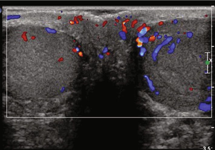
You can also read