Case Report Epstein-Barr Virus Mucocutaneous Ulcer: An Unexpected Diagnosis of a New Entity
←
→
Page content transcription
If your browser does not render page correctly, please read the page content below
Hindawi
Case Reports in Otolaryngology
Volume 2021, Article ID 9989756, 4 pages
https://doi.org/10.1155/2021/9989756

Case Report
Epstein–Barr Virus Mucocutaneous Ulcer: An Unexpected
Diagnosis of a New Entity

                                           1,2
          William Howden                         and Tony Kuo2
          1
              Discipline of Surgery, University of Sydney, Sydney, Australia
          2
              Department of Otolaryngology Head and Neck Surgery, Gosford Hospital, Gosford, Australia

          Correspondence should be addressed to William Howden; howdenwb@gmail.com

          Received 31 March 2021; Accepted 4 May 2021; Published 10 May 2021

          Academic Editor: Seckin Ulualp

          Copyright © 2021 William Howden and Tony Kuo. This is an open access article distributed under the Creative Commons
          Attribution License, which permits unrestricted use, distribution, and reproduction in any medium, provided the original work is
          properly cited.
          Epstein–Barr virus mucocutaneous ulcer (EBVMCU) is a new entity, only recently included in World Health Organisation
          classification of lymphoid neoplasms. Almost all cases described to date have been in patients with a predisposing risk factor of
          immunosuppression. This case presents a 21-year-old male admitted with tonsillitis and no overt immunosuppression, who is
          subsequently diagnosed with EBVMCU of likely iatrogenic origin.

1. Introduction                                                             Upon examination, he was afebrile on admission, with
                                                                       left-sided tender level II/III lymph nodes and normal range
Epstein–Barr virus mucocutaneous ulcer (EBVMCU) is a rare,             of motion of his neck. On direct and nasoendoscopic ex-
underdiagnosed, and newly recognised clinical entity, affecting         amination of his oropharynx, he was noted to have grade II
the sino-oral cavities, gastrointestinal tract, and skin. EBVMCU       bilaterally erythematous palatine tonsils, with a white
is a provisional entity in the 2016 Update of World Health             coating to the right inferior pole with mildly erythematous.
Organisation classification of lymphoid neoplasms. In the               On flexible nasoendoscopy, he had symmetrical and non-
literature to date, a vast majority of cases have been described in    oedematous arytenoids and epiglottis. He had no trismus,
immunosuppressed patients, usually as a result of autoim-              palatal oedema or petechiae, uvula deviation, or periorbital
mune, transplantation, or haematological malignancy therapy.           oedema. He was not examined for splenomegaly.
This report examines the course and treatment of an otherwise               His bloods revealed a lymphocytosis of 9.0 × 10⁹/L and a
well young male presenting clinically with tonsillitis, who was        C-reactive protein (CRP) of 10 mg/L. A serum monospot,
subsequently diagnosed with EBVMCU on further testing.                 added retrospectively to admission bloods, was positive.
                                                                            A computed tomography (CT) scan of his neck dem-
2. Case Presentation                                                   onstrated a 7 × 8 × 7 mm collection posterior to his left
                                                                       palatine tonsil. There was no evidence of deep neck space
A 23-year-old male presented to a district general hospital            involvement. This was not drained due to the absence of
with a 1-day history of progressive odynophagia, blood-                clinical features of peritonsillar abscess. The patient was
streaked sputum, unilateral neck pain and swelling, and                originally discharged with analgesia and advise. However, he
associated subjective fevers. He had no notable medical                represented the following day with worsening pain.
history including autoimmune disorders or cancer and                        He was admitted to the ward and treated with IV
denied any regular medications. He had smoked 10–20                    amoxicillin and clavulanic acid, analgesia, and IV
cigarettes per day for the past 5 years.                               dexamethasone.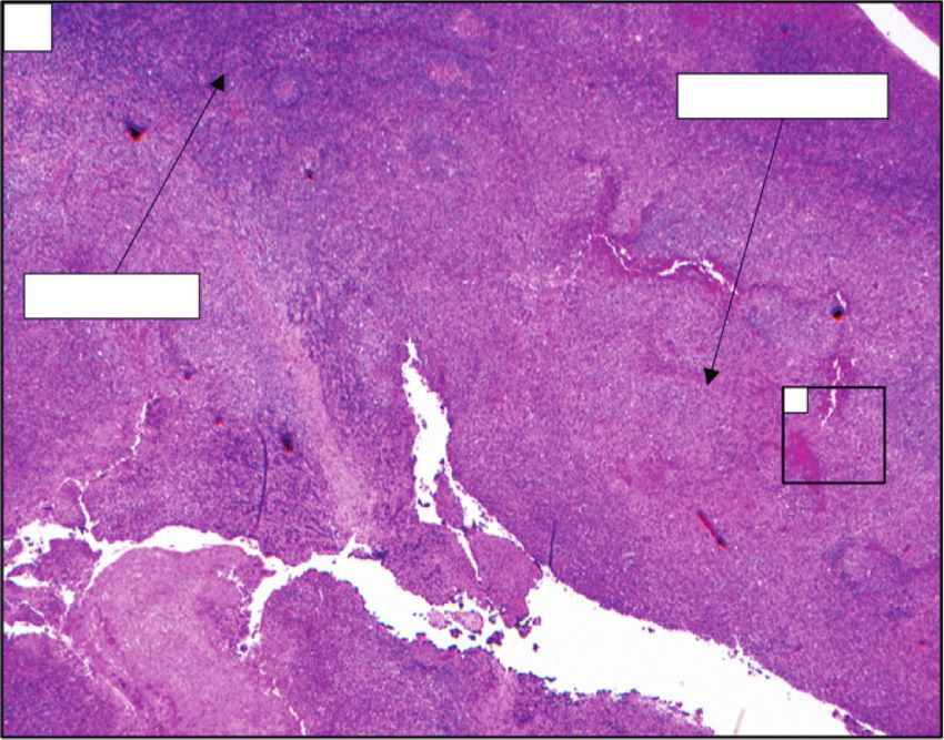
2                                                                                              Case Reports in Otolaryngology

     On day 3 of medical management, he began to spike             then thought to tip the delicate immunological balance,
fevers to 39°C and had ongoing, severe odynophagia.                allowing localised EBV-driven lymphoproliferation [5].
His lymphocytes increased to 11.9 × 10⁹/L. Due to radio-           Locations where EBV + B cells are abundant such as
logical collection and worsening pain, an emergency ton-           Waldeyer’s ring may be particularly prone to this dis-
sillectomy was subsequently performed, and the excisional          equilibrium [4, 6].
biopsy was sent for histopathological analysis.                        There remains no distinct diagnostic criteria for
     Following tonsillectomy, the patient remained in con-         EBVMCU and therefore requires correlation of clinical,
siderable postoperative pain, for which he was prescribed          histopathological, and immunophenotypic findings. Pa-
further dexamethasone. He was discharged on postoperative          tients generally present with localised, well-circumscribed
day 2.                                                             superficial ulcerations, with absence of a mass lesion. Serum
     Histopathology (Figure 1) demonstrated several shallow,       generally demonstrates a lymphocytosis and evidence of
sharply circumscribed ulcers with the ulcer base showing           EBV infection. Polymerase chain reaction (PCR) quantifi-
some larger, atypical cells including smudged cells with           cation of EBV-DNA in peripheral blood may also be useful,
variable nuclear size and shape. Immunohistochemistry              and the diagnosis of EBVMCU is questioned if high titres are
demonstrated positivity for CD-20, EBER, CD-30, MUM-1,             isolated [7].
and PAX-5. The slides were sent for expert opinion, and the            Microscopically, the superficial mucocutaneous lesions
diagnosis of EBV mucocutaneous ulcers confirmed.                    demonstrate a well-demarcated base with infiltration of
     At 6 months, there was no evidence of disease recurrence.     inflammatory cells, in particular reactive T lymphocytes.
                                                                   Histologically, the monoclonal EBVMCU immunoblasts
3. Discussion                                                      stain positive for CD-20, CD-30, EBER-1, MUM-1, OCT-2,
                                                                   and PAX-5. There is variability in staining for BCL-6, CD-
Epstein–Barr virus (EBV) mucocutaneous ulcer (MCU) is a            15, CD-45, and CD-79a. Usually, atypical Reed–Sternberg-
rare but underdiagnosed condition, presenting as solitary,         like cells demonstrating CD-15 and CD-30 coexpression
sharply demarcated ulcerations of the oral cavities, gastro-       while retaining PAX-5 positivity will be present in the
intestinal tract, and skin [1]. The disease is a relatively new    polymorphic infiltrate. The dense reactive lymphocytic in-
clinicopathological entity since its inclusion in the 2016 World   filtrates on the periphery of the lesion are usually rich in
Health Organisation (WHO) classification of lymphoid                CD3+ T cells [1–4].
neoplasms [2]. Due to its many histopathological similarities,         The distinction between EBVMCU and EBV + DLBCL is
it has previously been undifferentiated from the EBV-asso-          vital in order to prevent exposure of these patients from
ciated lymphoproliferative disorders (LPDs), in particular         unnecessary exposure to chemotherapeutic agents. Man-
EBV + diffuse large B-cell lymphoma (EBV + DLBL). This              agement of EBVMCU overwhelmingly favours conservative
distinction was made and is of particular importance given the     management strategies. In the age-related population,
drastic differences in outcomes favouring conservative man-         EBVMCU generally takes a self-limiting, indolent course
agement in EBVMCU in contrast to EBV + DLBCL [3, 4].               (96.6% spontaneous remission), while in those iatrogenically
These characteristics are summarised in Table 1.                   suppressed, a reduction in immunosuppression is usually
    The median age of patients with EBVMCU is 68.5 years,          sufficient (94.1%) to induce remission [5, 6]. Importantly,
with a female predilection (60%) [3]. A vast majority (90%)        the remainder can represent a progressive and debilitating
of cases are unifocal, most commonly involving in the              condition, necessitating aggressive medical therapy or ex-
mouth/oral cavity (58%), gastrointestinal tract (20%), skin        cision [8].
(19%), and rarely the sinonasal cavity (3%).                           The initial presentation was diagnosed and treated as a
    Almost all cases of EBVMCU reported to date possess at         presumed viral tonsillitis with bacterial superinfection. The
least one of the WHO-defined, predisposing risk factors of          patient discussed in the above report did not have any of the
immunosuppression (medication-induced, age-related                 WHO-defined risk factors for EBVMCU. Although he had a
immunosenescence, and primary and acquired immuno-                 moderate smoking history, the patient was otherwise young,
deficiency disorders) [3], which is proposed to contribute          fit, and healthy. Following histopathological diagnosis, the
significantly to its pathogenesis.                                  patient was counselled and referred for HIV screening and
    The pathogenesis of EBVMCU is not fully established.           immunoglobulin analysis. The patient did not follow-up on
After initial infection at an early age, EBV will continue to      this referral. However, his pretest probability was otherwise
infect B cells of most adults. Through complex mechanisms,         low. He had no other source of immunosuppression other
the virus then elicits the transformation and proliferation of     than 3 days of inpatient treatment with dexamethasone for
B cells. Physiologically, this characteristic propensity for of    severe pharyngitis. It is possible that the patient had
EBV to induce the proliferation of B cells is balanced by          EBV + pharyngitis, with the delicate immunomodulatory
complex immunologic interactions which are effective to             balance tipped in favour of EBV by this iatrogenic immu-
maintain EBV-infected cells at very low levels in immu-            nosuppression. The use of short courses of corticosteroids is
nocompetent individuals [5]. It is speculated either age or        recommended by current Australian guidelines for phar-
medication reduces immune surveillance to a level which is         yngitis unresponsive to simple analgesia [9] and is supported
only just sufficient to maintain the virus in its dormant state      by high quality evidence [10]. However, the decision should
[6]. Further exposure to an immunomodulating factor is             be balanced against its potential for immunosuppression.
Case Reports in Otolaryngology 3
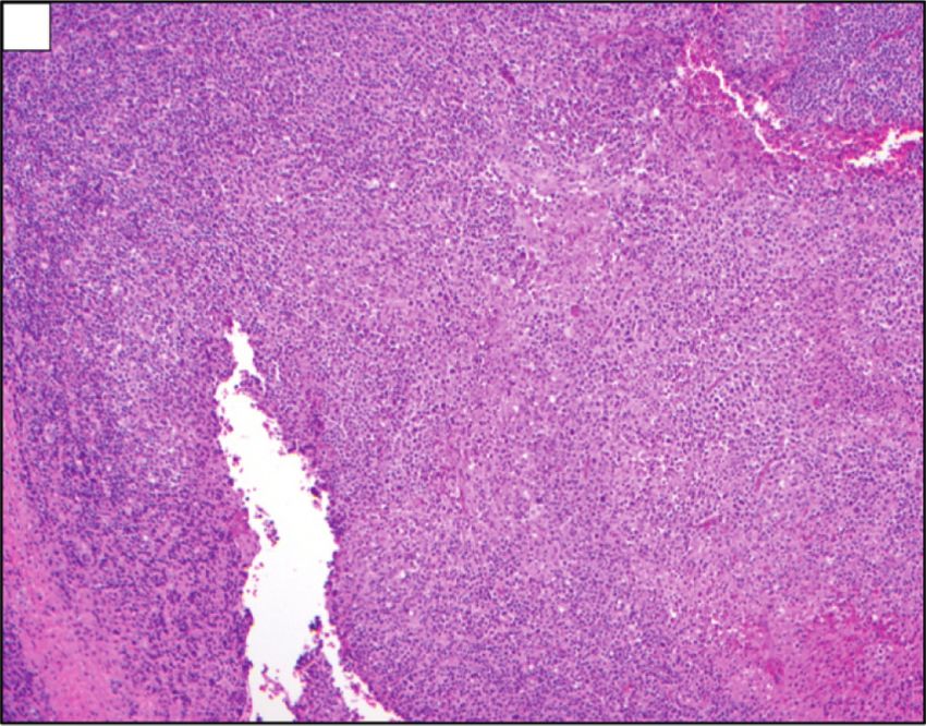
(a) (b)
Area of ulceration
Normal tonsil
B
(c) (d)
Larger immunoblasts
Centre of ulcer
(e) (f)
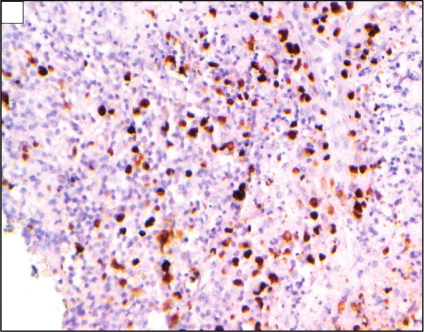
Figure 1: Histopathology demonstrating the normal tonsillar tissue with areas of deep ulceration at 4x (a) and 10x (b) magnification. At 40x
magnification, the large immunoblasts and the occasional Reed–Sternberg-like cells are demonstrated in the centre of the ulcer (c). The
larger immunoblasts demonstrated positive staining for CD-30 (d), EBERish (e), and MUM-1 (f ).
Table 1: Differentiating characteristics of EBV +ve DLBCL and EBVMCU.
EBV +ve DLBCL EBVMCU
Usually age-related immune somnolence or young patients
Population Usually elderly patients
with iatrogenic immunosuppression
Macroscopic Usually associated with mass lesion Sharply demarcated ulcers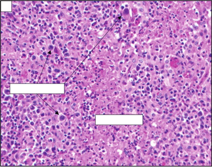
4                                                                                                  Case Reports in Otolaryngology

                                                        Table 1: Continued.
                                   EBV +ve DLBCL                                               EBVMCU
                 Mixed inflammatory infiltrate of lymphocytes, plasma     Mixed inflammatory infiltrate of lymphocytes, plasma cells,
                          cells, histiocytes, and eosinophils                         histiocytes, and eosinophils
Histopathology
                               EBV +ve large cells only                         Band of small T cells at the base of ulcer
                                                                                    EBV +ve in variety of cell sizes
Growth                               Aggressive                                                 Limited
Prognosis                               Poor                                                   Favourable
Treatment                     Chemoradiotherapy, surgical                                 Usually conservative

4. Conclusion                                                       [3] R. B. Sinit, K. L. Horan, R. K. Dorer, and D. M. Aboulafia,
                                                                        “Epstein-barr virus-positive mucocutaneous ulcer: case report
EBVMCU is a rare and newly described disorder. Given that               and review of the first 100 published cases,” Clinical Lym-
the condition can masquerade as conventional tonsillitis, it            phoma Myeloma and Leukemia, vol. 19, no. 2, pp. e81–e92,
may be commonly overlooked or misdiagnosed. It should be                2019.
included in the differential for severe tonsillitis, particularly    [4] J. K. Au, J. W. Said, A. R. Sepahdari, and M. A. John, “Head
                                                                        and neck Epstein-Barr virus mucocutaneous ulcer: case report
in those unresponsive to conventional medical management.
                                                                        and literature review,” The Laryngoscope, vol. 126, no. 11,
The use of dexamethasone in tonsillitis should be a carefully           pp. 2500–2504, 2016.
considered decision made in conjunction with a serological          [5] S. D. Dojcinov, G. Venkataraman, M. Raffeld, S. Pittaluga, and
testing.                                                                E. S. Jaffe, “EBV positive mucocutaneous ulcer-a study of 26
                                                                        cases associated with various sources of immunosuppression,”
Data Availability                                                       American Journal of Surgical Pathology, vol. 34, no. 3,
                                                                        pp. 405–417, 2010.
The data used to support the findings of this study are              [6] Y. Natkunam, J. R. Goodlad, A. Chadburn et al., “EBV-
available from the corresponding author upon request.                   positive B-cell proliferations of varied malignant potential,”
                                                                        American Journal of Clinical Pathology, vol. 147, no. 2,
                                                                        pp. 129–152, 2017.
Additional Points                                                   [7] M. Hart, B. Thakral, S. Yohe et al., “EBV-positive mucocu-
                                                                        taneous ulcer in organ transplant recipients,” American
Summary. (i) Epstein–Barr Virus (EBV) mucocutaneous ulcer               Journal of Surgical Pathology, vol. 38, no. 11, pp. 1522–1529,
(MCU) is a newly described, rare but underdiagnosed con-                2014.
dition. (ii) The condition has previously been reported only in     [8] T. K. Roberts, X. Chen, and J. J. Liao, “Diagnostic and
immunosuppressed individuals. (iii) EBVMCU can present as               therapeutic challenges of EBV-positive mucocutaneous ulcer:
a mimic of conventional tonsillitis. (iv) EBVMCU can occur as           a case report and systematic review of the literature,” Ex-
a result of acute steroid administration in an otherwise                perimental Hematology & Oncology, vol. 5, p. 13, 2016.
nonimmunosuppressed population. (v) In EBV +ve tonsillitis          [9] eTG Complete in Symptomatic Treatment for Sore Throat,
patients, the use of dexamethasone and other immunosup-                 Therapeutic Guidelines Ltd., West Melbourne, Australia,
                                                                        2019.
pressive agents should be carefully a considered decision. (vi)
                                                                   [10] B. Sadeghirad, R. A. C. Siemieniuk, R. Brignardello-Petersen
The use of EBV serology can aid in this decision-making.                et al., “Corticosteroids for treatment of sore throat: systematic
                                                                        review and meta-analysis of randomised trials,” BMJ, vol. 358,
Conflicts of Interest                                                   Article ID j3887, 2017.

The authors declare no conflicts of interest.

Acknowledgments
The authors would like to acknowledge Dr. Amalika Edir-
isinghe for her assistance with the interpretation and ac-
quisition of histopathological imaging.

References
 [1] U. Sundram, “Cutaneous lymphoproliferative disorders:
     what’s new in the revised 4th edition of the World Health
     Organization (WHO) classification of lymphoid neoplasms,”
     Advances in Anatomic Pathology, vol. 26, no. 2, pp. 93–113,
     2019.
 [2] L. Quintanilla-Martinez, “The 2016 updated WHO classifi-
     cation of lymphoid neoplasias,” Hematological Oncology,
     vol. 35, no. S1, pp. 37–45, 2017.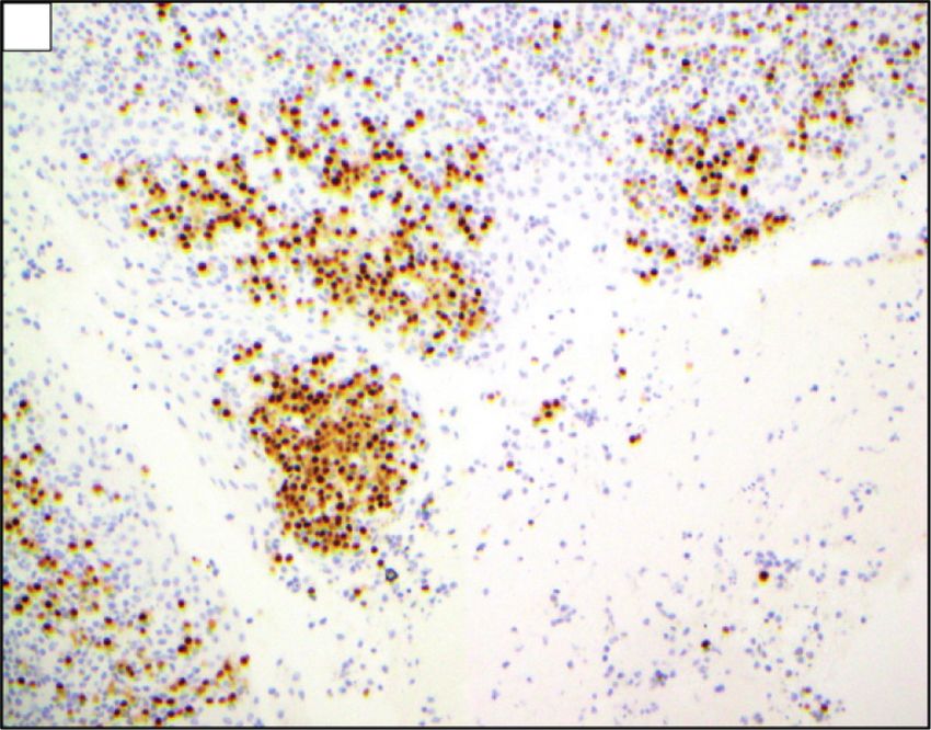
You can also read