Case Report Parapharyngeal teratoma in an adult: a case report and literature review
←
→
Page content transcription
If your browser does not render page correctly, please read the page content below

Int J Clin Exp Med 2019;12(1):1319-1322
www.ijcem.com /ISSN:1940-5901/IJCEM0074855

Case Report
Parapharyngeal teratoma in an adult: a case
report and literature review
Yu Cui1, Zhan-Peng Zhu2*, Li-Rong Bi3, Wan-Zhong Yin1*

Departments of 1Otolaryngology Head and Neck Surgery, 2Neurosurgery, 3Pathology, The First Hospital of Jilin
University, Changchun, Jilin, P. R. China. *Equal contributors.
Received February 23, 2018; Accepted October 9, 2018; Epub January 15, 2019; Published January 30, 2019

Abstract: We report an unusual case of a 61-year-old Chinese man who presented with a 15 years of foreign body
sensation and unclear speech, and 1 year of dysphagia. Radiographic imaging revealed a large space occupying
lesion of mixed fat density in the left parapharyngeal space, consistent with a teratoma. The mass was completely
resected with an intact capsule and characterized by a soft, irregular shape, and local bone-like stiffness. The
diagnosis of teratoma was confirmed by pathological examination. Microscopic examination showed a mesoderm
component, containing diffuse mature adipose tissue with distribution of sparse fibrous bands and mature bone
tissue with mature cortical bone and marrow. During 1 year of follow-up, the symptoms of pharyngeal foreign body
sensation and difficulty swallowing disappeared, with no hoarseness or aural fullness.

Keywords: Teratoma, parapharyngeal space, CT scan, pathological examination, surgical resection

Introduction                                                 foreign body sensation and unclear speech for
                                                             15 years as well as difficulty swallowing for 1
The incidence of teratoma is about one per                   year. A parapharyngeal mass was found on the
4,000 live births each year, only 3%-10% occur               left side. He had been treated by swelling punc-
in the head and neck region [1-4]. It most com-              ture and incision 15 years previously, and about
monly appears in the neck followed by naso-                  200 ml of bloody fluid had been removed. The
pharynx. It can be divided into three categories:            subjective symptoms were relieved, and the
mature teratoma, immature teratoma, and                      patient did not receive further treatment. One
malignant teratoma. The clinical manifesttions               year previously, the self-reported feeling of a
of benign teratoma are usually foreign body                  pharyngeal foreign body was aggravated again,
sensation and affected pronunciation. Head                   accompanied by difficulty swallowing and mild
and neck CT scan can help to locate and diag-                left aural fullness. The patient had no fever,
nosis the tumor, that is, a lesion can be highly             headache, sore throat, dyspnea, laryngeal
suspected of being a teratoma when the tumor                 spasm, or hoarseness. He also had no history
presents with a clear boundary and mixed sig-
                                                             of hypertension, diabetes, or heart disease.
nal involving both fat and calcification. The
therapeutic principle for a benign teratoma in               Physical examination revealed that the soft pal-
the parapharyngeal space is complete resec-                  ate and lateral wall of the pharynx on the left
tion. This study reports a case of a mature tera-            side were upheaved obviously close to the mid-
toma in the parapharyngeal space treated in                  line, and the uvula was slightly tilted to the
our department, with a review of the anatomi-                right. The mass extended from the top of the
cal location, pathology characteristics, clinical            nasopharynx to the lower level of the tonsil and
manifestation, imaging examination, and treat-               was soft with no tenderness. The left eardrum
ment principles of parapharyngeal space tera-
                                                             was compressed, but not perforated. The right
toma.
                                                             eardrum was normal. The results of a bilateral
Case report                                                  Rinne test were: air conduction > bone conduc-
                                                             tion; those on the Weber test were: left; pure
A 61-year-old male patient was admitted to our               tone listening examination showed mildly con-
hospital complaining of a feeling of swallowed               ductive deafness in the left ear. Enhanced com-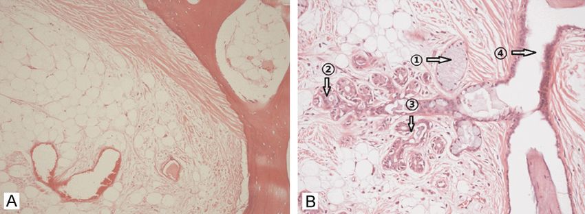
Adult mature parapharyngeal teratoma

Figure 1. CT scans of the lesion before surgery. Axial (A), sagittal (B) and coronal (C) demonstrated a large space
occupying lesion (arrow) with mixed fat density in the left pharyngeal space, considered a teratoma.

                                                                                 After suturing the cervical
                                                                                 incision, a small amount of
                                                                                 blood clot was observed in
                                                                                 the mouth. After the blood
                                                                                 clot was absorbed, a small
                                                                                 break about 1 cm long on the
                                                                                 lateral side of the left palatal
                                                                                 tongue arch was sutured.
                                                                                 During follow-up, the symp-
                                                                                 toms of pharyngeal foreign
                                                                                 body sensation and difficulty
                                                                                 swallowing disappeared, with
                                                                                 no hoarseness or aural
Figure 2. The relationship of the tumor and the artery. CT scans showing a       fullness.
mixed density, space occupying lesion (arrow) located on the left side of the
pharynx, pushing the left internal and external carotids to the left rear.      Microscopic observation sho-
                                                                                wed a mesoderm component,
puted tomography (CT) of the neck revealed a                  containing diffuse mature adipose tissue with
large space occupying lesion of mixed fat den-                distribution of sparse fibrous bands and mature
sity in the left pharyngeal space, consistent                 bone tissue with mature cortical bone and mar-
with a teratoma (Figure 1). The cervical vascu-               row (Figure 4A). The entoderm component had
larized enhanced CT (CTA) indicated a mixed                   complete salivary glands, including mucinous
density occupying lesion on the left side of the              gland vesicles, serous gland vesicles, mixed
pharynx, pushing the left internal and external               adenoids, and ducts (Figure 4B). The diagnosis
carotids to the left rear (Figure 2).                         of mature teratoma was made.

After complete preoperative examination, the                  Discussion
patient underwent resection of the left pharyn-
geal space neoplasm under general anesthesia                  The paralopharyngeal space is a potential gap
from the external cervical approach (Figure                   presenting as an inverted cone-shape between
3A). The mass envelope was complete. The                      the medial pterygoid, the deep parotid gland,
tumor was separated along the envelope care-                  and lateral pharyngeal wall. The parapharyn-
fully, and no adhesion was found between the                  geal space extends from the temporal and
mass and internal jugular artery and vein. After              sphenoid bones of the skull base down to the
cutting off part of the styloid process, the mass             hyoid bone. Its medial boundary is the upper
was completely removed with an intact capsule                 pharyngeal constrictors, and its lateral bound-
and characterized by a soft, irregular shape,                 ary is the mandibular ramus. It is located after
and local bone-like stiffness (Figure 3B).                    the cervical vertebrae and before the pterygo-

1320                                                                Int J Clin Exp Med 2019;12(1):1319-1322
Adult mature parapharyngeal teratoma

                                                                                   teratomas are composed of
                                                                                   undifferentiated tissue and
                                                                                   usually have a high potential
                                                                                   for malignancy [8]. Mature
                                                                                   teratomas are also referred to
                                                                                   as dermoid cysts, which usu-
                                                                                   ally have a smooth and intact
                                                                                   envelope. Teratoma is a true
                                                                                   neoplasm that may include
                                                                                   tissues from all three blasto-
                                                                                   derms and develop indepen-
                                                                                   dently of the host. Histo-
                                                                                   pathologically, the capsule
Figure 3. Imaging of the tumor. Intraoperative image of the tumor with intact      wall contains squamous epi-
membrane (A). The mass was completely removed with an intact capsule
                                                                                   thelium and its appendages,
and characterized by a soft, irregular shape, and local bone-like stiffness (B).
                                                                                   such as hair follicles, seba-
                                                                                   ceous glands, and sweat
                                                                                   glands [9, 10]. In addition,
                                                                                   capsule contents contain
                                                                                   mature nerve tissue derived
                                                                                   from ectoderm, such as glial
                                                                                   tissue, as well as fat, carti-
                                                                                   lage, and smooth muscle tis-
                                                                                   sues from mesoderm and
                                                                                   ectoderm. They are consid-
                                                                                   ered to grow from misplaced
                                                                                   embryonic, pluripotent germ
Figure 4. H&E staining of the tumor slice. (hematoxylin and eosin staining         cells that lose impact during
×100) Mesoderm component containing mature adipose tissue diffused                 embryologic development [4].
distributing sparse fibrous bands and mature bone tissue with mature corti-
cal bone and marrow (A). (hematoxylin and eosin staining ×200) Ectoderm
component showing complete ① mucinous gland vesicles, ① serous gland                Most teratomas reported in
vesicles, ③ mixed adenoids and ④ ducts (B).                                         the literature occurred in the
                                                                                    sacrococcygeal region, gonad,
                                                                 and other sites such as the mediastinum and
mandibular raphe. It is divided into the presty-                 retroperitoneum. Most teratomas occurring in
loid and poststyloid spaces bound by the sty-                    the pediatric populations are found within the
loid process and its adhesion structures, such                   pelvis and are typically benign in nature. Head
as the stylohyoid muscle and stylopharyngeal                     and neck teratomas occurring in newborns and
muscle. A tumor occurring in parapharyngeal                      infants mainly causes respiratory distress [11],
space is rare, accounting for about 0.5% of                      and those in adults may present as asymptom-
head and neck tumors [5]. The location of a                      atic throat and neck neoplasms. Cases of huge
tumor in different compartments of this space                    and benign tumors are usually associated with
usually indicates the potential histologic etiolo-               sleep apnea and difficulty breathing and eat-
gies of the lesion. A tumor in the prestyloid                    ing. To date, fewer than 40 cases of head and
space is often derived from a salivary gland,                    neck teratomas in adults have been reported in
such as the parotid pleomorphic adenoma [6].                     the literature. In the present case, the diagno-
Tumors in the poststyloid space are mostly                       sis and treatment of the patient was a process
paraganglioma, schwannoma, and other rare                        lasting more than 10 years, and the tumor
malignant lesions.                                               caused obvious pharynx foreign body sensa-
                                                                 tion and affected his pronunciation, which con-
Teratomas originate from totipotential stem                      formed to the clinical manifestations of a
cells [7]. According to their histological fea-                  benign teratoma.
tures, they can be divided into three categories:
mature teratoma, immature teratoma, and                          Head and neck CT examination can help to
malignant teratoma. Immature and malignant                       locate a tumor in the pharynx. In addition, the

1321                                                                   Int J Clin Exp Med 2019;12(1):1319-1322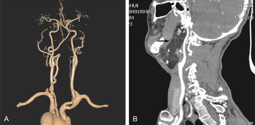
Adult mature parapharyngeal teratoma

CT manifestation of a teratoma has important        Address correspondence to: Dr. Wan-Zhong Yin,
diagnostic value; that is, a lesion can be highly   Department of Otolaryngology Head and Neck
suspected of being a teratoma when the tumor        Surgery, The First Hospital of Jilin University, 71
presents with a clear boundary and mixed sig-       Xinmin Street, Chaoyang District, Changchun
nal involving both fat and calcification [4].       130021, Jilin, P. R. China. Tel: +86-15804300621;
However, lipoma and liposarcoma with radio-         E-mail: yinwanzhong88@hotmail.com; Zhan-Peng
logic evidence of fat lesions should be carefully   Zhu, Department of Neurosurgery, The First Hospital
distinguished. Intravenous contrast is also         of Jilin University, Changchun, Jilin, P. R. China.
commonly used together with CT to help dem-         E-mail: 282324491@qq.com
onstrate the displacement of the carotid artery.
A teratoma typically presents local low attenua-    References
tion representing fat, foci of high attenuating
                                                    [1]  Gullane PJ, Lampe HB and Slinger R. Erosive
calcifications, and diffuse heterogeneous areas
                                                         parapharyngeal space teratoma. J Otolaryngol
with soft tissue densities. Moreover, since mag-         1986; 15: 317-321.
netic resonance imaging (MRI) provides better       [2] Handler SD and Raney RB Jr. Management of
soft tissue resolution in the head and neck, it          neoplasms of the head and neck in children. 1.
has more advantages for clarifying the surgical          Benign tumors. Head Neck Surg 1981; 3: 395-
resection range compared with CT.                        405.
                                                    [3] Holt GR, Holt JE and Weaver RG. Dermoids and
Conclusions                                              teratomas of the head and neck. Ear Nose
                                                         Throat J 1979; 58: 520-531.
The therapeutic principle for a benign teratoma     [4] Smirniotopoulos JG and Chiechi MV. Terato-
in the pharyngeal space is complete resection.           mas, dermoids, and epidermoids of the head
An intraoral or transmandibular approach can             and neck. Radiographics 1995; 15: 1437-
be selected according to the infiltration of the         1455.
tumor site and the surrounding tissues involved.    [5] Hughes KV 3rd, Olsen KD and McCaffrey TV.
                                                         Parapharyngeal space neoplasms. Head Neck
The parapharyngeal space tumor specimen
                                                         1995; 17: 124-130.
should not be taken out through the mouth to
                                                    [6] Shin JH, Lee HK, Kim SY, Choi CG and Suh DC.
avoid damage to blood vessels and nerves. In             Imaging of parapharyngeal space lesions: fo-
addition, it may destroy the integrity of the            cus on the prestyloid compartment. AJR Am J
membrane, leading to the dissemination of                Roentgenol 2001; 177: 1465-1470.
tumor cells. This patient had received intraoral    [7] Benson RE, Fabbroni G and Russell JL. A large
incision drainage several years previously,              teratoma of the hard palate: a case report. Br J
which was risky. Moreover, the local integrity           Oral Maxillofac Surg 2009; 47: 46-49.
was damaged, resulting in mucosal damage in         [8] Tobias S, Valarezo J, Meir K and Umansky F.
the scar adhesion during resection. The trans-           Giant cavernous sinus teratoma: a clinical ex-
cervical approach has the advantages of full             ample of a rare entity: case report. Neurosur-
                                                         gery 2001; 48: 1367-1370; discussion 1370-
exposure and reducing blood vessel and nerve
                                                         1361.
damage. Therefore, it is frequently used for the
                                                    [9] Mamoon N, Jaffri SA, Ilahi F, Muzaffar K, Iqbal
detection, diagnosis, and treatment of benign            Y, Akhter N, Nasir H and Ahmad IN. Yolk sac
tumors in the parapharyngeal space. The range            tumour arising in mature teratoma in the para-
of resection should be expanded when the                 pharyngeal space. J Pak Med Assoc 2011; 61:
tumor presents malignant potential. Once diag-           1025-1027.
nosed with an immature or a malignant terato-       [10] Punch GE, Sniezek JC, Berkey BD and Peter-
ma, patients commonly need postoperative                 mann GW. A benign, mature, parapharyngeal
adjuvant radiotherapy and chemotherapy. Our              teratoma presenting in an adult. Radiol Case
patient showed no evidence of recurrence after           Rep 2007; 2: 46.
1 year of follow-up.                                [11] Saing H, Lau WF, Chan YF and Chan FL. Para-
                                                         pharyngeal teratoma in the newborn. J Pediatr
Disclosure of conflict of interest                       Surg 1994; 29: 1524-1525.

None.

1322                                                      Int J Clin Exp Med 2019;12(1):1319-1322
You can also read