AVIAN MEDICINE: PRINCIPLES AND APPLICATION - ANATOMY OF THE UMBRELLA COCKATOO ACETATE OVERLAY STRUCTURE IDENTIFICATION SYSTEM
←
→
Page content transcription
If your browser does not render page correctly, please read the page content below
ANATOMY OF THE
     UMBRELLA COCKATOO

 ACETATE OVERLAY STRUCTURE
   IDENTIFICATION SYSTEM
                 from

AVIAN MEDICINE:
PRINCIPLES AND APPLICATION

  RITCHIE, HARRISON AND HARRISON

         © 1994 Wingers Publishing
         Lake Worth, Florida 33463
               800-946-4782
            FAX (407) 641-0234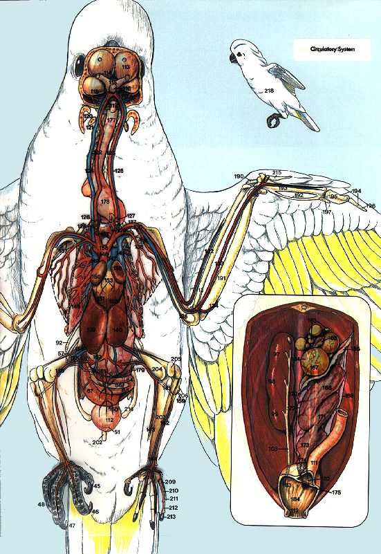
Anatomy of the
Umbrella Cockatoo
Anatomic Illustrations by
Lesley E. Sealing
n artist’s concept of the lateral and ventrodorsal
A views of the anatomy of a cockatoo are presented
in a clear overlay system. When two or more
pages are viewed together, the drawings represent the
relative position and relationship of important anatomic
structures. By inserting white paper behind a page, that
page can be viewed separately.
This format is particularly useful as a reference to
general anatomic sites during radiographic, endoscopic
or necropsy evaluation of companion birds. Additionally,
the format should provide an easy-to-visualize method
for discussing a patient’s problem and management
techniques with clients.
Most of the illustrations were developed from dissec-
tions and radiographs. The primary radiographic model
was an average-sized female Umbrella Cockatoo. The
bird was believed to be a normal, healthy individual
based on clinical assessment, diet evaluation, hema-
tologic and biochemical laboratory test values, radio-
graphic interpretations and results of bacteriologic,
parasitic, chlamydial and viral testing. Other illustra-
tions, such as those of the central and peripheral nerv-
ous systems, were adapted from the domestic fowl, and
parameters for the cervicocephalic air sac system and
sinuses were adapted from descriptions in the literature
combined with dissections of cockatoos. Organ size and
location should be expected to vary with the species as
well as with the gender, age, reproductive status,
prandial state and presence of disease condi-
tions in individual birds. Additionally, artistic
liberties were taken in order to depict the
most logical representation of the sys-
tems. To simplify the illustrations,
some body parts are shown on
only one side of the figure.
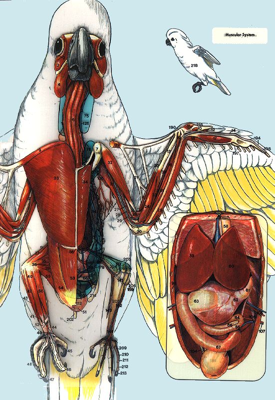
1. Forehead 16. Alular remiges
2. Cere 17. Ventral antebrachial
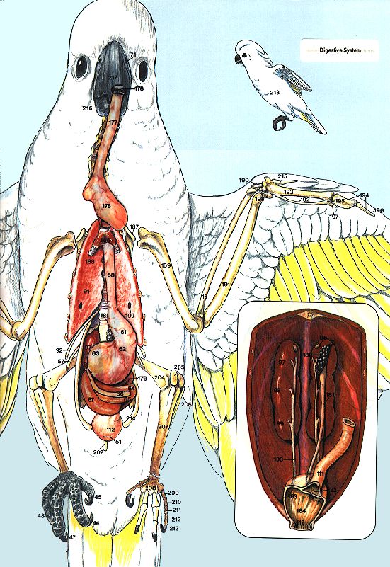
3. Naris coverts
4. Rhinotheca 18. Ventral minor coverts
5. Gnathotheca 19. Ventral major coverts
6. Lore 20. Primary remiges (10)
7. Crest 21. Secondary remiges (12)
8. Crown 22. Thigh
9. Postorbital region 23. Shank lateral region (leg)
10. Maxillary and mandibular 24. Tarsometatarsus
malar region (cheek) 25. Shoulder region
11. Dorsal neck region 26. Dorsal major caudal coverts
12. Submalar region (chin) 27. Prolatal region
13. Ventral neck region (throat) 28. Shank anterior region
14. Lateral neck region 29. Postventer region
15. Proventer region (breast) 30. Tail, ventral region
31. Rectrices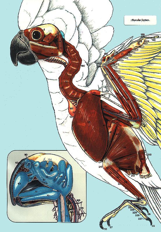
Lateral View
Muscular System 60. A. and V. metatarsalis 105. Ulnar artery 166. Lumbar plexus
dorsalis 106. Left thyroid gland 167. Sacral plexus
1. M. orbicularis palpebrarum 61. M. gastrocnemius pars later- 107. Left parathyroid gland 168. Pudendal plexus
2. Sclerotic ossicle alis 108. Left ultimobranchial gland 169. Caudal plexus
3. M. adductor mandibulae 109. Right brachiocephalic trunk 170. Ischiatic nerve (branches to
■ Inset: Infraorbital Sinus 110. Area of pectoralis muscle fibular and tibial nerves)
externus
4. External ear canal and Cervicocephalic Air 111. Left cranial vena cava 171. Intestines
5. M. branchiomandibularis Sac 112. Pulmonary trunk 172. Pancreas
6. M. intermandibularis ven- 62. Nares 113. Auricle of left atrium 173. Obturator nerve
tralis (mylohyoideus) 63. Rostral diverticulum of in- 114. Left pulmonary veins 174. Femoral nerve
7. Zygomatic arch fraorbital sinus (IS) 115. Left pulmonary arteries
116. Right hepatic portal veins ■ Inset: Cut-away of Skull
8. M. rectus capitis ventralis 64. Maxillary chamber of IS
pars lateralis 65. Preorbital diverticulum of IS 117. Left lobe of liver 175. Cranial nerve III
9. M. rectus capitis ventralis 66. Infraorbital diverticulum of IS 118. Thoracic aorta 176. Cranial nerve IV
pars medialis 67. Suborbital chamber of IS 119. Left pectoral artery and vein 177. Cranial nerve V
10. M. longus colli ventralis 68. Postorbital diverticulum of IS 120. Intercostal arteries 178. Cranial nerve VI
11. M. sternohyoideus 69. Preauditory diverticulum of IS 121. Celiac artery 179. Cranial nerve VII
12. M. biventor cervicis 70. Mandibular diverticulum of IS 122. Cranial mesenteric artery 180. Cranial nerve IX
13. M. tensor propatagialis 71. Cranial portion 123. Caudal vena cava 181. Cranial nerve X
14. M. flexor alulae cervicocephalic air sac 124. Left external iliac artery 182. Cranial nerve XI
15. M. abductor alulae 72. Cervical portion 125. Left femoral artery 183. Cranial nerve XII
16. M. adductor alulae cervicocephalic air sac 126. Left internal iliac artery 184. Supraorbital nerve
17. Radius 127. Median caudal artery 185. Lacrimal gland nerve
18. M. flexor digitorum 128. Left external iliac vein 186. Sphenopalatine ganglion
Respiratory System 129. Left ischiatic vein 187. Chorda tympani
superficialis
19. M. flexor digitorum 130. Left ischiatic artery 188. Nasopalatine nerve
73. Larynx
profundus 131. Left internal iliac vein
74. Laryngeal mound
20. Ulna 75. Cervical air sac
132. Left caudal tibial artery Skeletal
21. M. interosseus ventralis 133. Left cranial tibial artery and Urogenital System
76. Trachea
22. Fascial plane overlying M. 134. Left dorsal metatarsal artery
77. Pneumatic diverticulum of
flexor digiti minoris clavicular air sac into clavicle ■ Inset: Digestive 189. Vertebral canal
23. M. ulnometacarpalis dorsalis 78. Pneumatic diverticulum of 190. Cervical vertebra
24. M. ulnometacarpalis ventralis Portion of Head
clavicular air sac into scapula 191. Clavicle
25. M. extensor longus digiti ma- 135. Palatine salivary glands (me- 192. Radial carpal bone
79. Pneumatic diverticulum of
joris dial) 193. Alular digit
clavicular air sac into coracoid
26. M. extensor metacarpi ra- 136. Angularis oris salivary 194. Major metacarpal bone
80. Pneumatic diverticulum of
dialis glands 195. Minor metacarpal bone
clavicular air sac into
27. M. pronator profundus 137. Maxillary salivary glands 196. Major digit
humerus (light blue)
28. M. pronator superficialis 138. Roof of oropharynx 197. P1
81. Clavicular air sac (blue)
29. M. flexor carpi ulnaris 139. Tongue 198. P2
82. Pneumatic diverticulum of
30. M. brachialis 140. Rostral mandibular salivary 199. Minor digit
clavicular air sac into
31. A. radialis glands 200. Ulnar carpal bone
sternum (light blue)
32. A. ulnaris 141. Lingual salivary glands 201. Humerus (partially shown)
83. Cranial thoracic air sac (stri-
33. V. basilica 142. Caudal mandibulary sali- 202. Thoracic vertebra
ated)
34. M. triceps brachii vary glands 203. Synsacrum
84. Caudal thoracic air sac
35. M. biceps brachii 143. Choanae (not seen) 204. Cranial division of kidney
(light blue)
36. Medianoulnaris nerve 144. Sphenopterygoid salivary 205. Middle division of kidney
85. Ventral hepatic peritoneal
37. Clavicle (furcula) glands 206. Caudal division of kidney
cavity (stippled)
38. M. pectoralis superficialis 145. Esophageal opening 207. Vertebral ribs
86. Pneumatic diverticulum of
39. Keel projecting from ster- 146. Cricoarytenoid salivary 208. Uncinate process
abdominal air sac into fe-
num glands 209. Sternal ribs
mur (light blue)
40. M. serratus superficialis 147. Esophagus 210. Caudal vertebrae
87. Abdominal air sac (blue)
41. M. intercostales externi 88. Lung (impression of 6th rib) 211. Pygostyle
42. M. latissimus dorsi 89. Syrinx Central Nervous System 212. Cloaca
43. M. expansor secundariorum and Digestive System 213. Rectum
44. M. iliofibularis ■ Inset: Cut-away of Skull 214. Left adrenal gland
45. M. levator caudae 90. Cere 148. Cerebral hemisphere 215. Left testicle
46. M. flexor cruris medialis 91. Nasal cavity 149. Optic lobe 216. Ductus deferens
47. M. depressor caudae 92. M. genioglossus 150. Cerebellum 217. Cranial cnemial crest
48. M. iliotibialis cranialis 93. Cranial nerve II (optic) 151. Medulla oblongata 218. Ischium
49. M. iliotibialis lateralis 94. Antevestibular recess 152. Cranial nerve I 219. Fibula
50. M. pubo-ischio-femoralis 153. Pituitary gland 220. Tibiotarsus
pars lateralis 221. Pubis
51. M. tibialis cranialis
Circulatory System 154. Spinal cord
155. Cervical spinal nerve 222. Tarsometatarsal 2,3,4
52. M. fibularis longus 95. Left internal carotid artery 156. Cervical esophagus 223. P1
53. M. flexor perforans et perfo- 96. Left external carotid artery 157. Crop 224. P2
ratus digiti III 97. Left jugular vein 158. Brachial plexus 225. P3
54. M. flexor perforans et perfo- 98. Left brachiocephalic trunk 159. N. radialis 226. P4
ratus digiti II 99. Left subclavian artery 160. Thoracic esophagus 227. P5
55. M. extensor digitorum longus 100. Left axillary artery and vein 161. Intercostal spinal nerve
56. Digit 1 101. Brachial artery 162. Proventriculus
57. Digit 2 102. Superficial ulnar artery 163. Isthmus
58. Digit 3 103. Radial artery 164. Ventriculus
59. Digit 4 104. Recurrent ulnar artery 165. Medianoulnar nerve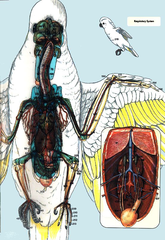
winga1.tif
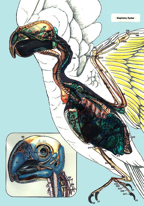
winga1_.tif
winga2.tif
winga2_.tif
winga3.tif
winga3_.tif
winga4.tif
winga4_.tif
winga5.tif
Ventrodorsal View
Muscular System ■ Inset: Gastrointestinal Tract 107. Left ischiatic artery and vein ■ Inset: Mature Ovary
58. Thoracic esophagus 108. Caudal mesenteric artery 163. Immature follicles
1. M. orbicularis palpebrarum 59. Right lobe of liver and vein 164. Mature follicle
2. Sclerotic ossicle 60. Left lobe of liver 109. Left internal iliac artery 165. Post-ovulatory follicle (calix)
3. M. adductor mandibulae 61. Proventriculus 110. Median caudal artery 166. Infundibulum
externus 62. Isthmus 111. Rectum 167. Stigma
4. M. intermandibularis (mylo- 63. Ventriculus 112. Cloaca 168. Magnum
hyoideus) 64. Descending duodenum 169. Dorsal ligament of oviduct
5. Zygomatic arch 65. Ascending duodenum Circulatory and Central 170. Oviductal blood vessels
6. M. sternohyoideus 66. Pancreas Nervous System 171. Isthmus
7. Coracoid 67. Ascending loop of colon 172. Ventral ligament of oviduct
8. Clavicle (furcula) 113. Cerebral hemisphere 173. Uterus
9. M. tensor propatagialis 114. Cranial nerve II 174. Vagina
10. M. flexor alulae Respiratory System
115. Optic chiasm 175. Sphincter vaginae
11. M. abductor alulae 116. Pituitary gland
12. M. adductor alulae 68. Preorbital diverticulum of in-
fraorbital sinus (IS) 117. Cerebellum Digestive System
13. Radius 118. Optic lobe
14. M. flexor digitorum profundus 69. Infraorbital diverticulum of IS
70. Rostral diverticulum of IS 119. Spinal cord 176. Tongue
15. M. flexor digitorum 120. Mandible
superficialis 71. Nares 177. Cervical esophagus
72. Maxillary chamber of IS 121. Hyoid bone 178. Ingluvies (crop)
16. M. extensor longus digiti ma- 122. Right internal carotid artery
joris 73. Larynx 179. Supraduodenal loop
74. Mandibular diverticulum of 123. Left internal carotid artery
17. M. interosseus ventralis
IS 124. Right jugular vein ■ Inset: Immature Ovary
18. M. ulnometacarpalis dorsalis 125. Left jugular vein 180. Ovary
19. M. ulnometacarpalis ventralis 75. Cervical portion of cervico-
cephalic air sac (light blue) 126. Right thyroid gland 181. Oviduct
20. M. extensor metacarpi ra- 127. Left thyroid gland 182. Coprodeum
dialis 76. Trachea
77. Cervical air sac (blue) 128. Right parathyroid gland 183. Urodeum
21. Tensor propatagialis pars 129. Left subclavian artery and 184. Proctodeum
brevis tendon 78. Clavicular air sac (blue)
79. Pneumatic diverticulum of vein
22. Tensor propatagialis pars 130. A. and V. axillaris
longus tendon clavicular air sac into clav- Skeletal and
icle (dashed outline) 131. A. collateralis radialis
23. M. pronator profundus 132. A. and V. radialis Urogenital Systems
24. M. pronator superficialis 80. Pneumatic diverticula of cla-
vicular air sac into the cora- 133. A. and V. ulnaris
25. M. flexor carpi ulnaris 185. Cervical vertebra
coid (dashed outline) 134. A. and V. recurrent ulnaris
26. M. brachialis 186. Syrinx
81. Right cranial thoracic air 135. V. collateralis ulnaris
27. A. radialis 187. Scapula
sac (striated) 136. A. pectoralis
28. A. ulnaris 188. Primary bronchus
82. Left cranial thoracic air sac 137. Area of pectoral muscles
29. V. basilica 189. Left humerus
(striated) (dashed outline)
30. Medianoulnar nerve 190. Radial carpal bone
83. Right caudal thoracic air sac 138. Auricle of left atrium
31. M. triceps brachii 191. Ulna
(light blue) 139. Right lobe of liver
32. M. biceps brachii 192. Minor metacarpal bone
84. Left caudal thoracic air sac 140. Left lobe of liver
33. M. pectoralis superficialis 193. Major metacarpal bone
(light blue) 141. Left popliteal artery and vein
34. M. supracoracoideus 194. Major digit
85. Pneumatic diverticula of cla- 142. Left cranial tibial artery and
35. M. ambiens 195. P1
vicular air sac into the vein
36. M. femorotibialis internus 196. P2
humerus (dashed outline) 143. Left caudal tibial artery and
37. M. iliotibialis cranialis 197. Minor digit
86. Right ventral hepatic perito- vein
38. Area of M. ulnometacarpus 198. Ulnar carpal bone
neal cavity (stippled) 144. Left dorsal metatarsal ar-
ventralis 199. Ostium for caudal thoracic
87. Left ventral hepatic perito- tery and vein
39. M. pubo-ischio-femoralis air sac
neal cavity (stippled) 145. Left cranial vena cava
pars medialis 200. Synsacrum
88. Right abdominal air sac 146. Aorta
40. M. tibialis cranialis 201. Ischium
(blue) 147. Pulmonary trunk
41. M. fibularis brevis 202. Pygostyle
89. Left abdominal air sac (blue) 148. Pulmonary artery and vein
42. M. fibularis longus 203. Coccygeal vertebrae
90. Pneumatic diverticula of ab- 149. Right cranial vena cava
43. M. flexor perforans et perfo- 204. Femur
dominal air sacs into the fe- 150. Auricle of right atrium
ratus digiti III 205. Patella
mur (dashed outline) 151. Right ventricle
44. M. extensor digitorum lon- 206. Fibula
152. Left ventricle
gus ■ Inset: Male Urogenital System 207. Tibiotarsus
153. Coronary arteries and veins
45. Digit 1 208. Tarsometatarsus 2,3,4
91. Lung 154. Right femoral artery and
46. Digit 2 209. P1
92. Rib 7 vein
47. Digit 3 210. P2
93. Caudal vena cava 155. Right external iliac artery
48. Digit 4 211. P3
94. Aorta and vein
49. A. and V. metatarsalis dor- 212. P4
95. Left testicle 156. Right ischiatic artery and
salis 213. P5
96. Left adrenal gland vein
50. M. gastrocnemius (medial 214. Pubic bone
97. Cranial division of kidney 157. Right ultimobranchial gland
head) 215. Alular digit
98. Middle division of kidney 158. Right and left brachio-
51. Vent 216. Rostrum mandibulare
99. Caudal division of kidney cephalic trunks
52. Extensor retinaculum (gnathotheca)
100. Left common iliac vein 159. Left fibular artery
53. M. rectus abdominis 217. Rostrum maxillare (rhino-
101. Left external iliac artery 160. Left internal iliac artery
54. M. obliquus abdominis exter- theca)
and vein 161. Caudal mesenteric artery
nus 218. Miniature lateral perspec-
102. Right caudal renal vein and vein
55. M. flexor cruris medialis tive of ventrodorsal model.
103. Right ureter 162. Right caudal renal vein
56. Sternum 104. Right vas deferens
57. Rib 8 (dotted area = ribs and 105. Left femoral artery and vein
sternum) 106. Ilioishiatic foramenwingb1.tif
wingb1_.tif
wingb2.tif
wingb2_.tif
wingb3.tif
wingb3_.tif
wingb4.tif
wingb4_.tif
wingb5.tif
Lateral View
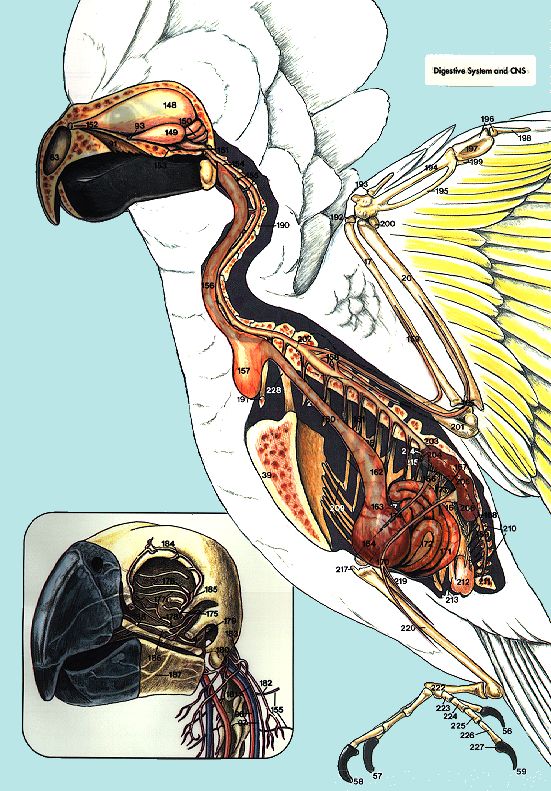
Muscular System The muscles of the trunk, neck, head tion of the pectoralis muscle is indicated with dotted
and appendages are displayed. The vessels of the great- lines for comparative purposes. Other dotted areas indi-
est clinical importance and their relationship to the cate that vessels are passing under or into anatomic
muscles and bones are also shown. Stubs of the rachis structures.
from the primary and secondary flight feathers are de- The digestive portion of the head is depicted in the
picted from their origin on the dorsal surface of the wing. inset, including the tongue, palate, esophagus and sali-
The inset shows a composite drawing of information vary glands.
available concerning the sinuses and cervicocephalic air Digestive System and CNS The lateral orientation of the
sac system of psittacine birds. digestive system and portions of the central nervous
Respiratory System A composite drawing provides the system are depicted. The humerus is represented with
clinician with insight into the avian respiratory system. a dotted line for orientation purposes.
The ventral hepatic peritoneal cavity is also represented The inset shows an enlargement of a representation
in this view. This layer has been specially designed so of the orientation of the nerves with respect to the bones
that all underlying structures are clearly visible. The of the head and neck. Note the location and degree of
individual layer can be segregated for study by placing innervation in the beak.
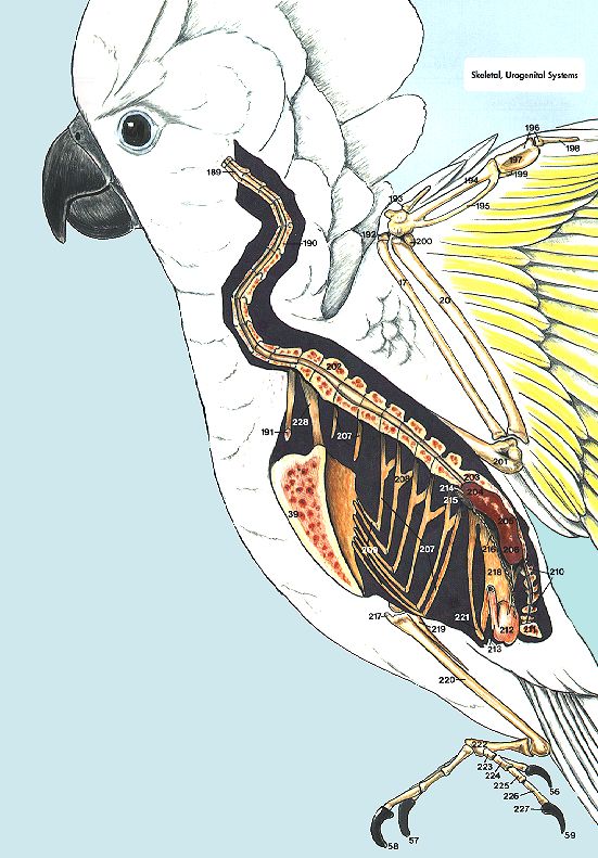
a sheet of white paper under the acetate. Skeletal, Urogenital Systems The skeletal and urogenital
The relationship of the bones of the head and sinuses systems are superimposed over the exterior of the bird.
in an Umbrella Cockatoo are shown in the inset. The lateral body wall has been darkened to enhance the
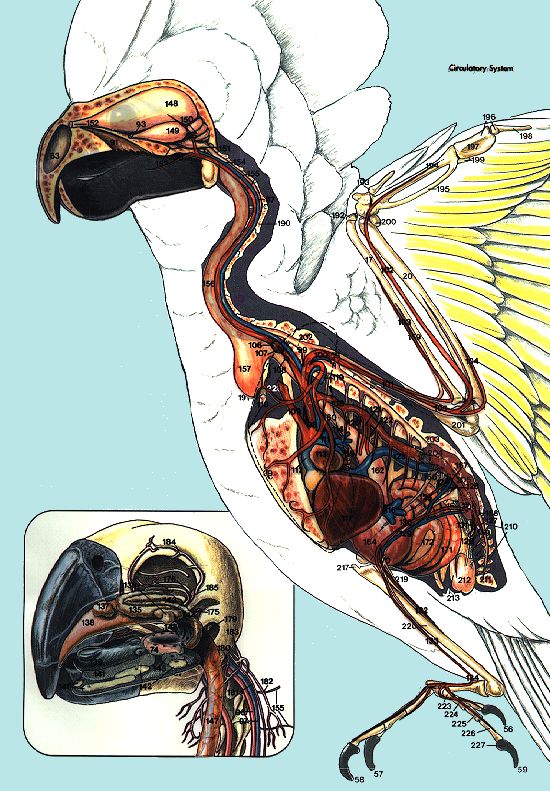
Circulatory System This layer depicts the clinically rele- color of overlying anatomic structures. The bird has been
vant portions of the circulatory system, along with the transected sagittally to allow visualization of the spinal
liver, thyroid and parathyroid glands. The relative posi- cord and kidneys.
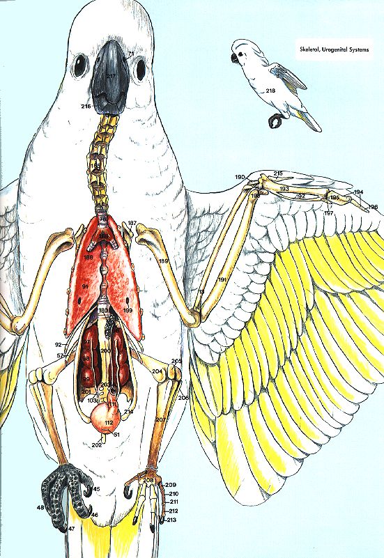
Ventrodorsal View
The ventrodorsal view illustrates a live bird in nor- Circulatory System The complex system of vessels has
mal perching position (the femur, tibiotarsus, tarsus been simplified in order to facilitate identification of
and metatarsus have been foreshortened); therefore, the those structures that are most clinically applicable for
drawing does not represent radiographic positioning of venipuncture, cannulation and surgery.
the legs. The primary emphasis is on the torso. The position of the lungs and pectoral muscles are
Muscular System The superficial pectoralis muscle has represented by dotted lines. This layer includes the
been removed from the left side of the bird to expose part liver, thyroid glands, parathyroid glands and ultimo-
of the coracoid bone and the clavicle. The tendon of the branchial glands (for position only, as the parathyroid
supercoracoidius is barely visible. The left portion of the and ultimobranchial glands are difficult to visualize. A
abdominal muscle has also been removed. It should be transected view of the brain, skull, mandible and hyoid
noted that the pectoralis muscle fills the space between bones are visible in this view.
the coracoids, holding the crop centrally and away from The position of the mature ovary and oviduct are illus-
the bones. The cranial extension of this muscle is clear trated in the inset.
in photographs, but has not been accurately depicted in Digestive System The esophagus and crop are shown as
many previous drawings of Psittaciformes. The rachis of solid structures for clarity. In reality, these organs are
the transected primary and secondary feathers are de- thin, translucent membranes. The break in the thoracic
picted from their origin on the dorsal surface of the wing. esophagus indicates the point where the organ courses
The inset is an enlargement of the abdominal cavity dorsally to the syrinx and primary bronchi.
depicting the relationship of the liver, thoracic esopha- The inset shows a view of the urogenital system of a
gus, proventriculus, ventriculus and intestines. developing female. The cloaca is opened ventrally to
Respiratory System The lungs were not included in the reveal the positions of the rectum, ureters and oviduct.
ventrodorsal drawing of the respiratory system so that Skeletal, Urogenital Systems Shown are the skeleton and
the relative position of the air sacs could be clearly dorsal body wall of a female cockatoo with the heart,
depicted. For clarity purposes, the trachea is depicted in liver and gastrointestinal tract removed. An end-on view
an unnatural position lying over the esophagus and crop. of the ribs is provided for reference purposes. Note the
The artist’s concept of a composite of information on melanistic ovary, which is common in cockatoos.
pneumatized avian bones is also provided. The ventral
hepatic peritoneal cavity and cranial thoracic air sacs
have been visually enhanced with textures for improved
visualization.
Nomenclature References
A transection of a male cockatoo is shown in the inset.
Baumel JJ (ed): Nomina Anatomica Avium. New York, Academic
The liver and gastrointestinal tract have been removed Press, 1979.
to reveal the organs associated with the dorsal body King AS, McLelland J: Form and Function in Birds. Vols 1-4.
wall. Note the melanistic testicles, which commonly New York, Academic Press, 1979, 1981, 1985, 1989.
occur in cockatoos.You can also read