Elevated Fetal Middle Cerebral Artery Peak Systolic Velocity: Anemia if Not; Then What?
←
→
Page content transcription
If your browser does not render page correctly, please read the page content below
ID: IJARS/2014/9825:2020                                                                                                       Case Report

                                                      Elevated Fetal Middle Cerebral
Radiology Section

                                                       Artery Peak Systolic Velocity:
                                                         Anemia if Not; Then What?
Sushil. G. Kachewar, Smita Balwant Sankaye

ABSTRACT                                                                             uterine state of its being. Hypertrophic cardiomyopathy in
It is now a globally accepted and time tested fact that                              this neonate of a diabetic mother was found to be the cause
elevated fetal middle cerebral artery peak systolic velocity                         of this temporary increase in blood velocity. The raised
as seen on color Doppler is a non-invasive indicator of fetal                        values eventually normalized by the 10th day of life by which
anemia. Cases describing causes of raised fetal middle                               time the cardiac status of the neonate had improved and
cerebral artery peak systolic velocities in the absence of                           stabilized following appropriate medications. Thus fetal
moderate to severe fetal anemia are very rare.                                       anemia alone is not the exclusive cause of elevated fetal
                                                                                     middle cerebral artery peak systolic velocities. Elevated
We describe one such rare case of a nearly full term male
                                                                                     velocities in absentia of fetal anemia should prompt an
of a diabetic mother whose middle cerebral artery peak
                                                                                     active search for other possible causes, one of which is as
systolic velocities were persistently raised in the absence of
                                                                                     mentioned in this report.
any fetal anemia; both in the intra as well as the early extra

                                                        Keywords: Cardiomyopathy, Colour doppler ultrasound, Diabetic mother, Fetus

Case Report                                                                          were thick. The fetal cardiac interventricular septum too was
A 26-years-old pregnant lady who was diagnosed to have non                           thickened [10 mm]. There was however no abnormal pleural,
insulin dependent diabetes mellitus was referred for obstetric                       pericardial or peritoneal free fluid.
ultrasound in Rural Medical College, Loni, Maharashtra, India.                       Due to the prenatal sonographic suggestion of severe fetal
She was a primi gravida and had no other significant illnesses                       anemia and the presence of polyhydramnios in this known
in the past, especially pertaining to her cardiovascular system.                     diabetic mother, lower segment caesarean section was
Her blood sugar was under control on oral hypoglycemics                              immediately performed [Hence we could not obtain multiple
alone before as well as during the pregnancy. She had been                           measurements that are recommended to determine the trend
on oral hypoglycemisc for almost last two years.                                     of increasing values] and a 3999 grams male child was born.
Obstetric ultrasound was performed after informed written                            A pan systolic murmur and a bluish tinge to the child’s overall
consent as per the existing law of this land. Ultrasound revealed                    appearance were noted. Pulse Oximeter demonstrated a
a single intrauterine live gestation of average gestational age                      saturation of only 76% without oxygen and that of 91% with
35 weeks. This patient presented to us for the first time. She                       100% oxygen. The neonate was shifted to neonatal intensive
had no earlier obstetric ultrasound reports. As color doppler                        care unit (NICU) for further management.
demonstrated an abnormally elevated fetal Middle Cerebral                            The fetal blood sugar level was 56gm/dl. On delivery the
Artery Peak Systolic Velocity [Table/Fig-1] of 70 cm/s that                          neonate had a hemoglobin value of 15.6 gm/dl. On day 2 and
was more than 1.5 times the MoM value for that particular                            day 3 it was 15.2 and 16.4 gm/dl respectively. Total Bilirubin
GA [41 +/- 8 cm/s] by local as well as international standards                       on day 1 was 1.9 and that on day 3 was 5.9. The mother was
[1-6]; a diagnosis of severe fetal anemia was suggested.                             O Rh positive and the child`s blood group was B Rh Positive.
Each wave was tall and belonged to KG Type III waveform                              Follow up ultrasound on Day 1 following birth; still demonstrated
as per the Kachewar Gandage classification system for types                          elevated fetal Middle Cerebral Artery-Peak Systolic Velocity in
of fetal MCA velocity waveform patterns [7]. In addition, as                         absence of fetal anemia. Plain Radiograph of Chest Antero
the Amniotic Fluid Index [AFI] was 22 cm the diagnosis of                            Posterior view [CXR-AP] showed cardiomegaly [Table/
polyhydramnios too was mentioned. A Grade III placenta with                          Fig-3]. Heart size on the chest radiograph was larger than
normal placental thickness (3.2 cm) was seen. Soft tissues of                        normal (cardiothoracic ratio > 0.65). Cardiac ultrasound
the fetal thoracic wall [Table/Fig-2] as well as the abdomen                         demonstrated asymmetric septal hypertrophy [12 mm] and

International Journal of Anatomy, Radiology and Surgery, 2014 Dec, Vol-3(4): 15-18                                                                       15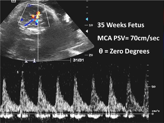
Sushil. G. Kachewar and Smita Balwant Sankaye, Hypertrophic Cardiomyopathy Causing Fetal Anemia http://ijars.jcdr.net
marked concentric left ventricular hypertrophy [Table/Fig-4], setting of Hypertrophic Cardiomyopathy and not fetal anemia
indicative of Hypertrophic Cardiomyopathy. He was put on was responsible for the rise in fetal Middle Cerebral Artery-
intravenous injection Lasix 3 mg every 8 hourly, i.v. Dopamine Peak Systolic Velocity values.
and Dobutamine @ 10-15 µl/kg/hour and Tablet Propranolol
2.5 mg every 12 hourly. Over the next few days his bluish body Discussion
tinge and pan systolic murmur disappeared. Pulse Oximeter Several studies [1-5] across the globe have proved it time
demonstrated a saturation of 92-96% without oxygen. and again that by measuring the peak of the systolic velocity
of fetal Middle Cerebral Artery waveform as seen on colour
Repeat CXR – AP on Day 10 of life showed reduced and
Doppler, a non-invasive diagnosis of fetal anemia can be
normalized size of cardiac silhouette [Table/Fig-5] and
reached. Infact, severe fetal anemia can be reliably diagnosed
ultrasound [Table/Fig-6] demonstrated reduced thickening of
if this raised fetal Middle Cerebral Artery-Peak Systolic Velocity
interventricular septum [6 mm] as well as the left ventricle.
values are 1.5 times the Multiples of Median (MoM) value for
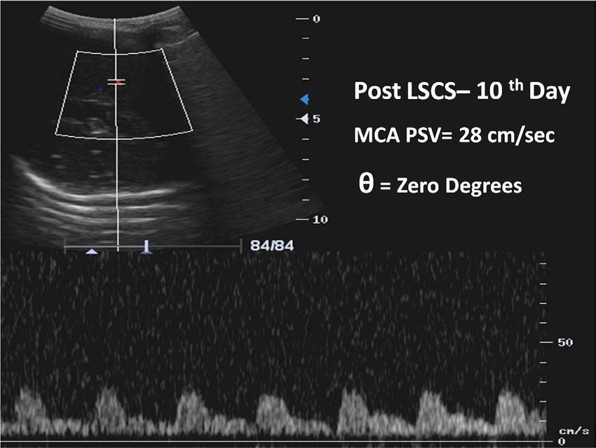
Middle Cerebral Artery-Peak Systolic Velocity of the neonate
that particular gestational age (GA) of the fetus. It is believed
[Table/Fig-7] demonstrated reduction in the values [28cm/s]
that the low viscosity of the fetal blood when it is anemic, due
as compared to the intrauterine measurement. The pattern
to any cause; leads to an increase in the cardiac output which
of waveform too resembled a normal third trimester type
ultimately manifests as raised peak velocity.
waveform i.e. KG Type IC as per the Kachewar Gandage
classification system for types of fetal Middle Cerebral Artery Apart from a rare case report [6] describing alpha thalassemia
velocity waveform patterns [7]. as a possible cause of raised fetal Middle Cerebral Artery-
Peak Systolic Velocity in the absence of fetal anemia, no
As the neonate was hale and hearty, he was discharged and
other causes have been documented to the best of our
called for follow-up every 3 months for the upcoming year. Thus,
knowledge.
in this case the diagnosis of Hypertrophic Cardiomyopathy was
reached on imaging studies that backed the clinical findings Hence we report this curious case of an infant of a diabetic
and it was postulated that increased velocity of blood ejected mother, who during the intranatal period as well as during
in the aorta due to increased left ventricular contractility in the the early period following birth had elevated Middle Cerebral
Artery-Peak Systolic Velocity more than 1.5 times the MoM
[Table/Fig-1]: Color Doppler Ultrasound showing raised Fetal MCA-PSV in 35 Weeks Fetus
[Table/Fig-2]: Grey Scale Ultrasound showing axial section of thorax of 35 Weeks Fetus. Thickening of soft tissues of thorax (arrows) and
thickened interventricular septum (*) are seen
[Table/Fig-3]: Day 1-CXR-AP. Chest X-ray showing significant cardiomegaly (cardiothoracic ratio > 0.65). Hypertrophic cardiomyopathy in an
infant of diabetic mother
[Table/Fig-4]: Day 1 - Grey Scale Ultrasound showing axial section of thorax. Thickened interventricular septum (*) and left ventricle are still seen
[Table/Fig-5]: Day 10-CXR-AP shows reduced size of cardiac silhouette with a normal cardio- thoracic ratio < 0.65
[Table/Fig-6]: Day 10 - Grey Scale Ultrasound of axial section of thorax showing reduced thickening of interventricular septum and left
ventricle
16 International Journal of Anatomy, Radiology and Surgery, 2014 Dec, Vol-3(4): 15-18http://ijars.jcdr.net Sushil. G. Kachewar and Smita Balwant Sankaye, Hypertrophic Cardiomyopathy Causing Fetal Anemia
There is a rare case report [6] describing an abnormal increase
in fetal Middle Cerebral Artery-Peak Systolic Velocity without
anemia on cordocentesis; even when Hydrops was present in
a fetus with alpha-thalassemia. This is because HbBarts that
is present in such patients has high affinity for oxygen which
therefore prevents its release at tissue level; thereby inducing
relative hypoxia. In order to compensate for this, cardiac output
is increased as well as the peripheral resistance is reduced
in Middle Cerebral Artery and this ultimately manifests as
increased fetal Middle Cerebral Artery-Peak Systolic Velocity.
Thus the amount as well as type of Hb; both determines the
manifestation of anemia and Middle Cerebral Artery-Peak
Systolic Velocity changes [7]. Cardiomegaly and elevated
intracardiac blood velocities, along with elevated Middle
Cerebral Artery Doppler flow, as an indirect result of increased
[Table/Fig-7]: Color Doppler Ultrasound of neonatal head showing cardiac output, was detected. Multiple logistic regression
normalized MCA-PSV at 10 Days after Birth analysis revealed that both fetal hemoglobin and oxygen
content were the factors that contributed to the increase in
value for that particular GA; and yet was non anemic. The the Middle Cerebral Artery-Peak Systolic Velocity [7].
raised MCA-PSV values ultimately normalized by the tenth
To the best of our knowledge, abnormally elevated fetal
day of birth. Hypertrophic Cardiomyopathy that is known to
Middle Cerebral Artery-Peak Systolic Velocity and the
occur in the fetus of a diabetic mother was found to be the
occurrence of Kachewar Gandage Type III waveforms have
cause of this curious occurrence.
never been reported without fetal anemia. The present case
A global search for a non-invasive method to determine fetal report is the first to successfully document that elevation of
anemia zeroed in on the utility of measuring the fetal Middle fetal Middle Cerebral Artery-Peak Systolic Velocity is possible
Cerebral Artery-Peak Systolic Velocity. Replication of this due to cardiomyopathy that sometimes leads to enhanced left
method was found to be simple, efficient as well as reliable ventricular contractility which pumps more blood in the aorta
and hence has been widely accepted [1-4] and is being with increased force that ultimately manifests as abnormal
actively practised. Off late, loco-regional differences between increase in the fetal Middle Cerebral Artery-Peak Systolic
the standard values for each geographic locality have been Velocity.
described [2-6]. The shape as well as the pattern of MCA
There is an increased risk of Hypertrophic Cardiomyopathy in
waveform also aid in diagnosing fetal hypoxia usually due to
infants of diabetic mothers [8, 9]. Such infants usually present
fetal anemia as has been described by Type III waveforms in
with cardio respiratory distress and a disproportionate septal
the Kachewar Gandage classification system for types of fetal
hypertrophy. The spectrum of Hypertrophic Cardiomyopathy
Middle Cerebral Artery velocity waveform patterns [7].
can vary and may manifest as an incidental finding on
The lowered viscosity of anaemic blood coupled with echocardiography or as congestive heart failure. It has been
increased cardiac output to maintain adequate oxygen, at reported that symptomatic Hypertrophic Cardiomyopathy
least to vital organs results in elevated fetal Middle Cerebral occurs in 12.1% of infants of diabetic mothers and
Artery-Peak Systolic Velocity [1] and Type III Middle Cerebral echocardiography surveillance has placed this figures at
Artery waveforms [2] when the fetal haemoglobin is reduced. 30%. Initially, the left ventricular mass and contractility are
The increase in this Middle Cerebral Artery-Peak Systolic increased [10, 11]. Maternal hyperglycemia during the third
Velocity beyond 1.5 times MoM is indicative of severe fetal trimester leads to fetal hyperinsulinemic, anabolic status, the
anaemia [1, 2, 4]. result of which manifests as hypertrophy of septum as well as
Anemia in a fetus occurs when there is an inadequate number ventricular wall.
or quality of red blood cells in the fetal circulatory system. Fetuses of diabetic mothers, who are diagnosed to have
Hemolytic causes of fetal anemia can be grouped into cardiomyopathy, in some cases demonstrate normal or slightly
Immune related disorders like Rh- Isoimmunization, ABO and
increased cardiac systolic function that manifests as slightly increased
Anti Kell Antibodies. Non Immune hemolytic causes include
doppler velocity through the aorta. Hypertrophic Cardiomyopathy
Red Cell Enzyme or Membrane Deficiency. Fetal anemia may
also occur due to blood loss as in Fetomaternal Hemorrhage or is usually benign and manifests clinically as systolic murmur and
Twin-twin transfusion. Miscellaneous conditions like Placental transitory cardiomegaly. All symptoms usually regress spontaneously
Chorioangioma and Congenital Infections like Parvovirus B19 within a few weeks. Sometimes, overt congestive heart failure
or Cytomegalovirus too can cause fetal anemia. All these develops, and is seen along with tachypnea, tachycardia, gallop
conditions can ultimately manifest as elevated fetal Middle rhythm and hepatomegaly [10, 11].
Cerebral Artery-Peak Systolic Velocity. All surviving cases with maternal diabetes have been found
International Journal of Anatomy, Radiology and Surgery, 2014 Dec, Vol-3(4): 15-18 17Sushil. G. Kachewar and Smita Balwant Sankaye, Hypertrophic Cardiomyopathy Causing Fetal Anemia http://ijars.jcdr.net
to progressively normalize after 3–6 months. This explains as the Hypertrophic Cardiomyopathy regresses. An active
the finding of raised Middle Cerebral Artery-Peak Systolic search should therefore be carried out in all fetuses that
Velocity that was seen in utero and in early neonatal period in have abnormally elevated fetal Middle Cerebral Artery-Peak
the present case that eventually normalized by day 10. Like Systolic Velocity so as to label them as severly anemic, but
in a reported study [9], present study too had asymmetric have no anemia on cordocentesis; to find underlying cardiac
left ventricular hypertrophy and a nondilated left ventricle as abnormalities like Hypertrophic Cardiomyopathy for earlier
the hallmark of HOCM. Echocardiography in Hypertrophic diagnosis and timely management.
Cardiomyopathy demonstrates intraventricular septum
thickening of mean 4.77 mm when compared with those born References
to non-diabetic mothers 2.5 mm [11]. [1] Nardozza LM, Simioni C, Garbato G, Araujo Júnior E, Guimarães Filho
HA, Torloni MR, et al. Nomogram of fetal middle cerebral artery peak
As the natural history of Hypertrophic Cardiomyopathy is systolic velocity at 23-35 weeks of gestation in a Brazilian population:
that of spontaneous regression of symptoms and septal pilot study. J Matern Fetal Neonatal Med. 2008; 21(10):714-18.
[2] Tan KB, Fook-Chong SM, Lee SL, Tan LK. Foetal peak systolic velocity in
hypertrophy, supportive care, with fluid restriction, diuretics the middle cerebral artery: an Asian reference range. Singapore Med J.
and oxygen, help such neonates to tide over this crisis [9, 10]. 2009; 50(6):584-86.
Digoxin and inotropes are contraindicated as they increase [3] Kachewar SG, Gandage SG, Kulkarni DS.A local Indian scenario of
left ventricular outflow tract obstruction. Beta adrenergic fetal middle cerebral artery peak systolic velocities. Japanese Journal of
Radiology. 2011; 29: 725-29.
blocking agent like propranolol is found to be very effective [4] Kachewar SG, Gandage SG, Pawar HJ. A prospective cross-sectional
as it decreases heart rate, left ventricular contractility and wall study of fetal middle cerebral artery peak systolic velocity in normal obstetric
stress, and a total relief of symptoms has been reported in 30% population attending an Indian Medical College. Japanese Journal of
Radiology. 2012; 30:575-81.
of affected infants. Surgery has a role only when the septal
[5] Kachewar SG. Non-invasive diagnosis of fetal anemia:Multicentric nationwide
hypertrophy and outflow tract obstruction cause significant input for a worldwide output. Diagn Prenat. 2013; 24:141-47.
symptoms despite medical therapy. Catheter alcohol ablation [6] Maguire K, Johnson A, Ou CN, Lantin RL and Moise KJ. Elevated middle
and surgical septal myotomy or myomectomy are some of the cerebral artery peak systolic velocity without fetal anemia in a case of
homozygous α–thalassemia-1. Prenatal Diagnosis 2008; 28: 72–74.
options. It is also believed that the severity of Hypertrophic [7] Kachewar SG, Gandage SG. (2012). A classification of patterns of
Cardiomyopathy can be reduced by, appropriate diabetic Fetal Middle Cerebral Artery Velocity Waveforms as seen on Doppler
management in every pregnancy itself. Ultrasound. Japanese Journal of Radiology. 2012; 30:582-88.
[8] Picklesimer AH, Oepkes D, Moise KJ Jr, Kush ML, Weiner CP, Harman
Conclusion CR, Baschat AA. Determinants of the middle cerebral artery peak systolic
velocity in the human fetus.
All fetuses with abnormally elevated fetal Middle Cerebral Am J Obstet Gynecol. 2007; 197(5):526.e1-4.
Artery-Peak Systolic Velocity values do not necessarily have [9] Shiraz A. Maskatia. Hypertrophic Cardiomyopathy: Infants, Children, and
fetal anemia. Hypertrophic Cardiomyopathy in the fetus as Adolescents. Congenital Heart Disease. 2012; 7:84–92.
[10] Narchi H and Kulaylat N. Heart disease in infants of diabetic mothers.
well as a neonate of a diabetic mother too can give rise to Images Paediatr Cardiol. 2000; 2(2): 17–23.
raised fetal Middle Cerebral Artery-Peak Systolic Velocity [11] Deorari AK, Saxena A, Singh M, Shrivastava S. Echocardiographic
values. This elevation is temporary and normalizes as soon assessment of infants born to diabetic mothers. Arch Dis Child. 1989;
64:721–24.
AUTHOR(S): Professor, Department of Radio-Diagnosis, Rural Medical
1. Dr. Sushil. G. Kachewar College, PIMS (DU), Pravara Medical Trust, At Post-Loni,
2. Dr. Smita Balwant Sankaye Ta- Rahata, District- Ahmednagar, Maharashtra- 413 736,
India.
PARTICULARS OF CONTRIBUTORS: Mobile- 0091-9921160357, Telephone- 0091-2422271810
1. Professor, Department of Radio-Diagnosis, Rural Fax- 0091-2422271529,
Medical College (RMC), PIMS (DU), Loni, India. Email- sushilkachewar@hotmail.com
2. Assistant Professor, Department of Pathology,
SKNMC & GH,PUNE, India. Financial OR OTHER COMPETING INTERESTS:
None.
NAME, ADDRESS, E-MAIL ID OF THE
CORRESPONDING AUTHOR:
Date of Publishing: Dec 01, 2014
Dr. Sushil Ghanshyam Kachewar,
18 International Journal of Anatomy, Radiology and Surgery, 2014 Dec, Vol-3(4): 15-18You can also read