Sample requirements for TSE testing and confirmation - EURL guidance - APHA Science Services
←
→
Page content transcription
If your browser does not render page correctly, please read the page content below
Sample requirements for TSE testing and confirmation
                – EURL guidance.
BACKGROUND

The first stage of all the current TSE diagnostic or screening tests involves the
sampling of the central nervous system at the level of the brainstem, and the
subsequent examination of the sampled tissue for the presence of disease-
specific PrP using immunochemical methods.

As new, atypical, forms of disease have been identified in cattle (H-BSE and
L-BSE) and sheep (atypical scrapie) it is becoming apparent that the
cerebellum is also a key area for robust confirmation and classification of
these variants.

PrP has proved to be the most consistent marker for all known forms of TSE,
being present in the CNS of all recognised clinically suspect TSE cases, and it
has been shown experimentally that demonstrable accumulations of PrP arise
in the CNS (and in a more variable way the lymphoreticular system) in
advance of any clinical disease. It is thus a useful marker in pre-clinical
animals, as well as in those presenting with overt disease.

The brain consists of multiple interrelated but anatomically and functionally
distinct areas, and disease related PrP accumulation shows distinct
anatomically-specific trophisms which result in clear-cut patterns of PrP
accumulation (Fig 1). These patterns are specific both in end-stage disease,
and through the pathogenesis of each form of TSE.

Fig 1 anatomically-specific tropisms which result in clear-cut patterns of PrP
accumulation

Sampling Guidance Document v2 September 2013                             Page 1 of 11
TSE EURL Reviewed: January 2018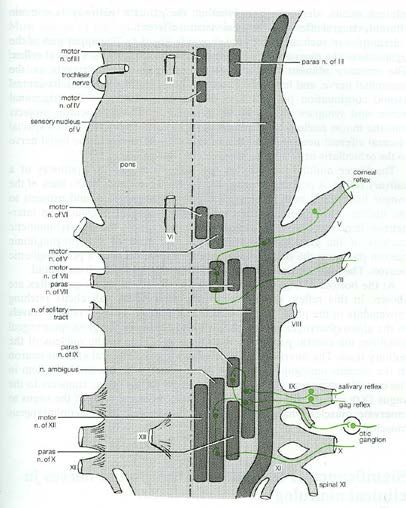
SPECIFIC SAMPLING REQUIREMENTS
(to fulfil the current statutory requirements as laid down in Annex X to
regulation (EC) No 999/20001)
These guidelines are based on the approaches recommended in the OIE
manual chapters for BSE and scrapie
http://www.oie.int/fileadmin/Home/eng/Health_standards/tahm/2.04.06_BSE.pdf
http://www.oie.int/fileadmin/Home/eng/Health_standards/tahm/2.07.13_SCRAPIE.pdf
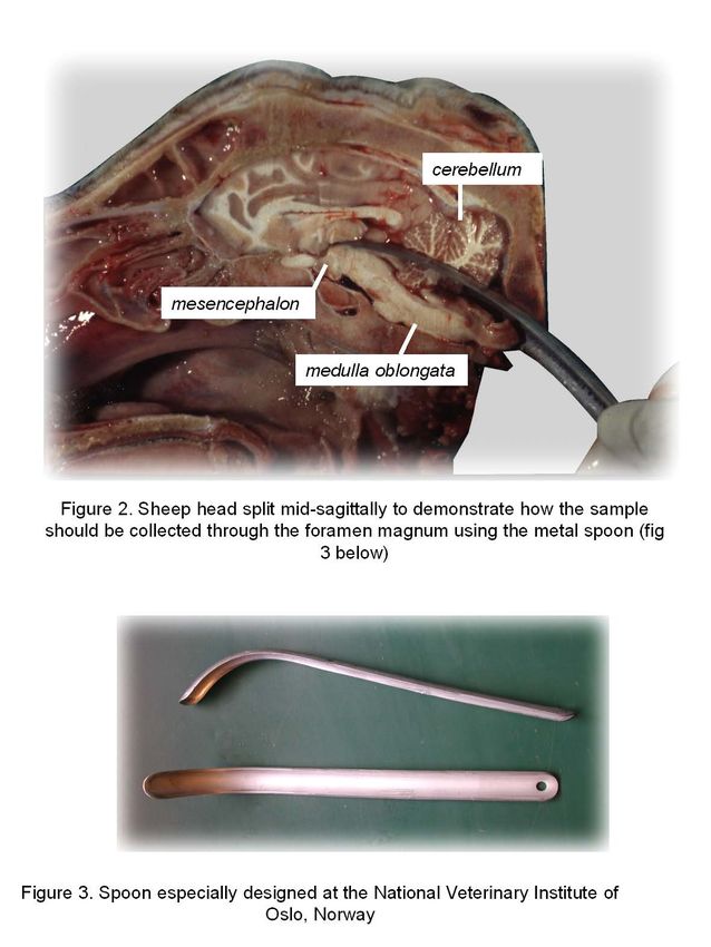
The minimum sampling requirement for any animal from either source
population is the brainstem (at the level of the obex). In addition, for small
ruminants it is advised that part of the cerebellum is also collected. Both of
these brain areas can be accessed through the foramen magnum using a
proprietary sampling spoon (see below for sampling methods).
Cerebellum may also be a useful sample to assist with confirmation of the
classification of atypical bovine TSE 1.
The quantity of tissue taken for testing should be sufficient to provide the
following:
• A hemisection 2 of fresh brainstem at the level of the obex, for the
initial rapid test (or a full transverse section immediately adjacent to
the obex, or equivalent sample taken unilaterally using one of the
commercially available sampling devices).
• A fixed cross-section, or hemi-section2 of brainstem at the level of
the obex for confirmatory IHC. (Ideally this sample would be fixed
immediately, but for practical reasons it is appropriate to keep the
sample chilled, and fix it on receipt of a positive rapid test. In some
countries it is established practice to freeze material prior to fixation.
While this does not detract from the subsequent immunoreactivity of
positive cases, it can significantly compromise the tissue morphology,
making the contextual interpretation of negative or equivocal
immunostains difficult or impossible.)
1Konold T, Bone GE, Clifford D, Chaplin MJ, Cawthraw S, Stack MJ, Simmons MM (2012) Experimental
H-type and L-type bovine spongiform encephalopathy in cattle: observation of two clinical syndromes
and diagnostic challenges. BMC Vet Res 8:22
2 Historically, a full transverse cross-section of the brainstem at the level of the obex has been
recommended for histopathological confirmation. With the advent of rapid molecular techniques the
need to apply multiple tests in a single animal has led to competition between tests for the optimal early
diagnostic sites at the obex. Hemisection of the brainstem at the level of the obex will result in loss of
the ability to assess the symmetry of lesions, but the need for such assessment is less if
immunohistochemistry is used. If this approach is adopted however, it becomes critical to ensure that
the target site is not compromised. The dorsal nucleus of the vagus nerve (the optimal target area for
most cases of scrapie) is small, and lies close to midline
Sampling Guidance Document v2 September 2013 Page 2 of 11
TSE EURL Reviewed: January 2018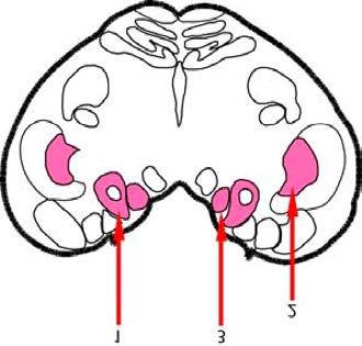
•   A fixed section of cerebellum, or sufficient fresh cerebellum to
        conduct an OIE-SAF Western blot. This may be required for the
        confirmation of atypical scrapie.

    •   Sufficient remaining fresh-frozen medullary tissue (adjacent to the
        obex) and fresh-frozen cerebellum to provide fresh material for
        primary molecular testing (discriminatory WB) and possibly a
        range of secondary and tertiary testing (5-10 g whenever possible).

    •   brain tissue from other areas in addition to the brainstem and
        cerebellum are always an advantage, as are lymphoreticular tissues
        when available. Although not essential for statutory purposes, they will
        be important in characterising the scrapie strain present if results differ
        from the expected confirmation at the level of the obex.

BRAINSTEM AND CEREBELLUM SAMPLING METHODS

Whole brain removal is ideal. However, this is not always practical or
achievable and the following approaches outline how to remove the
brainstem and cerebellum through the foramen magnum.

Any method that produces                the   entire   brainstem     intact   and
uncontaminated may be used.

The samples should be collected in a manner that prevents any risk of
contamination between successive samples.

APHA Method

•   Place the disarticulated head upside down on table with the foramen
    magnum facing the operator. Remove any blood clots obscuring the view
    of the brainstem, and identify the dura mater. Use a clean pair of scissors
    to reflect the dura mater and expose the brain stem.
•   Gently hold the brain stem as close to the end as possible with a
    new/clean pair of forceps and move the brainstem to the side to visualise
    the cranial nerves. Insert the scissors into the foramen magnum and cut
    through the cranial nerves (VII –XI). Do this on both sides of the
    brainstem, taking care not to damage it.
•   Using the forceps, very gently pull the brainstem until it is straight and
    insert the spoon above the brainstem with the cutting edge of the blade

Sampling Guidance Document v2 September 2013                         Page 3 of 11
TSE EURL Reviewed: January 2018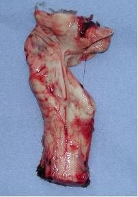
facing down. Keep the blade of the spoon 3 against the bone above as it is
    inserted. Insert to the level of the notch of the spoon –approximately 8-10
    cm. When in position, point the cutting edge of the blade downwards by
    lifting the handle upwards to sever the brainstem.
•   Cut through the cerebellar peduncles by moving the handle from side to
    side.
•   After completing the cutting, carefully withdraw the spoon and insert it with
    the blade pointing downwards on top of the brainstem until it enters to a
    depth of 8-10cm. Gently move the blade side to side and downwards,
    cutting through the rostral medulla (avoiding rotational movement as this
    will damage the obex).
•   Gently pull with the forceps and using the spoon as a scoop pull as much
    brainstem as possible out of the skull. If resistance is encountered,
    continue to cut through the brainstem with a side to side and downward
    motion of the handle.
•   With the head still upside down on the table, look downwards through the
    foramen magnum and identify the cerebellum.             Insert the spoon
    underneath the cerebellum, and lever it upwards into the centre of the
    space vacated by the brainstem. If the cerebellum is not moving freely,
    loosen it by moving the spoon gently around underneath it. Lift the
    cerebellum towards the foramen magnum using the spoon to support and
    guide, and gently take hold of the cerebellum through the foramen with the
    forceps. Remove the cerebellum through the foramen magnum.

3Sheep brainstem sampling spoons are available commercially from :
Vet Way Ltd, 1&2 Buccaneer Court, Airfield Business park, Elvington, York, YO41 4AU.
Fax: 01904 607601

Sampling Guidance Document v2 September 2013                                       Page 4 of 11
TSE EURL Reviewed: January 2018
Norwegian method

Sampling Guidance Document v2 September 2013   Page 5 of 11
TSE EURL Reviewed: January 2018
ISSUES RELATED TO SUBSAMPLING FOR SCREENING TESTS

Rapid tests protocols for active surveillance of healthy slaughter and/or at risk
populations generally require a specific weight of brainstem tissue, taken at
the level of the obex, to be presented to the test.

In cattle BSE the pattern appears to be highly consistent, with early changes
appearing first in certain nuclei in the brainstem at the level of the obex.
Following experimental oral challenge with BSE, the earliest visible PrP
accumulation is consistently seen (using immunohistochemistry (IHC)) in the
nucleus of the solitary tract, with involvement of the adjacent dorsal motor
nucleus of the vagus nerve (DNV) and the nucleus of the spinal tract of the
trigeminal nerve (V) following soon after. The vestibular nuclei in the rostral
medulla may also become involved at an early stage. The IHC patterns
observed in many ‘early’ field cases (detected through both active and
passive surveillance) support the consistent early involvement of these areas
in natural disease. These areas are recognised as the diagnostic target areas
for sampling (Fig 4)

Fig 4                                Fig 5

In advanced clinical cases, all the grey-matter areas within the brainstem
become affected to some extent (Fig 5).

Sampling may be achieved by taking a cross-sectional slice of
brainstem.

This approach may give rise to some health and safety concerns depending
on the type of blade used, and it also requires that each sample is weighed
before use, which is time-consuming. This has led to the development of safer
plastic sampling tools, many of which offer the additional advantage of
collecting a measured volume of tissue, thereby dispensing with the need to
weigh each sample before testing.

Sampling Guidance Document v2 September 2013                       Page 6 of 11
TSE EURL Reviewed: January 2018
Another hypothetical drawback of the transverse slice approach is the
possibility that a full cross-sectional sample, if taken from an animal with very
restricted PrP distribution, might suffer a ‘dilution effect’ from the non-involved
areas which would compromise the sensitivity of the test. This has led to the
development of increasingly popular sampling techniques in which a
graduated syringe is used to take a longitudinal ‘core’ sample of brainstem,
focussing on the target areas.

Sampling ‘tools’

The EURL has a role in evaluating and approving rapid test kits for use within
the EU. Sampling tools are sometimes offered as part of a commercial test
package, and as such have to be evaluated as fit for purpose, in particular to
ensure that any tissue disruption as a result of sampling does not compromise
the suitability of material for confirmatory testing should it be required.

In principle these tools take a core of grey matter from the obex region of the
brainstem thereby reducing any dilution effect of the peripheral white matter,
and increasing the sensitivity of the test. If used correctly, and with a clear
understanding of the three-dimensional anatomy of the TSE target areas
these tools perform adequately, and dispense with the need to assess the
weight of each individual sample, with considerable savings in time. However,
there are a number of potential drawbacks with this technique that users
should be aware of, and conduct appropriate monitoring checks.

These drawbacks can be broadly divided into two categories:

Inadequately sampled brainstem delivered to the laboratory.

This can be a problem, especially when dealing with fallen stock, where
material may be significantly autolysed before sampling takes place. In such
cases, the obex region may be damaged or incomplete in some way (Fig 6) or
the sample may be so distorted or autolysed that anatomical orientation is not
possible (Fig 7).

In these cases it will not be possible to take an anatomically targeted sample.
This cannot be avoided, but it should be recorded, to assist with interpretation
of the resulting test results, and to feed back to the personnel collecting the
samples that such poor quality samples should be avoided whenever
possible.

Sampling Guidance Document v2 September 2013                         Page 7 of 11
TSE EURL Reviewed: January 2018
Fig 6
a) Sampling damage                        b) Autolysis – obex identifiable, but
 – obex incomplete                               tissue integrity compromised

Fig 7
Distortion of brainstem – obex not identifiable

Sampling Guidance Document v2 September 2013                       Page 8 of 11
TSE EURL Reviewed: January 2018
This problem affects both tissue slice and sampling tool samples.

Inappropriately targeted sample in early/ pre-clinical disease

To avoid compromising the sensitivity of the test, the sampler must ensure
that the relevant TSE target areas are adequately represented, in addition to
the sample being of a consistent and appropriate weight for the test.

The nucleus of the solitary tract, which is the earliest site of PrP accumulation
at the obex in pre-clinical disease in cattle, runs rostro-caudally throughout the
caudal brainstem [1,2], but in its most caudal portion (caudal to the obex) it
runs more medially, immediately adjacent to the spinal canal, and dorsal to
the parasympathetic nucleus of the vagus nerve.

One potential danger with syringe-type ‘core’ extractor samples taken from
variable distances caudal to the obex is that the resulting sample may be too
caudal to incorporate the nucleus of the solitary tract at the entry point, and
may potentially miss the target areas more rostrally if the sampling tract veers
laterally or ventrally (Fig 8).

Fig 8

                                      Inappropriate sample site
                                      using a sampling syringe.
                                      (Blue circles represent the
                                      position of the nucleus of
                                      the solitary tract)

This presents a potential sensitivity problem, especially in active surveillance
cases where the PrP accumulation is likely to be very focal, and anatomically
targeted to the nucleus of the solitary tract, the dorsal motor nucleus of the
vagus nerve and the nucleus of the spinal tract of the trigeminal nerve
(Figures 3,9). Such cases may be missed by any sampling method that takes

Sampling Guidance Document v2 September 2013                        Page 9 of 11
TSE EURL Reviewed: January 2018
a longitudinal core which, if inaccurately directed, might not contain the target
areas.
Fig 9
Towards front
of brain
Maximum
acceptable
sampling range
Approx 1cm
in cattle atlas
Towards spinal cord
7
Robust training and great care is needed in the application of this type
of sampling method to ensure that the initial rapid test is not
compromised by the collection of a sample which does not represent
the desired target area.
All of the above issues apply also to sheep, with the added complication that
PrP distribution patterns vary more than in cattle, and the physical size of the
brainstem is smaller than in cattle.
The principal target area (the dorsal nucleus of the vagus) lies very close to
midline, and any approach which relies upon hemisectioning through the
midline has little tolerance for inaccuracy.
Accepting that this should be a secondary option only, where the obex is
not identifiable, other parts of the brain stem/cervical spinal cord can be tested
using approved rapid tests, even though their approval is currently specific to
testing of the obex. In this case a positive result is valid and should be
reported as such. Negative results must however be reported with the caveat
that optimal tissues were not available for testing. The alternative option is to
Sampling Guidance Document v2 September 2013 Page 10 of 11
TSE EURL Reviewed: January 2018report such tissues as “no test” i.e. un-testable as the target tissue is not available. ACTION ADVISED Each NRL should ensure that all local sampling instructions contain appropriate detailed reference to the cross-sectional and longitudinal anatomy of the structures which require to be targeted. Additionally, instructions must be included on how to sample material which is not optimally collected/oriented or properly identifiable at an anatomical level when it is not possible to correctly position the sampling tool, or accurately identify the obex. It is advised that the accuracy of sampling is monitored by the NRL by review of a randomly-selected proportion of negative cases in addition to any positives which are referred for confirmation. References 1. Yoshikawa,T. (1968). Atlas of the Brains of Domestic Animals. The Pennsylvania State University Press, University Park and London. 2. Lignereux,Y. (1986) Atlas stereotaxique de l’encephale de la vache frisonne. PhD Thesis, l’Universite Paul Sabatier de Toulouse. Sampling Guidance Document v2 September 2013 Page 11 of 11 TSE EURL Reviewed: January 2018
You can also read