A transfer-learning approach for lesion detection in endoscopic images from the urinary tract
←
→
Page content transcription
If your browser does not render page correctly, please read the page content below
A transfer-learning approach for lesion detection
in endoscopic images from the urinary tract
Jorge F. Lazo1,2 , Sara Moccia3,4 , Aldo Marzullo5 , Michele Catellani6 ,
Ottavio de Cobelli6 , Benoit Rosa2 , Michel de Mathelin2 , Elena De Momi1
1 Department of Electronics, Information and Bioengineering, Politecnico di Milano, Milan, Italy
2 ICube, UMR 7357, CNRS-Université de Strasbourg, Strasbourg, France
3 The BioRobotics Institute, Scuola Superiore Sant’Anna, Pisa, Italy
4 Department of Excellence in Robotics and AI, Scuola Superiore Sant’Anna, Pisa, Italy
5 Department of Mathematics and Computer Science, University of Calabria, Rende (CS), Italy.
arXiv:2104.03927v1 [eess.IV] 8 Apr 2021
6 Istituto Europeo di Oncologia (IRCCS), Milan, Italy
Abstract— Ureteroscopy and cystoscopy are the gold stan- diagnosis and treatment [3]. The procedures are carried out
dard methods to identify and treat tumors along the urinary with the visual guidance of an endoscopic camera [4] in most
tract. It has been reported that during a normal procedure a cases using White Light Imaging (WLI) or Narrow Band
rate of 10-20 % of the lesions could be missed. In this work we
study the implementation of 3 different Convolutional Neural Imaging (NBI) [5] .
Networks (CNNs), using a 2-steps training strategy, to classify The clinical challenge for BC is to reduce the high recur-
images from the urinary tract with and without lesions. A total rence rate with values of 75% [6] and the miss rate, reported
of 6,101 images from ureteroscopy and cystoscopy procedures to be between 10-20% [7]. Computer-aided diagnosis (CAD)
were collected. The CNNs were trained and tested using transfer systems for endoscopic image analysis have recently shown
learning in a two-steps fashion on 3 datasets. The datasets
used were: 1) only ureteroscopy images, 2) only cystoscopy to be able to support clinicians diagnosis capabilities during
images and 3) the combination of both of them. For cystoscopy endoscopic procedures [8]. Deep Learning (DL) methods and
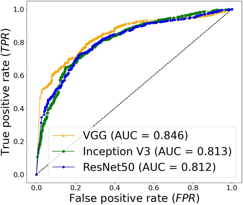
data, VGG performed better obtaining an Area Under the ROC specifically Convolutional Neural Networks (CNNs) have
Curve (AUC) value of 0.846. In the cases of ureteroscopy and become in the last years the standard for image analysis in
the combination of both datasets, ResNet50 achieved the better endoscopic image data [9].
results with AUC values of 0.987 and 0.940. The use of a
training dataset which comprehends both domains results in Few methods have been proposed for the detection and
general better performances, but performing a second stage of classification of tumors in the urinary system. This might
transfer learning achieves comparable ones. There is no single be due to the lack of publicly available, annotated datasets.
model which performs better in all scenarios, but ResNet50 is Shkolyar et al. [10] propose a model based on CNNs for
the network that achieves the better performances in most of papillary and flat bladder tumor detection. Yang et al. [11]
them. The obtained results open the opportunity for further
investigation with a view for improving lesion detection in compared 3 different CNNs architectures as well as the DL
endoscopic images of the urinary system. platform EasyDL, training the models with WL cystoscopy
Clinical relevance — A computer-assisted method based on images and showing that the use of DL-based methods can
CNNs could lead to a better early and adequate detection of achieve accuracy values comparable to the ones of a surgical
tumor-like lesions in the urinary tract. This could support expert. Ikeda et al. [12] propose the use of transfer learning
surgeons to perform better follow-ups and reduce the high
recurrence rates present on this disease. using GoogLeNet for the classification of endoscopic frames
with and without nonmuscle-invasive BC. However, all these
I. INTRODUCTION studies focus only on the bladder, missing the chance of
Urinary tract cancer is common and comprises differ- validating their results in the whole urinary tract.
ent types of lesions ranging from small benign tumors to As an initial step in the development of a fully automated
aggressive neoplasms with high mortality. In 2020, this CAD system to identify lesions in the urinary tract, we study
disease had 159,120 patients affected in the United States [1]. the implementation of 3 CNNs. Considering the disparity
Urothelial carcinoma can occur in kidney pelvis, ureters, in terms of occurrence of lesions in different organs in the
urinary bladder, and urethra. The predominant urinary tract urinary system, and the existing limitations in data collection
malignancy is Bladder Cancer (BC), which represents more and sharing, we study to which extent it is possible for a
than 90% of the cases [2]. CNN to make inferences when it is trained on a similar
Ureteroscopy (URS) and Cystoscopy (CYS) dare the main domain, and if the use of transfer learning is suitable to
techniques for upper tract urothelial cancer (UTUC) and BC achieve a better generalization in a different domain.
We propose a two-steps transfer learning strategy where
This work was supported by the ATLAS project. This project has received
funding from the European Union’s Horizon 2020 research and innovation initially the CNNs are trained separately on ureteroscopy and
programme under the Marie Skłodowska-Curie grant agreement No 813782. cystoscopy data. After evaluating each network on its own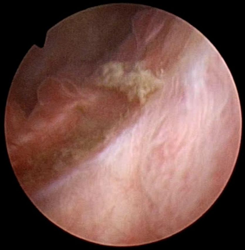
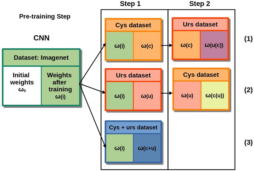
(a) (b)
Fig. 2: Training strategy implemented for the 3 training cases. Each sub-
table represents: [starting weights (loaded ones) | ”output” weights (weights
after training)]
TABLE I: Dataset of the images collected. Two types of procedures
were considered: ureteroscopy (urs) and cystoscopy (cys), and two light
modalities; White Light Imaging (WLI) and Narrow Band Imaging (NBI).
cases lesion no lesion
NBI WLI NBI WLI Total
(c) (d) cys 11 337 906 298 1,346 2,887
urs 13 28 1801 227 1158 3,214
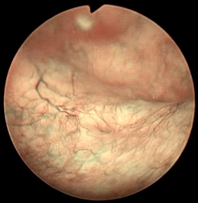
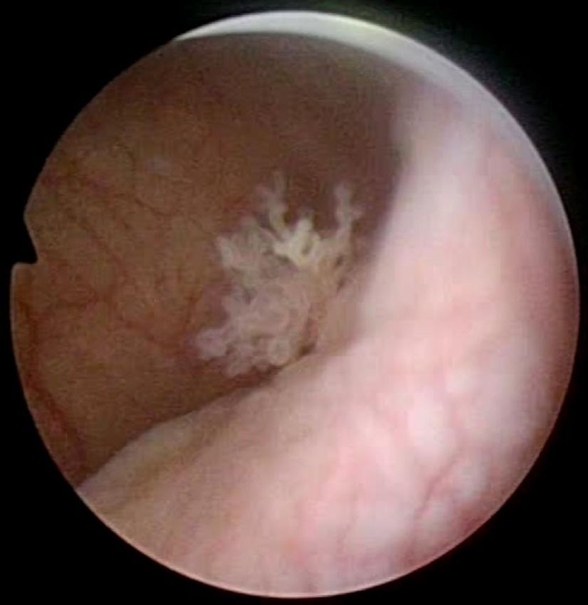
Fig. 1: Samples of endoscopic images in different organs in the urinary Total 24 3,072 3,029 6,101
system. (a)-(b) images from the bladder. (c)-(d) images from the ureter and
the kidney pelvis. (a), (c) correspond to samples of healthy tissue. (b), (d)
samples of images with lesions.
• Inception V3 uses an architectural block called incep-
tion module, it consists of convolutional kernels with
different sizes (1x1, 3x3 and 5x5) that are connected
domain and the other domain in which it was not originally
in parallel. The use of different kernel sizes allows the
trained (e.g. to identify lesions on cystoscopy images when
identification of image features at different scales [14].
the networks have been trained on ureteroscopy ones) we
• ResNet50 was presented during the ILSCRC 2015
perform a second training stage. We change the training data
classification challenge obtaining the 1st place. The
to a different domain i.e., the networks originally trained with
core idea of this model is the use of residual blocks
cystoscopy are then trained with ureteroscopy images and
to deal with the vanishing gradient problem, these are
the networks originally trained with ureteroscopy are trained
formed by 3 convolutional blocks in which a short skip
with cystoscopy images. We compare this approach to the
connection is placed between the input and the output
one of training the networks initially with both datasets.
of each residual block [15].
Determining to which extend the former approach is valid
of relevance, since full annotated datasets are not always On top of the last layer of the networks 3 Fully Connected
available. (FC) layers, of size 2048, 1024 and 2, were added. For the
first 2 FC layers, the activation function was ReLu while in
II. PROPOSED METHODS the case of the last layer sof tmax was used.
In this work, we investigated the use of transfer learning
using different CNN architectures trained on data from A. Dataset
bladder and upper urinary tract. Three state state-of-the- For this study, we extracted frames from videos of 24
art architectures built under different paradigms for image patients undergoing cystoscopy and/or ureteroscopy proce-
classification, and whose weights after being trained on dures using WLI and NBI. Of these videos, 13 correspond
Imagenet dataset are publicly available, were considered. to patients undergoing ureteroscopy and 11 cystoscopy. The
• VGG-16 is a 16-layer CNN model which has a feed- composition of the dataset is summarized in Table I. The
forward architecture consisting of 13 convolutional lay- images in the dataset corresponds to endoscopic images
ers and 5 max-pooling layers [13]. The architecture of different organs in the urinary tract including urethra,
starts with a convolutional layer with 64 kernels and bladder, ureter and kidneys.
this number is doubled after each pooling operation The extracted images were labeled into two classes (with
until it reaches 512 kernels. The pooling layers are lesion, and without lesion) under the supervision of an expert
placed after selected convolutional layers in order to surgeon. Some of the images include several artifacts such as
reduce dimension in the activation maps and hence of blood, floating debris, specular reflections, bubbles, motion
the number of parameters that the CNN needs to learn. blur, as shown in Fig. 1, which are common to appear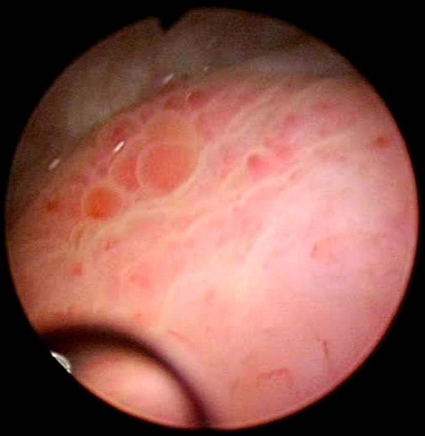
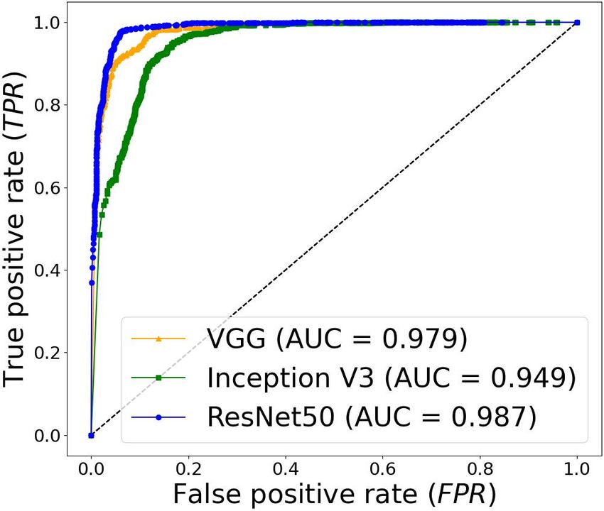
(a) Cystoscopy dataset                       (b) Ureteroscopy dataset                (c) Ureteroscopy + cystoscopy dataset
    Fig. 3: ROC curves obtained with the different models in the 3 datasets. The networks were trained and tested on data of the same domains.

in clinical practice and were included in the dataset to
have a realistic approach. The videos were acquired from
the European Institute of Oncology (IEO) at Milan, Italy
following the ethical protocol approved by the IEO and in
accordance with the Helsinky Declaration.
B. Training
   Transfer learning was implemented in a two-steps fash-
ion. A diagram depicting the training strategy is shown in
Fig. 2. The following 3 training scenarios were performed.
1) During step 1, the CNN was trained only on cystoscopy
data and tested on cystoscopy and ureteroscopy data. During
step 2, only ureteroscopy images were used for training, and
tested again in both cystoscopy and ureteroscopy images.
2) The same procedure was carried out but inverting training               Fig. 4: Comparison of the AU C values obtained for each of the models
data; during the first step ureteroscopy images were used for              trained. (orange: VGG16, green: Inception V3, blue: ResNet50). a) Results
                                                                           of step 1 (see Fig. 2), when testing models on data of a different domain
training and during the second stage only cystoscopy ones.                 than the one for which they were trained on (i.e. predictions of cys data
3) Training was performed with cystoscopy and ureteroscopy                 when trained on urs images, predictions of urs data when trained on cys).
images in one single step and the test was done using both                 b) Results of step 2, test data is of the same domain as the one of the second
                                                                           training. c) Separate results for the cystoscopy and ureteroscopy data on step
types of images.                                                           1 case (3).
   For the 3 methods, at step 1 the initial weights of the
networks were the ones obtained by previously training the
CNN on the Imagenet dataset ω(i) as depicted in Fig. 2.                    Area Under the Curve (AUC) of the Receiver Operating
At the second step, for case 1) the initial weights were the               Characteristic (ROC) curve. Where T P and T N are the
ones obtained by training the network in cystoscopy data                   number of endoscopic images correctly classified as having
ω(c); for case 2) the initial weights of the networks were                 lesions and no lesions, respectively, F N and F P are the
the ones obtained by training the network in ureteroscopy                  amount of images missclassified.
images ω(u).
   At both steps the CNNs were initially re-trained for                                                III. RESULTS
5 epochs with a learning rate of 0.0001. Subsequently,                        The ROC curves obtained with each of the models on
the weights from all the layers except the last 4 were                     different datasets during the first step are shown in Fig. 3. In
frozen. These layers were trained for 30 epochs using Adam                 cystoscopy images the model that performed better is VGG
optimization algorithm with a learning rate of 0.001. A                    with an AUC value of 0.846. In the case of ureteroscopy
3-fold cross validation strategy was used to estimate the                  images, and the dataset composed by the combination of
performances of the models at each step.                                   ureteroscopy and cystoscopy images, the model that obtains
   The models were implemented using Keras with Ten-                       the better results is ResNet50 with AUC values of 0.987
sorFlow as backend in Python 3.6 trained on a NVIDIA                       and 0.938 respectively. Results comparing the performance
GeForce RTX 280 GPU.                                                       before and after the second transfer learning step are shown
                                                                           Fig. 4.
C. Performance Metrics                                                        After completing Step 1 (see Fig. 2), Inception V3 is
   The performance evaluation of each of the tested CNNs                   the model with best performances on data from a different
was done by computing the True Positive Rate: T P R =                      domain than the one it was originally trained with. It obtains
   TP                                          FP
T P +F N , the False Positive Rate: F P R = F P +T N and the               an AUC value of 0.895 when it is trained on cystoscopy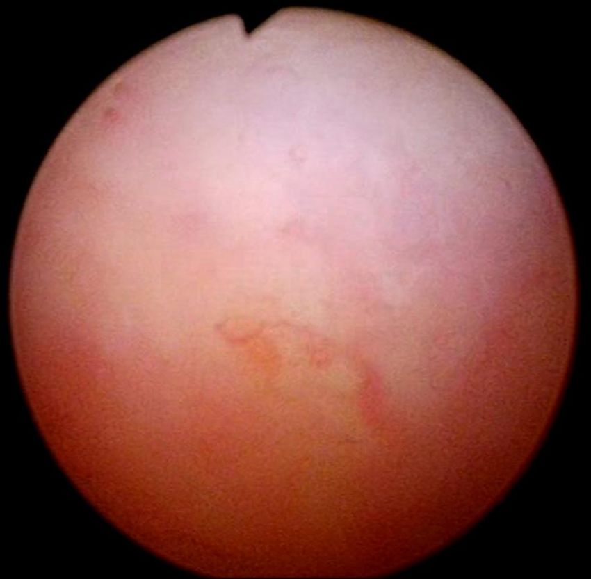
classification of tissue which can differentiate between tumor
                                                                                and other non-tumor kind of lesions.
                                                                                                     ACKNOWLEDGMENT
                                                                                   This work was supported by the ATLAS project. This project has received
                                                                                funding from the European Union’s Horizon 2020 research and innovation
                                                                                programme under the Marie Skłodowska-Curie grant agreement No 813782.
             (a)                    (b)                    (c)
                                                                                                             R EFERENCES
                                                                                 [1] R. L. Siegel, K. D. Miller, and A. Jemal, “Cancer statistics, 2020,”
                                                                                     CA: A Cancer Journal for Clinicians, vol. 70, no. 1, pp. 7–30, 2020.
                                                                                 [2] I. Kausch, M. Sommerauer, F. Montorsi, A. Stenzl, D. Jacqmin,
                                                                                     P. Jichlinski, D. Jocham, A. Ziegler, and R. Vonthein, “Photodynamic
                                                                                     diagnosis in non–muscle-invasive bladder cancer: a systematic review
                                                                                     and cumulative analysis of prospective studies,” European Urology,
                                                                                     vol. 57, no. 4, pp. 595–606, 2010.
             (d)                    (e)                    (f)                   [3] M. Cosentino, J. Palou, J. M. Gaya, A. Breda, O. Rodriguez-Faba,
                                                                                     and H. Villavicencio-Mavrich, “Upper urinary tract urothelial cell
Fig. 5: Image samples where a class activation heat-map was added to                 carcinoma: location as a predictive factor for concomitant bladder
show the regions of the images that the models recognized as lesions.                carcinoma,” World Journal of Urology, vol. 31, no. 1, pp. 141–145,
(a)-(c) correspond to images from bladder whereas (d)-(c) correspond to              2013.
images from the upper urinary tract. The images (a),(d) show samples of          [4] S. E. Wason and S. W. Leslie, “Ureteroscopy,” StatPearls, 2020,
predictions using as training data cystoscopy images. (b),(e) show samples of        (Accessed 29-11-2020), https://pubmed.ncbi.nlm.nih.gov/32809391/.
predictions using as training data ureteroscopy images. (c),(f) show samples     [5] C. Zheng, Y. Lv, Q. Zhong, R. Wang, and Q. Jiang, “Narrow band
of predictions of a combination of both datasets for training.                       imaging diagnosis of bladder cancer: systematic review and meta-
                                                                                     analysis,” BJU international, vol. 110, no. 11b, pp. E680–E687, 2012.
                                                                                 [6] R. J. Sylvester, A. P. Van Der Meijden, W. Oosterlinck, J. A. Witjes,
                                                                                     C. Bouffioux, L. Denis, D. W. Newling, and K. Kurth, “Predicting
images and tested on ureteroscopy ones, and 0.783 when                               recurrence and progression in individual patients with stage Ta T1
                                                                                     bladder cancer using eortc risk tables: a combined analysis of 2596
trained on ureteroscopy and tested on cystoscopy images.                             patients from seven eortc trials,” European Urology, vol. 49, no. 3,
   The models that show a better improvement from the first                          pp. 466–477, 2006.
to the second transfer learning step are VGG in cystoscopy                       [7] R. Chou, S. Selph, D. I. Buckley, R. Fu, J. C. Griffin, S. Grusing, and
                                                                                     J. L. Gore, “Comparative effectiveness of fluorescent versus white light
data, changing from a AUC value of 0.691 to 0.834 and                                cystoscopy for initial diagnosis or surveillance of bladder cancer on
ResNet50 in ureteroscopy images improving from an initial                            clinical outcomes: systematic review and meta-analysis,” The Journal
AUC value of 0.897 to 0.979 after the second step. Some                              of Urology, vol. 197, no. 3, pp. 548–558, 2017.
                                                                                 [8] C. C. Poon, Y. Jiang, R. Zhang, W. W. Lo, M. S. Cheung, R. Yu,
samples images of the predictions in the test dataset, with                          Y. Zheng, J. C. Wong, Q. Liu, S. H. Wong et al., “AI-doscopist:
the heap-map obtained using Grad-CAM for class activation                            a real-time deep-learning-based algorithm for localising polyps in
visualization [16] to show the regions of the image that the                         colonoscopy videos with edge computing devices,” NPJ Digital
                                                                                     Medicine, vol. 3, no. 1, pp. 1–8, 2020.
model recognized as part of the lesions, are shown in Fig. 5.                    [9] I. Patrini, M. Ruperti, S. Moccia, L. S. Mattos, E. Frontoni, and
                                                                                     E. De Momi, “Transfer learning for informative-frame selection in
                                                                                     laryngoscopic videos through learned features,” Medical & Biological
                                                                                     Engineering & Computing, pp. 1–14, 2020.
            IV. DISCUSSION & CONCLUSION                                         [10] E. Shkolyar, X. Jia, T. C. Chang, D. Trivedi, K. E. Mach, M. Q.-H.
                                                                                     Meng, L. Xing, and J. C. Liao, “Augmented bladder tumor detection
   All the networks performed better on detecting lesions on                         using deep learning,” European Urology, vol. 76, no. 6, pp. 714–718,
ureteroscopy than cystoscopy images. However, this might                             2019.
                                                                                [11] R. Yang, Y. Du, X. Weng, Z. Chen, S. Wang, and X. Liu, “Automatic
be related to the fact that the former dataset is bigger and                         recognition of bladder tumours using deep learning technology and its
has more patient cases than the later one.                                           clinical application,” The International Journal of Medical Robotics
                                                                                     and Computer Assisted Surgery, p. e2194, 2020.
   For the models tested, the performance was higher when                       [12] A. Ikeda, H. Nosato, Y. Kochi, T. Kojima, K. Kawai, H. Sakanashi,
the networks were trained on cystoscopy images and tested                            M. Murakawa, and H. Nishiyama, “Support system of cystoscopic
on ureteroscopy images than the opposite scenario. Con-                              diagnosis for bladder cancer based on artificial intelligence,” Journal
                                                                                     of Endourology, vol. 34, no. 3, pp. 352–358, 2020.
sidering that the dataset of ureteroscopy is larger than the                    [13] K. Simonyan and A. Zisserman, “Very deep convolutional networks
ureteroscopy one, a possible explanation to this result might                        for large-scale image recognition,” arXiv preprint arXiv:1409.1556,
be related to the anatomical properties of the upper urinary                         2014.
                                                                                [14] C. Szegedy, V. Vanhoucke, S. Ioffe, J. Shlens, and Z. Wojna, “Re-
tract and the bladder. While one is a tubular structure which                        thinking the inception architecture for computer vision,” in IEEE
in case of lesions might look like an obstruction, in the                            Conference on Computer Vision and Pattern Recognition, 2016, pp.
bladder the tissue has more visual diversity.                                        2818–2826.
                                                                                [15] K. He, X. Zhang, S. Ren, and J. Sun, “Deep residual learning for image
   The use of a training dataset which comprehends both                              recognition,” in Proceedings of the IEEE Conference on Computer
domains resulted in better performances, as expected, but                            Vision and Pattern Recognition, 2016, pp. 770–778.
performing a second stage of transfer learning achieves com-                    [16] R. R. Selvaraju, M. Cogswell, A. Das, R. Vedantam, D. Parikh, and
                                                                                     D. Batra, “Grad-cam: Visual explanations from deep networks via
parable performances. In general ResNet50 is the network                             gradient-based localization,” in Proceedings of the IEEE international
that achieves the better performances in both scenarios.                             conference on computer vision, 2017, pp. 618–626.
Future work will include pathological confirmation of the
type of tissue as ground truth to perform a more detailed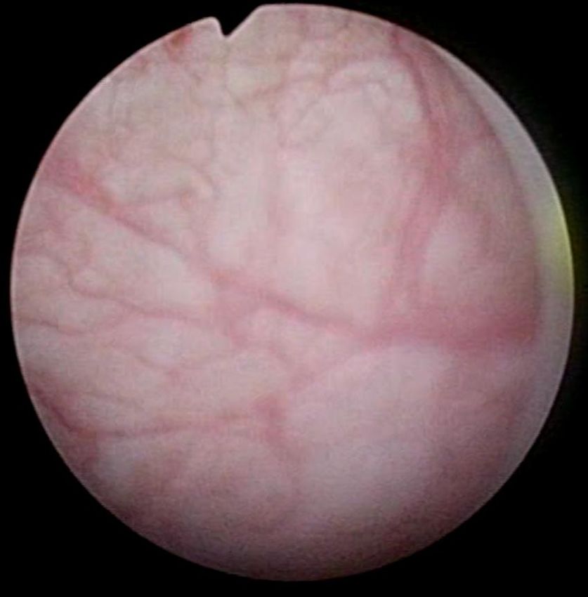
You can also read