A 46-year-Oldman with Gynaecomastia and Cirrhosis Hepatis Child Plug A. A case Report
←
→
Page content transcription
If your browser does not render page correctly, please read the page content below
Journal of Endocrinology, Tropical Medicine, and Infectious Disease (JETROMI) Vol. 03, No. 2, 2021 / 66-71

A 46-year-Oldman with Gynaecomastia and Cirrhosis
Hepatis Child Plug A. A case Report
Syarifah Dian*1, Melati Silvani2
1
  Department of Internal Medicine, Faculty of Medicine, University Sumatera Utara, Medan, North
Sumatera, Indonesia
2
  Division of Endocrinologist, Department of Internal Medicine, Faculty of Medicine, University
Sumatera Utara, Medan, North Sumatera, Indonesia

               ABSTRACT
               Background: Gynecomastia is defined as the presence of excessive breast tissue in males,
               which can appear unilateral or bilateral. Bilateral prepubertal gynecomastia in the absence of
               endocrine abnormalities is extremely rare, with only a few cases in literature.
               Case Presentation: A 46-year-old man presented with a 2 years history of bilateral breast
               enlargement and pain. There is heartburn, shrinking testicles, and decreased sexual drive.
               Vital examination showed normal. BMI : 24. 0 kg/m². Head dan neck: normal. On the
               chest both breasts are enlarged as avocados, palpable time around areola, chewy contingency,
               press pain, galactorrhea negative. Lung and cast are normal. In the abdominal: hepar, lien
               and renal are not palpable, and ascites are not found. There is an atrophy of testicles. Previous
               history of hepatic cirrhosis with Esophageal Varicose Veins caused by Hepatitis B.
               Laboratory examination Estradiol 98.71 pg/mL (range:
Journal of Endocrinology, Tropical Medicine, and Infectious Disease (JETROMI) Vol. 03, No. 2, 2021 67
ABSTRAK
Latar Belakang: Ginekomastia didefinisikan sebagai terdapat jaringan payudara yang
berlebihan pada laki-laki, yang dapat muncul unilateral atau bilateral. Ginekomastia
prepubertal bilateral tanpa adanya kelainan endokrin sangat jarang terjadi, terdapat hanya
beberapa kasus dalam literatur.
Presentasi Kasus: Seorang pria berusia 46 tahun mengalami pembesaran payudara
bilateral selama 2 tahun dan juga merasa sakit. Ada mulas, testis menyusut, dan penurunan
gairah seksual. Pemeriksaan vital menunjukkan normal. Didada kedua payudara membesar
seperti buah alpukat, sekitar areola menghitam, konsistensi kenyal, nyeri, galactorrhea (-).
Paru dan jantung. normal. Pada abdominal: hepar, lien dan ginjal tidak teraba, dan ascites
(-). Ada atrofi testis. Sebelumnya riwayat sirosis hati dengan varises Esopagus yang
disebabkan oleh Hepatitis B. IMT: 24,0 kg/m². Pemeriksaan laboratorium Estradiol 98,71
pg/mL (rentang: < 62), testosteron: 8,66 ng/mL (rentang: 3,0-10,6), LH 5,53 mlU/mL, FSH
6,49 mlU/mL, Prolactin 5,08 ng/mL; Total T4 5,57 ng/dL , TSH 1,84 μIU/mL. SGOT 53 U/L,
SGPT 71 U/L, Hepatitis B (+), HBsAg Reaktif S/CO 414,46; HBeAg: Non Reaktif, DNA
HBV tidak terdeteksi dan albumin 3,6 g/dL. Fibroscan: Cirrhosis Hepatis. USG Abdominal:
Cirrhosis Hepatis. Gastroskopi: Esophageal Varises (F1, LI, CB) + Pan Gastropati.
Kesimpulan: Dilaporkan Seorang pria berusia 46 tahun dengan ginaekomastia bilateral
dan Cirrhosis Hepatis Child Plug A, diberikan pengobatan konservatif dengan tamoxiphen.
Kata Kunci: Ginekomastia, Hirosis Hepatis, tamoxiphen
Received 12 May 2021 | Revised 27 May 2021 | Accepted 31 May 2021
1 Introduction
Gynecomastia is defined as the benign enlargement of the male breast, usually, a result of stromal
proliferation and ductal hyperplasia. which can appear unilateral or bilateral. Bilateral
gynecomastia is frequently found in the neonatal period, early in puberty, and with increasing
age. Prepubertal bilateral gynecomastia in the absence of endocrine abnormalities is extremely
rare, with only a few cases in literature.[1]
Among the underlying causes, 3 groups of triggers are distinguished: physiologic, pathologic and
pharmacologic/toxic. Pathophysiologically, female hormone imbalance has been shown to trigger
glandular growth. This imbalance may in turn also be of pathologic or physiologic/idiopathic
origin. There is strong evidence for the stimulating effect of estrogen on breast tissue
development. Any disorder or medication leading to female hormone imbalance can trigger
gynecomastia. Endogenous diseases like hyperthyroidism, chronic liver disease, primary or
secondary gonadal failure, androgen resistance syndromes, medication, and lifestyle factors like
drug abuse represent typical causes of gynecomastia [2]. Classification of gynecomastia [3].
Terdiri dari Grade I: minimal hypertrophy 500 g breast tissue with
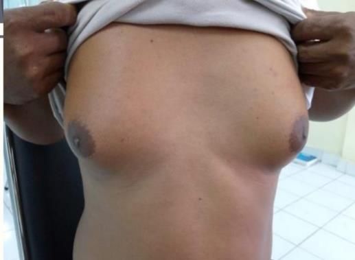
grade I ptosis and Grade IV. severe hypertrophy with grade II–III ptosis.Journal of Endocrinology, Tropical Medicine, and Infectious Disease (JETROMI) Vol. 03, No. 2, 2021 68 In most cases, gynecomastia is caused by short- term hormonal fluctuations that limit themselves physiologically, which can occur neonatally, during puberty, and in elderly men. In the neonatal period, a preliminary physiologic bilateral mammary glandular swelling is triggered by maternal placental estrogens and resolves within a few weeks after birth. In adolescence, physiologic pubertal gynecomastia can develop and may last up to 6 months. This is triggered by a relative estrogen excess, mainly due to peripheral aromatization of the testicular and adrenal androgens. Testicular testosterone production increases in late puberty and leads to a spontaneous regression of the gynecomastia [4]. Due to a decrease in testosterone production and an increase in sex hormone-binding globulin, gynecomastia is also common in elderly men. 2 Case report: An 46-year-old-man presented with a 2 years history of bilateral breast enlargement and pain. There is heartburn, shrinking testicles, decreased sexual drive, and no confidence. No history or sign of galactorrhea existed. Physical examination showed normal. BMI: 24.0 kg/m². Head dan Neck: normal. On the chest, both breasts are enlarged as avocados, palpable time around areola, chewy contingency, press pain, galactorrhea negative. Lung and cor is normal. In the abdominal: hepar, lien and renal is not palpable, and ascites are not found. gender: Male and there is atrophy of testicles. Previous history of hepatic cirrhosis with Esophageal Varicose Veins caused by Hepatitis B. Laboratory Results Urine, blood sugar and cholesterol are normal. Estradiol 98.71 pg/mL (high), Testosterone: 8.66 ng/mL (high), LH 5.53 mlU/mL, FSH 6.49 mlU/mL, Prolactin 5.08 ng/mL; Total T4 5.57 ng/dL , TSH 1.84 μIU/mL. SGOT 53 U/L, SGPT 71 U/L, Hepatitis B (+), HBsAg Reactive S/CO 414.46; HBeAg: Non Reactive, HBV DNA Undetectable and Albumin 3.6 g/dL. Mammae Ultrasound: Both mamae enlarge asymmetrically, left is larger than the right. Both mammae consist of fatty and fibroglandural components, but are more dominant fibroglandular in both mamaes especially the leftones (fig 1). There is no visible enlargement of the lymph nodes on both axillas. Impressions: Gynecomastia on the right and left mammae. Fibroscan: Sirosis Hepatis. Abdominal ultrasound: Hepar, irreguler surface, blunt edge, shrinking size, coarse homogeneous parenym. Conclusion: Cirrhosis Hepatis. Gastroscopy: Esophageal Varicose Veins (F1, LI, CB) + Pan Gastropathy. Diagnosis: gynaecomastia in the right and left mammae and Cirrhosis Hepatis Child Plug A, Hepatitis B, Varicose Veins of the Oesophagus. Diagnosis is bilateral gynaecomastia with Cirrhosis Hepatis Child Plug A, given conservative treatment with tamoxiphen for 1 month
Journal of Endocrinology, Tropical Medicine, and Infectious Disease (JETROMI) Vol. 03, No. 2, 2021   69

                                                    Figure 1

3    Discussion

Gynecomastia correlated directly with body mass index (BMI) and with age. Individuals with
more severe cirrhosis usually are very thin, although the BMI may be increased owing to the
pres¬ ence of ascites or peripheral edema. It has been suggested that gynecomastia in all subjects
is the result of a reduced circulating testosterone concentration, an elevated estrogen
concentration, or both resulting in an increase in the estrogen-testosterone ratio.[5] Men with
cirrhosis have decreased production of testosterone, increased production of androstenedione,
increased conversion of androstenedione to estrone, and increased levels of sex hormone-binding
globulin.[6] same in the control subjects and those with cirrhosis, even though the estrogen-
testosterone ratios were much lower in the controls.

In the case of true gynecomastia, clinical examination by palpation followed by ultrasound
examination and if necessary mammography reveal glandular tissue. Palpation of the breast
should be able to clinically distinguish fatty tissue from glandular tissue and should include a
more detailed assessment of the breast such as ptosis, skin excess, and nipple retraction. The
physical examination should also extend to testicular palpation in order to detect testicular
changes and atrophies at an early stage and to pave the way for further diagnostic measures. In
particular, in young men with a negative history and bilateral gynecomastia, the incidence of
testicular endocrine tumors is showing a significant increase [7]. The diagnosis of gynecomastia
must be confirmed histologically by a sonographically, showing a 90% sensitivity and specificity
[8]. Standard diagnostics include laboratory tests like human choric gonadotropin, luteinizing
hormone, thyroid-stimulating hormone, testosterone, and estradiol [9].

Conservative and Surgical Management
he therapy of gynecomastia may be conservative or surgical. Conservative therapy must be based
on the cause of the gynecomastia. Self-limiting physiologic forms of gynecomastia should be
followed up until resolved. In the case of persistence or progress, if necessary, laboratory
diagnostic tests should be repeated to detect hormonal processes as quickly as possible. A
Journal of Endocrinology, Tropical Medicine, and Infectious Disease (JETROMI) Vol. 03, No. 2, 2021 70
conservative therapy option is the administration of serum estrogen receptor modulators such as
tamoxifen. Tamoxifen at a daily oral dose of 20 mg for up to 3 months has shown good results in
randomized and non-randomized trials. and post-traumatic deep vein thrombosis, are rarely
reported [9]. The use of anastrozole, an aromatase inhibitor, is not recommended as it did not
show more effectiveness than placebo in boys with pubertal gynecomastia [11]. If conservative
pharmacologic therapy attempts are to be considered, it should be noted that hypertrophic
glandular tissue becomes irreversibly fibrotic by way of remodeling after no more than 12 months.
This fibrous tissue cannot be removed conservatively and proceeding with surgical management
must be considered.[12] The indication for surgical therapy is based on the patient’s suffering,
including psychosocial stress and pain as well as cosmetic distortion [13]. The aim of all surgical
procedures is to remove the hypertrophic fibrotic glandular tissue and to reestablish the male
breast shape. Therapeutic techniques include breast tissue resection, liposuction, and combined
techniques. From stage III onwards, reduction mammoplasty should be explored taking into
Regression of gynecomastia is seen in up to 80% of patients [14]. However, the available data on
tamoxifen stems from few studies with very small case numbers [15]. Adverse side effects,
including epigastric distress consideration the extent of the hypertrophic tissue and the expertise
of the surgeon [16]. For milder forms, the periareolar edge cut to the mastectomy can suffice.
4 Conclusion.
Reportedly A 46-year-old man presented with gynaecomastia bilateral with Cirrhosis Hepatis
Child Plug A, given conservative treatment with tamoxiphen for 1 month
REFERENCES
[1] Antonio Cutrupi, , M. Barbalace , C. Covani , S. Malavenda. Bilateral Gynecomastia: A
report of one case. Int J Ped & Neo Heal. vol.1, no,1, pp.04-06. 2016.
DOI:10.25141/2572-4355-2017-1.0004
[2] Johnson RE, Murad MH: Gynecomastia: phathophysiology, evaluation, and
management. Mayo Clin Proc vol.84:pp,1010–5. 2009
[3] Rohrich RJ, Ha RY, Kenkel JM, Adams WP Jr: Classification and management of
gynecomastia: defining the role of ultrasound-assisted liposuction. Plast Reconstr Surg;
vol,111:pp.909–23. 2003
[4] Nordt C, Divanta A: Gynecomastia in adolescents. Curr Opin Pediatr vol.20:pp.375–82.
2008
[5] Frantz AG, Wilson JD. Endocrine disorders of the breast. In: Wilson JD, Foster DW, eds.
Williams Textbook ofEndocrinology. Philadelphia, Pa: WB Saunders Co pp.410-16.
1985
[6] Kley, HK, Niederau C, Stremmel W, Lax R, Strohmeyer G, Kruskemper HL. Conversion
of androgens to estrogens in idiopathic hemochromatosis: comparison with alcoholic
liver cirrhosis. J Clin Endocrinol Metab vol.61:pp.1-6. 1985
[7] Bowers SP, Pearlman NW, McIntyre RC Jr, Finlayson CA, Huerd S: Cost-effective
management of gyneco- mastia. Am J Surg, vol.176, pp,638–41. 1998
[8] Morcos RN, Kizy T: Gynecomastia. When is treatment indicated? J Fam Pract vol.61,
pp.719–25. 2012
[9] Evans GF, Anthony T, Turnage RH, et al: The diagnos- tic accuracy of mammography in
the evaluation of male breast disease. Am J Surg vol.181:pp. 96–100. 2001Journal of Endocrinology, Tropical Medicine, and Infectious Disease (JETROMI) Vol. 03, No. 2, 2021 71
[10] Ting ACW, Chow LWC, Leung YF: Comparison of ta- moxifen with danazol in the
management of idiopathic gynecomastia. Am Surg vol.66:pp.38–40. 2000
[11] Plourde PV, Reiter EO, Jou HC, et al: Safety and efficacy of anastrozole for the treatment
of pubertal gynecomastia: a randomized,double-blind, placebo-controlled trial. J Clin
Endocrinol Metab vol.89: pp.4428-33. 2004
[12] Bannayan GA, Hajdu SI: Gynecomastia: clinicopathologic study of 351 cases. Am J Clin
Pathol vol.57:pp. 431-7. 1972
[13] Ordaz DL, Thompson JK: Gynecomastia and psychological functioning: a review of the
literature. Body Image vol.15:pp.141–148. 2015
[14] Hanavadi S, Banerjee D, Monypenny IJ, Mansel RE: The role of tamoxifen in the
management of gynaecomastia. Breast vol.15, no.79:pp.276–80. 2006
[15] Johnson RE, Kermott CA, Murad MH: Gynecomastia – evaluation and current treatment
options. Ther Clin Risk Manag vol.7:pp.145–8. 2011
[16] Wolter A, Scholz T, Diedrichson J, Liebau J: Surgical treatment of gynecomastia:
an algorithm (Article in German). Handchir Mikrochir Plast Chir vol.45:pp.73-
79. 2013You can also read