Expression and Significance of AQP3 in Cutaneous Lesions
←
→
Page content transcription
If your browser does not render page correctly, please read the page content below
Hindawi
Analytical Cellular Pathology
Volume 2021, Article ID 7866471, 6 pages
https://doi.org/10.1155/2021/7866471

Research Article
Expression and Significance of AQP3 in Cutaneous Lesions

          Dongfeng Niu , Yanhua Bai , Qian Yao , Wei Hou, Lixin Zhou, Xiaozheng Huang,
          and Chen Zhao
          Key Laboratory of Carcinogenesis and Translational Research (Ministry of Education), Department of Pathology, Peking University
          Cancer Hospital & Institute, Beijing, China

          Correspondence should be addressed to Dongfeng Niu; dongfengniu@foxmail.com

          Received 21 May 2021; Accepted 5 October 2021; Published 26 October 2021

          Academic Editor: Giovanni Tuccari

          Copyright © 2021 Dongfeng Niu et al. This is an open access article distributed under the Creative Commons Attribution License,
          which permits unrestricted use, distribution, and reproduction in any medium, provided the original work is properly cited.

          Aquaporin 3 (AQP3) is the membrane channel of water and involved in fluid homeostasis. The aim of this study was to reveal the
          expression and significance of AQP3 in cutaneous lesions. We analyzed AQP3 mRNA levels using RT-PCR in 311 cutaneous
          lesions and confirmed AQP3 expression in these lesions by immunohistochemistry. AQP3 mRNA was detected in normal
          epidermis, seborrheic keratosis, solar keratosis, Bowen’s disease, squamous cell carcinoma, eccrine poroma, apocrine
          carcinoma, and sebaceoma; however, AQP3 mRNA was absent in basal cell carcinoma, nevocellular nevus, or malignant
          melanoma. By immunohistochemistry, diffuse AQP3 expression was seen in all keratotic lesions including seborrheic keratosis,
          verruca vulgaris, molluscum contagiosum, solar keratosis, Bowen’s disease, and squamous cell carcinoma. Diffuse AQP3
          expression was also present in all extramammary Paget’s disease. No AQP3 staining was obtained in basal cell carcinoma.
          Positive AQP3 staining was seen in sweat gland tumors including hidradenoma, eccrine poroma, and apocrine carcinoma.
          Among sebaceous tumors, AQP3 expressed diffusely in all sebaceous hyperplasia and sebaceous adenoma, but not in sebaceous
          carcinomas. Only focal AQP3 staining was seen in nevocellular nevus and no AQP3 staining in melanoma. Our findings
          indicate the function of AQP3 maintained in most skin tumors. AQP3 may be used for differential diagnosis in skin tumors.

1. Introduction                                                        skin squamous cell carcinomas by immunohistochemical
                                                                       staining [7].
Aquaporin 3 (AQP3) is a water-transporting aquaglycero-                    Skin cancer is one of the most common human malig-
porin and plays a significant role in physiologic functions             nancies with increasing incidence worldwide [14–16]. Skin
of reabsorption and secretion in the kidney [1, 2], epidermis          cancer is mainly divided into two large groups: cutaneous
[3, 4], pancreas [5], prostate [6], etc. The AQP3 protein              melanoma and nonmelanoma skin tumor. Malignant mela-
expression was observed in some normal tissue such as pitu-            noma is one of the most aggressive human cancers and
itary cells, salivary gland, thymic epithelium, bronchial epi-         causes 75% mortality in all skin cancer in the United States
thelial cells and pulmonary alveolar epithelium, pancreatic            [17]. Nonmelanoma skin tumor accounts for nearly 95% of
islet, and squamous epithelium of the esophagus, uterine               cutaneous neoplasms, and the most common lesions include
cervix, skin [7], etc. Furthermore, AQP3 has been detected             basal cell carcinoma, squamous carcinoma, and sebaceous
in malignancy of several organs including the colon [8],               carcinoma [15]. Frequent exposure to ultraviolet radiation
ovary [9], breast [10], pancreas [11], and prostate [12].              or sunlight acts as the major factor for both groups of skin
Expression of AQP3 in these tumors was reported to corre-              cancer [14, 17]. With the development of clinical examina-
late with tumorigenesis, invasion, metastasis, and prolifera-          tion, such as dermoscopy and in vivo reflectance confocal
tion [8–13]. In a previous study, we found that AQP3                   microscopy, the diagnosis and differential diagnosis of skin
protein was widely distributed in neoplastic tissues including         cancer have become more precise.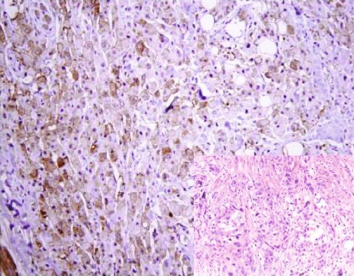
2 Analytical Cellular Pathology
In a recent study [7], we found that AQP3 protein was (SB), squamous cell carcinoma (SCC), Bowen’s disease
immunohistochemically diffusely positive in the cytoplasmic (BD), and apocrine carcinoma (AC) but not expressed in
membrane of normal cutaneous cells including squamous nevocellular nevus (NN), basal cell carcinoma (BC), and
epithelium, sudoriferous gland, sebaceous gland, and apo- malignant melanoma (MM). These results were in accor-
crine gland but not in melanocytes. We also found a high dance with our immunohistochemical observation.
positive expression of AQP3 in skin squamous carcinomas.
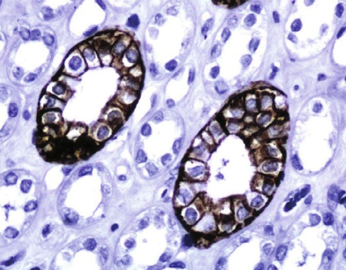
However, limited information on AQP3 expression in other 3.2. AQP3 Expression in Normal Control Tissue. We used the
skin tumors is available. In this study, we investigated the sections from normal kidney tissue as a positive control. The
expression and significance of AQP3 in a large series of skin expression of AQP3 was specifically localized in the cyto-
lesions, using RT-PCR and immunohistochemistry. plasmic membrane of the collecting duct cells of the kidney
(Figure 2(a)) and squamous cells of the skin (Figure 2(b)) as
2. Materials and Methods previously described [7].
2.1. Case Selection. Our study was approved by the local 3.3. AQP3 Expression in Benign Skin Lesions and Tumors.
research ethics committee of Peking University Cancer Hos- The immunohistochemical staining of AQP3 in skin lesions
pital & Institute. We included 311 surgically resected skin is listed in Table 1. Among the nonneoplastic skin lesions, all
lesions from the routine surgical pathology file of Peking cases of seborrheic keratosis (n = 14), verruca vulgaris
University Cancer Hospital & Institute, including 74 benign (n = 16), molluscum contagiosum (n = 5), and sebaceous
nonneoplastic lesions (14 seborrheic keratoses, 16 verruca hyperplasia (n = 13) showed diffuse staining for AQP3
vulgaris, 13 sebaceous hyperplasias, 5 molluscum contagio- (Figures 3(a)–3(d)). Among the 26 nevocellular nevi, 8
sum, and 26 nevocellular nevi), 40 benign skin tumors (7 showed focal AQP3 staining and the remaining 18 were neg-
hidradenomas, 7 eccrine poromas, 16 sebaceomas, and 10 ative. Among the benign skin tumors, positive AQP3 stain-
sebaceous adenomas), and 197 premalignant lesion and ing was seen in all 7 hidradenomas (all diffuse), 6/7 eccrine
malignant tumors (24 solar keratoses, 26 Bowen’s diseases, poromas (all diffuse), all 16 sebaceomas (4/16 focal, 4/16
43 squamous cell carcinomas, 32 basal cell carcinomas, 16 intermediate, and 8/16 diffuse), and all 10 sebaceous adeno-
extramammary Paget’ disease, 9 sebaceous carcinomas, 19 mas (all diffuse).
apocrine carcinomas, and 28 malignant melanomas). The 3.4. AQP3 Expression in the Premalignant Lesion and
cases are summarized in Table 1. The diagnosis of all these Malignant Skin Tumors. The immunohistochemical results
cases was confirmed by 2 pathologists (DN and YB). The of AQP3 staining in premalignant solar keratoses and malig-
blocks for the study also included normal tissues adjacent nant skin tumors are summarized in Table 1. All 24 solar
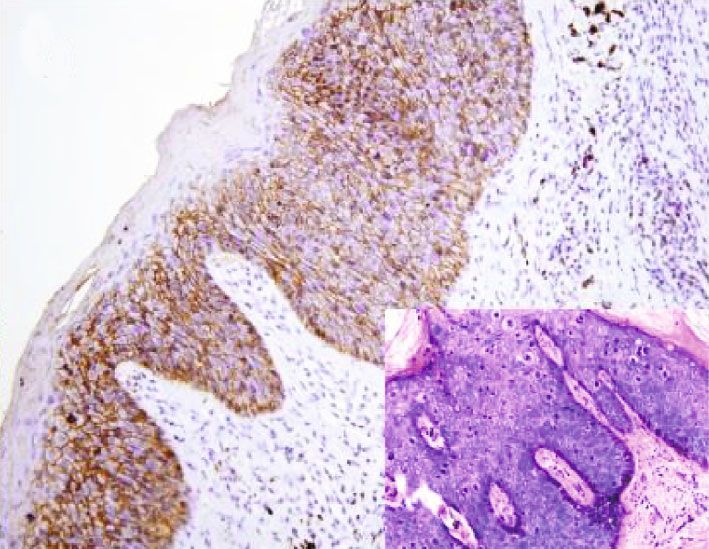
to the lesions. keratoses (Figure 4(a)) and 16 extramammary Paget’s dis-
2.2. RNA Extraction and RT-PCR. Total RNA was isolated eases (Figure 4(c)) showed diffuse AQP3 staining. Positive
from various formalin-fixed and paraffin-embedded skin AQP3 staining was seen in 26 Bowen’s diseases
lesion tissues with Recover ALL™ Total Nucleic Acid Isola- (Figure 4(b)) (2 focal, 24 diffuse), 43 squamous cell carcino-
tion Kit (Applied Biosystems, USA). We designed the spe- mas (Figure 4(d)) (3 focal, 7 intermediate, and 33 diffuse),
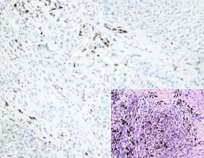
cific PCR primers targeted for AQP3 and PGK1 (as an and 19 apocrine carcinomas (Figure 4(e)) (3 intermediate,
internal control) as shown in Table 2. HotstarTaq DNA 16 diffuse). All 32 basal cell carcinomas (Figure 4(f)), 9 seba-
polymerase kit (Qiagen, USA) was performed for amplifica- ceous carcinomas (Figure 4(g)), and 28 malignant melano-
tion. PCR conditions were described previously [11]. mas (Figure 4(h)) were negative for AQP3 staining.
2.3. Immunohistochemical Staining. The immunohistochem- 4. Discussion
ical staining was performed as described previously [7]. The
rabbit polyclonal anti-AQP3 antibody was obtained from Aquaporin3 (AQP3), one of the aquaglyceroporins, is
Sigma (HPA014924, Sigma, USA, 1 : 4000 dilution). The responsible for transporting water and glycerol and main-
expression of AQP 3 was evaluated as negative (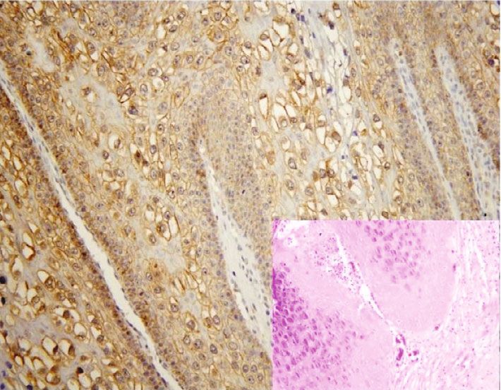
Analytical Cellular Pathology                                                                                                                                     3

                                      Table 1: Summary of immunohistochemistry for AQP3 in skin lesions.

                                                                                          N         Absent           Focal              Intermediate       Diffuse
                                                          Seborrheic keratosis            14          0               0                      0               14
                                                            Verruca vulgaris              16          0               0                      0               16
Nonneoplastic lesions                                    Sebaceous hyperplasia            13          0               0                      0               13
                                                        Molluscum contagiosum              5          0               0                      0                5
                                                          Nevocellular nevus              26         18               8                      0                0
                                                             Hidradenoma                  7           0               0                      0               7
                                                            Eccrine poroma                7           1               0                      0                6
Benign tumors
                                                              Sebaceoma                   16          0               4                      4                8
                                                          Sebaceous adenoma               10          0               0                      0               10
                                                             Solar keratosis              24          0               0                      0               24
                                                            Bowen’s disease               26          0               2                      0               24
                                                        Squamous cell carcinoma           43          0               3                      7               33
                                                          Basal cell carcinoma            32         32               0                      0                0
Premalignant lesion and malignant tumors
                                                             Paget’s disease              16          0               0                      0               16
                                                          Sebaceous carcinoma             9          9                0                      0                0
                                                          Apocrine carcinoma              19          0               0                      3               16
                                                          Malignant melanoma              28         28               0                      0                0
Absent, negative; mild, focal (1 to 9% of cells); moderate, intermediate (10 to 50%); diffuse (more than 50%).

                                                        Table 2: Primer pairs used in RT-PCR.

Target              Gene accession                                 Primer sequence                                   AT (°C)                     Product size (bp)
                                                    F: 5 ′ -GACAGAAGGAGCTGGTGTCC-3 ′                                   58                              199
AQP 3                 NM_004925
                                                   R: 5 ′ -AGAGTGACAGCAAAGCCAAAG-3 ′

                                                   F: 5 ′ -GCTGACAAGTTTGATGAGAAT-3 ′                                   58                              359
PGK1                  NM_000291
                                                   R: 5 ′ -AGGACTTTACCTTCCAGGAGC-3 ′                                            No sample
                                   Marker

                                                                                                                       RT (-)
                                            NST

                                                                           SCC

                                                                                                                MM
                                                  Sok

                                                                                               NN
                                                        Sek

                                                                                         AC
                                                                                  BD

                                                                                                      BC
                                                                     SB
                                                              EP

                          AQP3

                          PGK1

Figure 1: AQP3 mRNA level in skin normal tissue and lesions. RT-PCR showed AQP3 mRNA in normal squamous tissue (NST), solar
keratosis (SoK), seborrheic keratosis (SbK), eccrine poroma (EP), sebaceoma (SB), squamous cell carcinoma (SCC), Bowen’s disease
(BD), and apocrine carcinoma (AC), but AQP3 mRNA was absent in nevocellular nevus (NN), basal cell carcinoma (BC), and malignant
melanoma (MM).

from the lowermost layer of the epidermis and less com-                          noma, and sebaceoma all show retained expression of
monly from the outer root sheath of the pilosebaceous unit.                      AQP3, suggesting that loss of AQP3 may contribute to the
Basal cell carcinoma cells share many common features with                       carcinogenesis of sebaceous carcinoma.
follicular epithelium such as hair bulbs, follicular bulges, and                     The absent expression of AQP3 in basal cell carci-
follicular matrix cells [21]. In our previous study, we did not                  noma and sebaceous carcinoma also has some diagnostic
observe AQP3 expression in hair follicles [7]. No expression                     value. In clinical practice, sometimes, it is a big challenge
of AQP3 in basal cell carcinoma further confirms the hair                         to distinguish basal cell carcinoma from basaloid squa-
follicle origin of basal cell carcinoma. Sebaceous carcinoma                     mous cell carcinoma or metaplastic basal cell carcinoma
shows no expression of AQP3 whereas benign sebaceous                             from poorly differentiated squamous cell carcinoma.
lesions including sebaceous hyperplasia, sebaceous ade-                          AQP3 immunohistochemical staining is helpful in this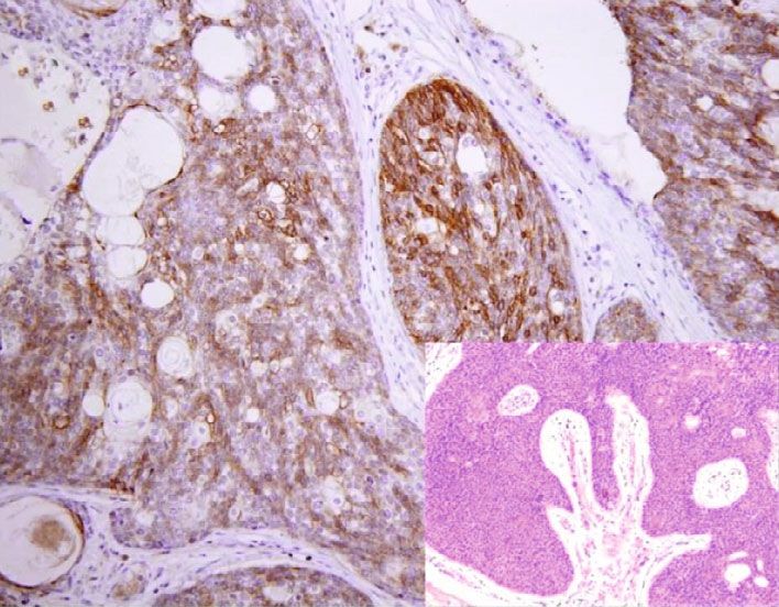
4                                                                                                      Analytical Cellular Pathology

                                         (a)                                              (b)

Figure 2: Immunohistochemical staining (immunoperoxidase staining in detail) of AQP3 in normal kidney and skin tissue, kidney tissue as
positive control. Immunoreactivity of AQP3 is shown in the cytoplasmic membrane of collecting ducts and squamous cell (magnification:
400x).

                                         (a)                                              (b)

                                          (c)                                             (d)

Figure 3: Expression of AQP3 in benign skin lesions and tumors. Distinctive AQP3 immunoreactivity is identified in molluscum
contagiosum (a), hidradenoma (b), eccrine poroma (c), and sebaceoma (d) with corresponding figure of HE staining on the lower right-
hand corner (magnification: (a, b) 400x; (c, d) 200x).

situation. Since both basal cell carcinoma and sebaceous             carcinomas [22, 23]. In our study, we found the diffuse
carcinoma show negative AQP3 staining, AQP3 is not                   positivity pattern of AQP3 expressed in all sebaceous
useful to distinguish basal cell carcinoma with sebaceous            hyperplasias and sebaceous adenomas, but not in seba-
differentiation from sebaceous carcinoma. As for seba-                ceous carcinomas. Therefore, AQP3 immunohistochemical
ceous tumors, which consist of sebaceoma, sebaceous ade-             staining may have some value for distinguishing benign
noma, and sebaceous carcinoma, their differential                     sebaceous tumors from malignant ones.
diagnosis depends mainly on histological architectural,                  It is not surprising that malignant melanoma shows no
cytological features, and mitotic rate. Only immunoreac-             expression of AQP3 as normal melanocytes within the epi-
tivity of p53 has been reported to act as a useful marker            dermis do not show AQP3 expression as demonstrated by
to distinguish between benign and malignant sebaceous                our prior study [7].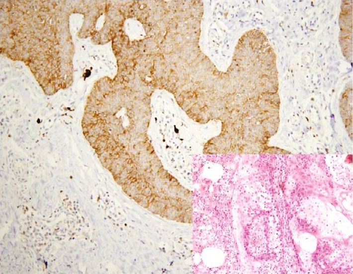
Analytical Cellular Pathology 5
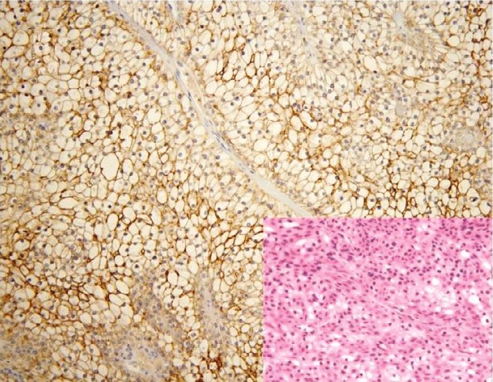
(a) (b)
(c) (d)
(e) (f)
(g) (h)
Figure 4: AQP3 expression in malignant skin tumors. Diffuse cytoplasmic membrane staining of AQP3 is identified in solar keratoses (a),
Bowen’s disease (b), Paget’s disease (c), squamous cell carcinoma (d), and apocrine carcinoma (e), while negative AQP3 staining is observed
in basal cell carcinomas (f), sebaceous carcinomas (g), and malignant melanomas (h) with corresponding figure of HE staining on the lower
right-hand corner (magnification: 200x).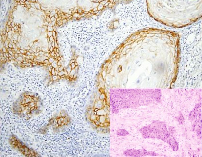
6                                                                                                            Analytical Cellular Pathology

5. Conclusion                                                            [8] A. Li, D. Lu, Y. Zhang et al., “Critical role of aquaporin-3 in
                                                                             epidermal growth factor-induced migration of colorectal car-
In summary, our results showed that AQP3 is expressed in                     cinoma cells and its clinical significance,” Oncology Reports,
most skin tumors except basal cell carcinoma, sebaceous car-                 vol. 29, no. 2, pp. 535–540, 2013.
cinoma, and malignant melanoma, reflecting the biological                 [9] J. H. Yang, C. X. Yan, X. J. Chen, and Y. S. Zhu, “Expression of
characteristic of skin tumors. AQP3 has some diagnostic                      aquaglyceroporins in epithelial ovarian tumours and their
utility in differential diagnosis of skin tumors.                             clinical significance,” The Journal of International Medical
                                                                             Research, vol. 39, no. 3, pp. 702–711, 2011.
Data Availability                                                       [10] Z. Zhu, L. Jiao, T. Li, H. Wang, W. Wei, and H. Qian, “Expres-
                                                                             sion of AQP3 and AQP5 as a prognostic marker in triple-
The data used to support the findings of this study are avail-                negative breast cancer,” Oncology Letters, vol. 16, pp. 2661–
able from the corresponding author upon request.                             2667, 2018.
                                                                        [11] W. Zou, Z. Yang, D. Li, Z. Liu, Q. Zou, and Y. Yuan, “AQP1
Conflicts of Interest                                                        and AQP3 expression are associated with severe symptoms
                                                                             and poor-prognosis of the pancreatic ductal adenocarcinoma,”
The authors declare that they have no conflict of interest.                   Applied Immunohistochemistry & Molecular Morphology,
                                                                             vol. 27, no. 1, pp. 40–47, 2019.
                                                                        [12] J. Bründl, S. Wallinger, J. Breyer et al., “Expression, localisation
Authors’ Contributions                                                       and potential significance of aquaporins in benign and malig-
Dongfeng Niu, Yanhua Bai and Qian Yao contributed                            nant human prostate tissue,” BMC Urology, vol. 18, no. 1, 2018.
equally to this work and are co-first author.                            [13] J. Chen, Z. Wang, D. Xu, Y. LIU, and Y. Gao, “Aquaporin 3
                                                                             promotes prostate cancer cell motility and invasion via extra-
                                                                             cellular signal-regulated kinase 1/2-mediated matrix
Acknowledgments                                                              metalloproteinase-3 secretion,” Molecular Medicine Reports,
                                                                             vol. 11, no. 4, pp. 2882–2888, 2015.
This work was supported by the National Natural Science
                                                                        [14] B. O. Cakir, P. Adamson, and C. Cingi, “Epidemiology and eco-
Foundation of China (No. 81301879, No. 81202114, and
                                                                             nomic burden of nonmelanoma skin cancer,” Facial Plastic Sur-
No. 81702839) and Beijing Municipal Administration of                        gery Clinics of North America, vol. 20, no. 4, pp. 419–422, 2012.
Hospitals Incubating Program (Code: PX 2018042).
                                                                        [15] L. E. Dubas and A. Ingraffea, “Nonmelanoma skin cancer,”
                                                                             Facial Plastic Surgery Clinics of North America, vol. 21, no. 1,
References                                                                   pp. 43–53, 2013.
                                                                        [16] A. Lomas, J. Leonardi-Bee, and F. Bath-Hextall, “A systematic
 [1] M. Echevarria, E. E. Windhager, S. S. Tate, and G. Frindt,
                                                                             review of worldwide incidence of nonmelanoma skin cancer,”
     “Cloning and expression of AQP3, a water channel from the
                                                                             The British Journal of Dermatology, vol. 166, no. 5, pp. 1069–
     medullary collecting duct of rat kidney,” Proceedings of the
                                                                             1080, 2012.
     National Academy of Sciences, vol. 91, no. 23, pp. 10997–
     11001, 1994.                                                       [17] J. A. Lo and D. E. Fisher, “The melanoma revolution: from UV
                                                                             carcinogenesis to a new era in therapeutics,” Science, vol. 346,
 [2] K. Ishibashi, S. Sasaki, K. Fushimi et al., “Molecular cloning
                                                                             no. 6212, pp. 945–949, 2014.
     and expression of a member of the aquaporin family with per-
     meability to glycerol and urea in addition to water expressed at   [18] A. Kakigi, M. Nishimura, T. Takeda, D. Taguchi, and
     the basolateral membrane of kidney collecting duct cells,” Pro-         R. Nishioka, “Expression of aquaporin1, 3, and 4, NKCC1,
     ceedings of the National Academy of Sciences, vol. 91, no. 14,          and NKCC2 in the human endolymphatic sac,” Auris Nasus
     pp. 6269–6273, 1994.                                                    Larynx, vol. 36, no. 2, pp. 135–139, 2009.
 [3] T. Ma, M. Hara, R. Sougrat, J. M. Verbavatz, and A. S. Verk-       [19] T. Litman, R. Sogaard, and T. Zeuthen, “Ammonia and urea
     man, “Impaired Stratum Corneum Hydration in Mice Lacking                permeability of mammalian aquaporins,” in Handbook of
     Epidermal Water Channel Aquaporin-3∗,” The Journal of Bio-              Experimental Pharmacology, pp. 327–358, Springer, 2009.
     logical Chemistry, vol. 277, no. 19, pp. 17147–17153, 2002.        [20] M. Zelenina, S. Tritto, A. A. Bondar, S. Zelenin, and A. Aperia,
 [4] R. Sougrat, R. Gobin, J. M. Verbavatz et al., “Functional               “Copper Inhibits the Water and Glycerol Permeability of
     expression of AQP3 in human skin epidermis and recon-                   Aquaporin-3∗,” The Journal of Biological Chemistry, vol. 279,
     structed epidermis,” The Journal of Investigative Dermatology,          no. 50, pp. 51939–51943, 2004.
     vol. 118, no. 4, pp. 678–685, 2002.                                [21] A. N. Crowson, “Basal cell carcinoma: biology, morphology
 [5] B. Burghardt, M. L. Elkjaer, T. H. Kwon et al., “Distribution of        and clinical implications,” Modern Pathology, vol. 19, Supple-
     aquaporin water channels AQP1 and AQP5 in the ductal sys-               ment 2, pp. S127–S147, 2006.
     tem of the human pancreas,” Gut, vol. 52, no. 7, pp. 1008–         [22] E. S. Cabral, A. Auerbach, J. K. Killian, T. L. Barrett, and D. S.
     1016, 2003.                                                             Cassarino, “Distinction of benign sebaceous proliferations
 [6] F. Umenishi, A. S. Verkman, and M. A. Gropper, “Quantita-               from sebaceous carcinomas by immunohistochemistry,” The
     tive analysis of aquaporin mRNA expression in rat tissues by            American Journal of Dermatopathology, vol. 28, no. 6,
     RNase protection assay,” DNA and Cell Biology, vol. 15,                 pp. 465–471, 2006.
     no. 6, pp. 475–480, 1996.                                          [23] S. C. Shalin, A. Sakharpe, S. Lyle, D. Lev, E. Calonje, and A. J.
 [7] D. Niu, T. Kondo, T. Nakazawa et al., “Expression of aquapo-            Lazar, “p53 staining correlates with tumor type and location in
     rin 3 in human neoplastic tissues,” Histopathology, vol. 61,            sebaceous neoplasms,” The American Journal of Dermato-
     no. 4, pp. 543–551, 2012.                                               pathology, vol. 34, no. 2, pp. 129–138, 2012, 136-138.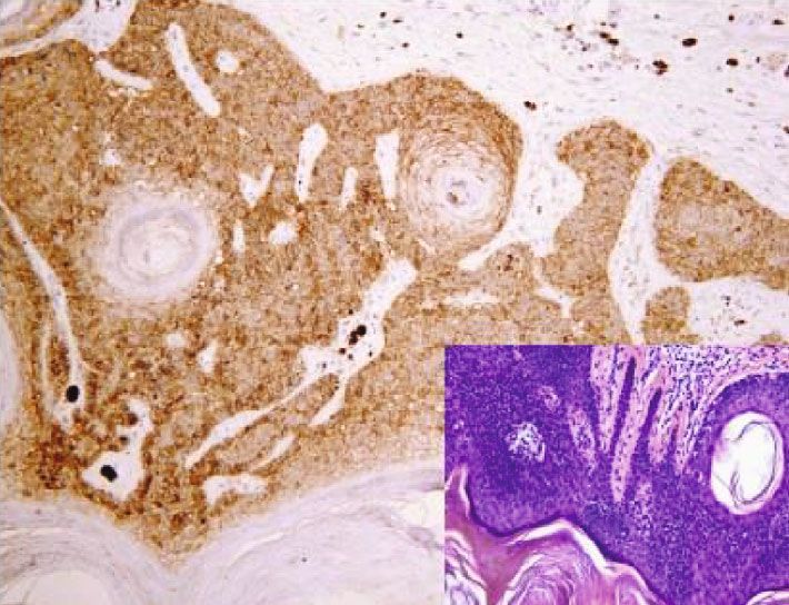
You can also read