Apoptosis in Tumor Progression From cell biology to therapy An example with Microtubule-Targeting Drugs - M2 onco 2011-2012
←
→
Page content transcription
If your browser does not render page correctly, please read the page content below
M2 onco 2011-2012

       Apoptosis in Tumor Progression
        From cell biology to therapy

An example with Microtubule-Targeting Drugs
             Diane BRAGUER

                     INSERM UMR911
Plan

                            Introduction

               mort versus survie et contexte du cancer

                Les 3 grandes voies de mort cellulaire

                             Apoptose
                             Autophagie
                              Nécrose

                            Mitochondrie

                           Fonctions
                Dynamique du réseau mitochondrial

                    Cytosquelette microtubulaire

                             Fonctions
                           Cibles des MTA

Relations microtubules - mitochondries au travers de notre expérience
Apoptosis and cancer : the genesis of a research field
                            Thomas G. Cotter

Nature reviews |Cancer 2009
No death without life : vital functions of apoptotic effectors
Apoptosis effectors exhibit vital functions that are predominantly involved in
                           the adaptation to stress
                                         Redox stress : AIF
                                    Metabolic stress : BH3-only
                                      DNA damage : Endo G
                                     Thermotolerance : Omit
                       Differentiation : caspases (inflammation, immunity)

    Defective ou inefficient apoptosis is an acquired hallmark of cancer cells
Tumor cells : genetic defects in progression of cell death that limit the clinical efficacy of the death
                    inducing agents  more focused in individualized therapies

          Therapy : to target the cell death pathways to render a cell once again sensitive

           Cell death pathways ubiquitinous but also differences in each type of cancer

                        3 highly conserved cell death pathways
                                   apoptosis autophagy necrosis
Apoptotic Cell

2 apoptotic
signaling pathways

Signal transduction pathways activated by TRAIL : apoptosis vs survival

                                                                                  Pavet oncogene 2010

                       Therapeutics: TRAIL receptor agonist antibodies
TRAILR1 (mapatumumab) TRAILR2 (lexatumumab and drozimumab) : few significant objective responses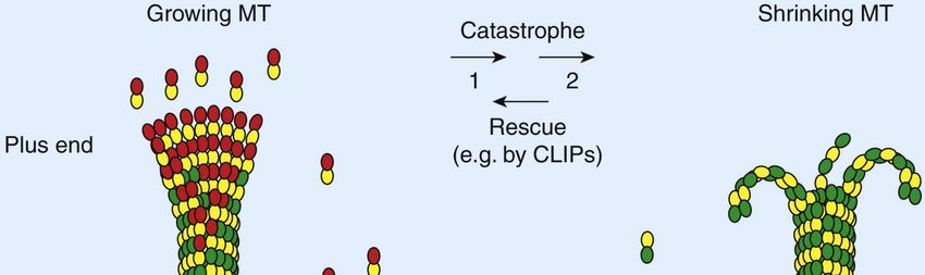
Bcl-2 protein family
                                                 Pore                Membrane
         phosphorylation sites                formation               anchor

        α1                 α2     α3    α4         α5     α6    α7 α8 α9
Bcl-2   BH4                 BH3              BH1               BH2     MA

                                 Hydrophobic pocket

                                 Bcl-2 homology domains (BH)

                                                                                                      Petros et al., 2000

                                                        BH3-only activators ( Bim, tBid, Puma) bind 5 antiapoptotic proteins
                                                         BH-only sensitizers : Bad and Bmf bind to Bcl2, BclXL and BclW
                                                                    Bik and Hrk bind to BclXL , BclW and A1
                                                                            Noxa bind to Mcl-1 and A1

                             Petros et al., 2000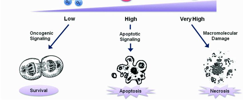
Activation of BH3-only proteins by different stimuli

                                                       2010
Overview of the three models of regulation of apoptosis by Bcl-2
                       family proteins

                                                                    Andrews DW, et coll (BBA) - Mol Cell Res
                                                                     Vol 1813,(4), April 2011, Pages 508-520
                                                                      Mitochondria: The Deadly Organelle

               Anti Bcl-2 : oblimersen, Gossypol, obatoclax : low efficacy
                         BH3 mimetics (ABT 737) : promising
Caspases
14 mammalian caspases
Synthesized as inactive precursors and upstream signals convert them
into mature proteases
Initiator caspases : long prodomains containing either a death effector domain (caspase-8 and
caspase-10) or a caspase recruitement domain (caspase-2 and caspase-9), activated via
oligomerization-induced autoprocessing
Effector caspases : short prodomains (caspase-3, caspase-6 and caspase-7), activated by
granzymeB or initiator of caspases
Caspase-4 and -12 : endoplasmic reticulum stress pathway
Proteolytic cleavage at Asp separating the large and small subunits
caspase-3 :capable of cleaving the DNA repair enzyme poly(ADP-ribose)polymerase (PARP) and the
inhibitor of caspase-activated DNAse (ICAD)
Could be considered to possess tumor suppressor function
Do their deregulation enhance tumorigenic potential ?Mammalian inhibitors of apoptosis (IAP) family
Mads Gyrd-Hansen and Pascal Meier, 2010Survivin
Survivin is a member of the IAPs family. It promotes cell survival through interference
with multiple cell cycle-related proteins such as Aurora B kinase.
Survivin also inhibits cell death through interference with both caspase-dependent and
-independent cell apoptosis. It may also play a role in the regulation of cancer cell
autophagy.
The physical association of XIAP with survivin was found to drive NFκB activation,
which in turn leads to increased autocrine production of fibronectin, signalling by β1
integrins and activation of the cell motility kinases focal adhesion kinase (FAK) and
SRC11. This results in tumour cell invasion in vitro and metastatic dissemination in
vivo. Importantly, the role of XIAP in regulating metastasis seems to be independent of
its ability to modulate cell survival through caspase inhibition.
At the clinical level, studies on clinical specimens have shown that survivin expression
is up-regulated in various human cancers and its up-regulation is associated with
tumour resistance to both chemotherapy and radiation therapy.
However, the development of survivin inhibitors is relatively slow as compared to
other therapeutic inhibitors for cancer treatment.Autophagy
A homeostatic cellular recycling mechanism that mediates removal of old or
dysfunctional proteins and organelles, and is particularly important for cell
survival during conditions of metabolic stress
A dual role in cancer: it can allow cancer cells to overcome metabolic stress
(hypoxia, lack of nutrients) or suppress tumor progression through
degradation of oncogenic proteins and cell death
Many anticancer drugs (such as inhibitors of mTORC1, the proteasome, or
histone deacetylases) induce autophagy; whether autophagy enhances their
antitumor properties or contributes to therapeutic resistance often remains
unclearAutophagy
Model of apoptosis regulation by Beclin 1 and its fragments

                                   R Kang et al, Cell Death and Differentiation (2011)
Necrosis
Krishna Biochem J 2011
Mitochondrial network

APOPTOTIC INTRINSIC PATHWAY
                                                                  METABOLISM
                                                               (ATP, respiration, ROS)
                      Bcl-2-like
                                       Bax-like

                                               caspase cascade
                                          activation…and apoptosis
                            BH3-only

MITOCHONDRIA DYNAMICS

                                                              Motility

              Fusion/Fission balance
Mitochondria in tumor cells
glycolysis
Bioenergetic index of the cell (BEC)
Indran et al, BBA 2011Mitochondria: promising targets for cancer chemotherapy
Regulation of mitochondrial metabolism by Bcl-2
               (non canonical activity)
Dynamique des mitochondries et morphologie
Fusion-fission and transport
3 GTPases de la famille des dynamines
DRP1 dynamin-related protein fission
MFN mitofusine 1 et 2 (mb externe) et OPA1 (optic atrophy 1, mb interne) fusion
Contrôle : fusion > fission mito filamenteuses
Inhibition fusion fragmentation = fission (ponctiforme)
Inhibition fission filamenteux ++ et interconnecté
Transport des mitochondries le long des microtubules
Intervention des protéines motrices
Fait intervenir fusion et fission
Transport d’ATP dans la celluleFusion mitochondriale
Permet l’échange de protéines, de complexes respiratoires et d’ADN
nécessaire à la stabilité de l’ADN mitochondrial (complémentation d’un
gène défectueux porté par une molécule d’ADN par un gène sain porté par
une autre molécule d’ADN)
Ne dépend pas du cytosquelette
Est abolie par dissipation du delta psy (apoptose)
Mb externe et interne fusionnent séparément
Utilisation de protéines fluorescentes photoconvertibles 2 types de fusion
Fusion instable : kiss and run
Fusion stable : mito allongéeFission mitochondriale (fragmentation) et apoptose
Nécessaire en fin de mitose
Associée à l’apoptose
Précède la libération de cyt c
Bcl-2 proapoptotiques sont impliquées
A.Savry, V.ReyHypoxia and mitochondria
Hypoxie induit elargissement des mitochondries due à une fusion anormale
(augmentation de l’expression de Mfn1), qui entraine une résistance à
l’apoptose et donc survie
Mazure et al, Bull Cancer 2011Microtubule cytoskeleton : an Integrator of signaling cascades
Mitosis
Cell proliferation
Cell polarity
Cell migration
Intracellular
transport
DifferenciationMicrotubule dynamics
Galjart 2010Regulatory coordination of Microtubule Associated Proteins
Structural MAPs (MAP1, MAP2, MAP4, Tau)
+ TIPs and co-polymerizing factors (EB1, CLIP 170, APC, CLASPs)
Dis1/TOG
Depolymerizing Kinesin-13 proteins
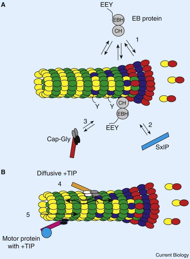
Stathmin KataninDelivery of +TIPs to microtubule ends
Classification        +TIP∗             Homologs†          Interaction with
                                                              other +TIPs
 ‘Core’ +TIPs     EB1-like proteins       Bim1 (Sc)        Most known +TIPs
                                          Mal3 (Sp)
CAP-Gly domain        CLIP-170          CLIP-190 (Dm)        EB1, CLIP-170,
                                          Bik1 (Sc)        CLIP-115, p150glued,
                                                           CLASP1,2, MCAK,
                                          Tip1 (Sp)
                                                                  LIS1

                      CLIP-115                –              EB1, CLIP-170,
                                                               CLASP1,2
                      p150glued          NudM (An)           EB1, CLIP-170
                                         Ssm4 (Sp)
  SxIP motif         CLASP1,2          Orbit/Mast (Dm)      EB1, CLIP-170,
                                          Stu1 (Sc)         CLIP-115, ACF7
                                         Peg1 (Sp)
                       APC               Kar9p (Sc)           EB1, MCAK
                       ACF7           Shot/Kakapo (Dm)       EB1, CLASP1,2

                       STIM1                 –                    EB1
                       MCAK             Klp10A (Dm)          EB1, CLIP-170,
                                         XKCM1 (Xl)           APC, Tip150
                       Tip150                –                 EB1, MCAK
                     Navigators              –                  Unknown
                    Melanophilin             –                    EB1
                      p140Cap                –                    EB1
                    CDK5RAP2                 –                    EB1
                   RhoGEF2 (Dm)              –                    EB1
                       DDA3                  –                    EB1
 TOG domain           Ch-TOG             Msps (Dm)                EB1
                                        XMAP215 (Xl)
                                          Stu2 (Sc)
 Not classified         LIS1             NudF (An)         CLIP-170, p150glued

                     NudA (An)        Dynein heavy chain     p150glued, LIS1
Microtubules are highly dynamic structures in living cells
It is not clear if microtubules serve as
molecular scaffolds for proteins to exert their activity or
                    if the proteins are
    sequestered by microtubules and therefore are
                  functionally inactive.
Microtubule-Targeting Agents are potent anticancer drugs
Drugs largely prescribed in adult and children cancers
Vinca alkaloids : vinblastine, vincristine, vinorelbine, vinflunine
Taxanes : paclitaxel, docetaxel, carbazitaxel, abraxane
Drug family in development :
Large reservoir of potentially therapeutic natural compounds
Small molecules in preclinical and clinical investigation
Drug combination schedules
Drug vectorisation
oral route with new pharmacokinetics profile
Novel therapeutic use
Limits :
Drug resistance
No tumor cell specificity
Neurotoxicity
Potent tools
for understanding the function of microtubule cytoskeleton in cancer developmentMicrotubule targeting agents (MTA)
Microtubule targeting agents (MTA)
Microtubule Targeting Agents (MTA) mechanism of action

  Inhibit microtubule dynamic instability

   Tubulin-GFP                              EB1-GFP

 Inhibit chromosome congression to metaphase plate

 Induce apoptosis via the mitochondrial pathway
MTAs induce the intrinsic apoptotic pathway
Mitochondrial morphology changes : increase in the cristae/matrix surface ratio
                           Control           Paclitaxel (2 hr)

                        0.07 ± 0.05              0.22 ±0.1       p < 0.0001
 Cytochrome c release from mitochondria to the cytosol

   Ctrl   4h     24 h     48h
               VFL IC70                                                       N. André
                                     24 h                                     B. Pourroy
MTAs induce the intrinsic apoptotic pathway
Paclitaxel mechanism of action in colon carcinoma cells
A.Gonçalves
Modification of microtubule network and mitotic block
Caspase activation independent of death receptor activation
Ψm concomitent with caspase-8 cleavage
Alteration of ∆Ψ
Caspase-3 activation and PARP cleavage
Involvement of mitochondria ?Mechanisms of drug resistance
Cellular efflux (MDR, MRP)
Clinical relevance ?
Limit diffusion in CNS
Binding on the target
Qualitative and quantitative alterations of tubulin-microtubule sytem
Β isotypes(betaIII), associated proteins such as MAP2, MAP4
(Β
Deficient induction of apoptosis
P53, Bcl2-family
Alterations in tumor cells that modulate microtubule functions
(protein, RNA, metabolism)
Others…Microtubule-mitochondria communications
MTA
Bim
Dynein
ROS
p53
Bax
bax
Bcl-2
bcl-2
Pro-apoptotic factorsP53 = gene suppresseur de tumeur
DNA damage activation des kinases de check points (ATM et ATR)
phosphorylation de p53 libération de sa liaison à mdm2 p53 active les check points
du cycle jusqu’à réparation, si pas possible induction de l’apoptose
P53 et apoptose
À la mitochondrie
Transcriptionnel Non transcriptionnel Interaction avec Bcl-2 donc antagonise
Inhibe survivine
bax, bid, puma les antiapoptotiques, et libère ainsi
Activation de Bax
les proapoptotiques
BrennerMTA and p53/Bcl-2 signaling
MTA
Dynein
p53
Bax
bax
Ctrl
bcl-2 Bcl-2
MTA
Pourroy, Carré, ReyMTAs and BCl-2
Diminution transcriptionnelle de Bcl-2 par les MTA
WB VRL (nM) 0 10 100
Bcl-2
actine
Q-RT-PCR Luciferase assay
Augmentation de la fixation de p53 sur le promoteur de Bcl-2 après traitement
(identification d’un nouveau site)
V.ReyBcl-2 down-regulation is associated with taxol and vinca resistance
in A2780 ovarian cells
120
Resistance (%)
Cell Mito 100 TC1-pUSE
Série1
80 *
WT TC1 WT TC1 * *
60 * Série2
TC1-Bcl-2
*
40
*
Bcl-2 20 Série3
TC1-Bcl-2∆loop
0
IC20 IC50 IC70
Resistance Resistance
A2780-wt A2780-TC1 Factor A2780-TC1-Bcl-2 Factor
TC1/wt TC1-Bcl-2/wt
IC20 20 nM 500 nM 25 30 nM 1,5
IC50 25 nM 20 µM 800 700 nM 28
IC70 30 nM 55 µM 1830 8 µM 270
Restoring Bcl-2 expression led to a significant increase in TC1 cell sensitivity to VFL
EsteveExpression de Bcl-2 : un facteur prédictif de réponse à la chimiothérapie ?
Bcl-2 dependent transcriptional regulation of Bim ?
Mitochondrial signaling related to microtubule cytoskeleton
        Mitochondrial ROS induce Bim translocation to mitochondria
                        and subsequent cell death

        Bim is involved in mitochontria fragmentation in Tax treated A549 cells
                Si ctrl            Si Bim

Taxol
                                                         Mechanism of Bim
                                                           translocation      ?
?
Bcl-2 nécessaire à la sensibilité des cellules A2780 au taxol ?
Bcl-2 : une cible mitochondriale du Taxol ?
Quels types de lignées/tissus ?
Interaction directe Bcl-2-Tax
Tax mime Nur77 récepteur d’une protéine capable de transformer Bcl-2 en « killer »
Bim plus important que Bcl-2 dans la réponse aux MTA ?
Etude Translationnelle ?
Autres rôles de Bim ?
migration ?MTA
Bim
Dynein
ROS
p53
Bax
bax
bcl-2 Bcl-2MTA
Bim
Dynein
ROS
p53
Bax
bax
Bcl-2
bcl-2
Pro-apoptotic factorsOlesoxime prevents
MTA Neurotoxicity
MTA + olesoxime
MTASelective preservation of EB comets in neuronal differentiated cells
Control olesoxime
vehicle
VCR
PTX PTX + olesoxime
VCR VCR + olesoximeOlesoxime prevents MTA-suppressed mitochondrial motility
**
Mitochondrial motility (µm/s) **
0,1
0,08
0,06
0,04
0,02
0
Control olesoxime MTA MTA +
olesoxime
Rovini A et al, bicohem Pharmacol 2010You can also read