The Resistance Mechanisms of Lung Cancer Immunotherapy - Frontiers
←
→
Page content transcription
If your browser does not render page correctly, please read the page content below
REVIEW
                                                                                                                                          published: 20 October 2020
                                                                                                                                      doi: 10.3389/fonc.2020.568059

                                              The Resistance Mechanisms of Lung
                                              Cancer Immunotherapy
                                              Fen Wang 1,2 , Shubin Wang 2 and Qing Zhou 1*
                                              1
                                               Guangdong Provincial Key Laboratory of Translational Medicine in Lung Cancer, Guangdong Provincial People’s Hospital,
                                              Guangdong Academy of Medical Sciences, School of Medicine, Guangdong Lung Cancer Institute, South China University
                                              of Technology, Guangzhou, China, 2 Shenzhen Key Laboratory of Gastrointestinal Cancer Translational Research,
                                              Department of Oncology, Cancer Institute of Shenzhen-PKU-HKUST Medical Center, Peking University Shenzhen Hospital,
                                              Shenzhen, China

                                              Immunotherapy has revolutionized lung cancer treatment in the past decade. By
                                              reactivating the host’s immune system, immunotherapy significantly prolongs survival
                                              in some advanced lung cancer patients. However, resistance to immunotherapy is
                                              frequent, which manifests as a lack of initial response or clinical benefit to therapy
                                              (primary resistance) or tumor progression after the initial period of response (acquired
                                              resistance). Overcoming immunotherapy resistance is challenging owing to the complex
                                              and dynamic interplay among malignant cells and the defense system. This review aims
                                              to discuss the mechanisms that drive immunotherapy resistance and the innovative
                                              strategies implemented to overcome it in lung cancer.
                                              Keywords: resistance mechanism, immunotherapy, PD-1/PD-L1, Immune check inhibitor, lung cance

                           Edited by:
                       Laura Mezquita,        INTRODUCTION
   Hospital Clínic de Barcelona, Spain
                        Reviewed by:          The discovery of the immune checkpoint inhibitors (ICIs), represented by the monoclonal
                        Ignacio Matos,        antibodies that block cytotoxic T−lymphocyte−associated protein 4 (CTLA-4), programmed
            University College London,        death protein 1 (PD-1), and programmed death protein ligand 1 (PD-L1), has revolutionized
                       United Kingdom         the therapeutic landscape of lung cancer. The significant survival benefit derived from ICI-
                            Félix Blanc,      containing treatment has established it as the mainstay first-line therapy in patients with advanced
      Institut Gustave Roussy, France
                                              or locally advanced non-small cell lung cancer (NSCLC) and extensive small-cell lung cancer
                  *Correspondence:            (SCLC). Unprecedented long-term clinical benefit or even, in some cases, a complete recovery has
                          Qing Zhou           been witnessed in lung cancer, particularly in patients with high PD-L1-expressing tumors (1–3).
                gzzhouqing@126.com
                                              Currently, investigations are under way aimed at integrating immunotherapy in the treatment of
                                              early-stage lung cancer.
                   Specialty section:
         This article was submitted to
                                                 However, most patients with NSCLC develop primary resistance during ICI monotherapy and
                    Thoracic Oncology,        only 15 to 20% achieve partial or complete response (3). Acquired resistance also occurs in initially
               a section of the journal       responding patients with advanced NSCLC treated with ICIs, after a median progression-free
                 Frontiers in Oncology        survival (PFS) of 4–10 months (4–9). The mechanisms of resistance to immunotherapy are not
            Received: 31 May 2020             yet fully understood, and methods to overcome them must be developed. Herein, we discuss the
      Accepted: 14 September 2020             pathways driving resistance to immunotherapy in lung cancer to help clinicians in their current
        Published: 20 October 2020            practice, as well as identify future research priorities and treatment strategies.
                            Citation:
         Wang F, Wang S and Zhou Q
  (2020) The Resistance Mechanisms            DIFFERENT SCHEMAS OF RESISTANCE TO IMMUNOTHERAPY
     of Lung Cancer Immunotherapy.
            Front. Oncol. 10:568059.          Unlike molecular targeted therapy and chemotherapy targeting tumor cells, immunotherapy targets
     doi: 10.3389/fonc.2020.568059            the immune system of the host by mobilizing the immune cells to recognize and eventually

Frontiers in Oncology | www.frontiersin.org                                        1                                        October 2020 | Volume 10 | Article 568059
Wang et al. Resistance Mechanisms of Immunotherapy
eliminate tumor cells. This mechanism of action determines the RESISTANCE MECHANISMS TO
complexity of the resistance mechanisms in immunotherapy. IMMUNOTHERAPY
Different mechanisms of immunotherapy resistance are listed
in Table 1. Underlying mechanisms of primary resistance span an extensive
In accordance with the timing of development, resistance range from tumor factors including genomic features,
can be considered as either primary, when no initial response transcriptomic signatures, and immune landscape, to host
or clinical benefit to the therapy is observed, or acquired, as factors. The potential mechanisms of acquired resistance at
disease progression occurs after an initial period of clinical benefit least partly overlap with those involved in primary resistance
(10). Clinically, 6-month treatment duration is adopted as a and mainly include loss of neoantigen and deficiency in
cutoff value (11). This classification schema correlates with real- presentation, loss of T-cell effector function, and up-regulation
time observations by clinicians and contributes to the clinical of alternate immune checkpoint receptors (10). Here, we
decision-making process in the absence of other information will discuss the mechanisms of resistance to immunotherapy
such as immune characteristics and tumor genetics. from tumor aspects (intrinsic and extrinsic mechanisms) and
Resistance is additionally classified as intrinsic or extrinsic host-related characteristics in order to avoid confusion and
to cancer cells. The former occurs in the tumor cell itself repetition (Figure 1).
and encompasses the inherent characteristics related to gene
expression, cell signaling, immune recognition, and DNA damage
response, whereas the latter is seen in the microenvironment Tumor Cell-Intrinsic Mechanisms
or systemic circulation throughout the T-cell bioactivation Genomic Features
process (12, 13). Low tumor mutation burden and neoantigen load
The cancer−immunity cycle is linked to immunotherapy Tumor-specific antigens are the key to activate T cells
resistance in another related schema (14). This classification to recognize tumor as foreign, which is the first step of
divides resistance from an immunological perspective into tumor-induced adaptive immune responses and immune-
immune desert (tumor fails to evoke an immune reaction), mediated tumor killing (15). These neoantigens, interestingly,
immune inflamed (tumor inhibits immune activities are derived from somatic mutations and contain new epitopes,
notwithstanding abundant immune cells infiltration), or and subsequently lead to tumor immunogenicity. Preclinical
excluded (tumor prevents immune cells infiltration in spite of and clinical studies have revealed that the response of
adequate immunogenicity) (13). neoantigen-specific effector T cell (Teff) paralleled tumor
It is noteworthy that the immune response is a continuous shrinkage (16–20).
and dynamic process rather than categorical (binary). Multiple With the improvement of sequencing techniques, it was
complex interactions, including immunologic, genomic, and found that nonsynonymous mutations can generate neoantigens
host characteristics and treatment interventions, rather than a that trigger cytotoxic responses against tumors (21, 22).
single, dominant determinant are involved in the resistance to Nonsynonymous mutation burden, rather than total mutation
immunotherapy. The fs can be overlapping or parallel in some burden of exons, was demonstrated to be more closely associated
cases despite the different timing of occurrence (11). with the clinical advantage of anti-PD-1 treatment, validating
the importance of neoantigens in dictating response (23). Tumor
mutation burden (TMB) is calculated as the total number of
TABLE 1 | Different schemas of resistance to immunotherapy. nonsynonymous mutations per DNA Megabase (Mb) (21, 24,
25). Low TMB, or low numbers of clonal neoantigens, presenting
Schemas Classifications Description
reduced tumor immunogenicity, is considered as a primary
Temporal Primary Lack of initial response or resistance marker to immunotherapy (15, 26).
perspective clinical benefit to therapy Clinically, low TMB or neoantigen load has correlated
Acquired Disease progression after with inferior response and poor PFS to monotherapy of
an initial period anti−PD1/PD-L1 antibodies in NSCLC (25, 27–30). However, it
(6 months) of clinical
fails to predict the clinical outcomes, in regard to overall survival
benefit
(OS) and combination regimens (31, 32). The influence on the OS
Spatial perspective Intrinsic Tumor-related resistance
by subsequent treatments and the additional complexities to the
Extrinsic Factors involved in
microenvironment or study of immunotherapy resistance added by combinations may
tumor-immunity cycle partly explain these controversial findings. Recently, a corrected
Immunological Immune inflamed Tumor inhibits immune TMB (cTMB) approach based on the adjustment of tumor
perspective activities notwithstanding purity was developed by Anagnostou and colleagues, which was
abundant immune cells identified on abundant tumor samples mined from The Cancer
infiltration
Genome Atlas (TCGA) and then confirmed in a patient cohort
Immune desert Tumor fails to evoke an
immunoreaction
received ICIs therapy. This cTMB more accurately predicted
Immune excluded Tumor prevents immune
the outcomes of immunotherapy, suggesting that the TMB in
cells infiltration in spite of samples with low tumor purity was mistakenly underestimated,
adequate immunogenicity which was especially important for metastatic NSCLC, because
Frontiers in Oncology | www.frontiersin.org 2 October 2020 | Volume 10 | Article 568059Wang et al. Resistance Mechanisms of Immunotherapy
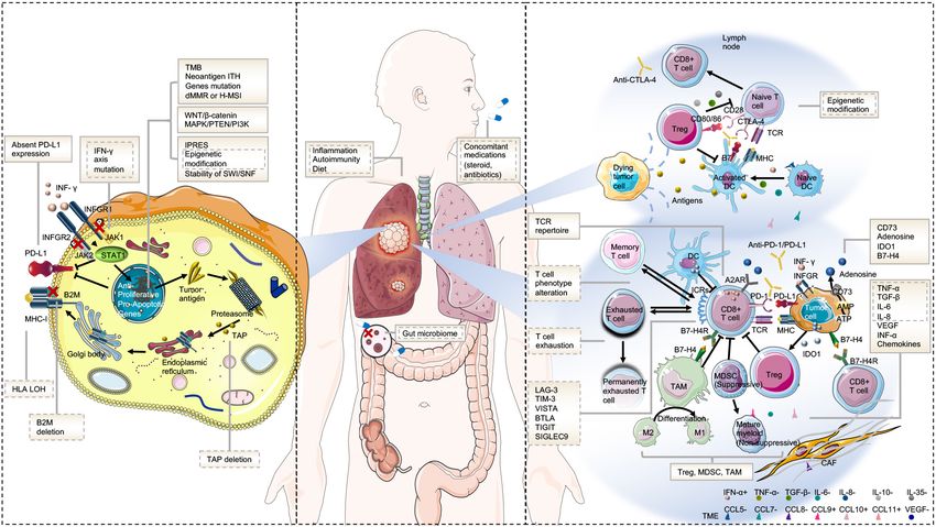
FIGURE 1 | Mechanisms of resistance to immunotherapy. (A) Tumor intrinsic mechanisms that are associated with resistance to immunotherapy include lack of
tumor immunogenicity (low TMB, heterogenous antigens, mutation of certain genes, and IPRES transcriptional signatures), deficiency in antigen presentation
(alterations in INF-γ signaling pathway, HLA LOH, B2M, and TAP deletion), aberrations in several signaling pathways (MAPK, PI3K, WNT, and IFN), and absent
PD-L1 expression. (B) Host-related characteristics that lead to primary or secondary resistance include the gut microbiome, diet, concomitant medications,
inflammation state, and autoimmunity. (C) Tumor extrinsic mechanisms involved in resistance to immunotherapy include T cell-related factors (alternative immune
checkpoints, T cell exhaustion and phenotype alteration, TCR repertoire, and epigenetic modification), immunosuppressive cells (Treg, MDSC, and M2-TAM), and
cytokines and metabolites (e.g., TGF-β, adenosine) released into the tumor microenvironment. Factors in the solid text boxes are involved in primary resistance,
whereas those in the dotted text boxes are involved in secondary or acquired resistance. Factors with solid and dotted dual text boxes are involved in both.
Cytokines with “+” and “-” represent positive and negative modulators to antitumor immune response, respectively. Abbreviations: TME, tumor microenvironment;
MHC, major histocompatibility complex; TCR, T cell receptor; Treg, regulatory T cell; MDSC, myeloid-derived suppressor cell; M2-TAM, type II tumor-associated
macrophage; ICR, immune checkpoint receptor; CAF, cancer-associated fibroblast; and IPRES, innate anti-PD-1 resistance.
the tumor purity of tissue samples obtained by bronchoscopy or clonality analysis in lung cancer data from TCGA and then
puncture biopsy was often limited (33). validated the approach in a cohort of NSCLC patients treated
The dilemma of insufficient tissue sample for TMB assessment with ICIs. Compared with high TMB alone, the combination of
in a considerable number of patients with NSCLC has given high TMB with low ITH seems to have a stronger association with
rise to the employment of peripheral blood TMB (bTMB) clinical benefit to ICIs in this population.
as a substitute predictor of response or resistance to ICIs in
NSCLC (34). In keeping with what was previously reported Aberrations in certain oncogene/tumor suppressor genes
in tissue, low bTMB evaluated by different plasma sequencing Aberrations in oncogenes and tumor suppressor genes can
assays was significantly correlated with poor survival or response regulate immune response by amending cytokine profile and
to immunotherapy in several retrospective and prospective immune cell composition and thus render tumor cells resistant
studies (35–37). or sensitive to ICIs.
Generally, alterations in oncogenic driver genes are
Increased neoantigen intratumor heterogeneity characterized as resistant markers to immunotherapy. Although
In addition to the TMB or the numbers of clonal neoantigens, epidermal growth factor receptor (EGFR) mutations and
increased neoantigen intratumor heterogeneity (ITH, defined as anaplastic lymphoma kinase (ALK) rearrangement tend to
relative fraction of subclonal neoantigens) can also impair the have high PD-L1 expression due to the activation of signaling
sensitivity to ICIs by elevating the likelihood of selection of pathways (39, 40), the low mutation or neoantigen load
subclones with poor immunogenicity (25, 38). The considerable (41), along with the following mechanisms, impairs the
variation of neoantigen heterogeneity was demonstrated by immunotherapy sensitivity in this group of patients with lung
McGranahan and colleagues in seven primary NSCLCs (25). On cancer. First, EGFR mutations have the potential to shape
average, 44% of heterogeneous neoantigens were reported only an inert immune environment by up-modulating a series of
in a subset of tumor regions. They conducted neoantigen and immune suppressors including inhibitory immune checkpoints
Frontiers in Oncology | www.frontiersin.org 3 October 2020 | Volume 10 | Article 568059Wang et al. Resistance Mechanisms of Immunotherapy
(e.g., PD-1 and CTLA-4), immunosuppressive cells recruitment and function (55, 56). Co-occurring KEAP1 and
(macrophages and regulatory T cells), and cytokines (like phosphatase and tensin homolog (PTEN) inactivation represent
TGF-β, IL-6, and IL-10) (42, 43). It has been reported that an immunologically “cold” tumor while concurrent mutations
activated EGFR cascade was associated with elevated T-cell in KEAP1 and STK11 leads to absence of pro-cancerogenic
exhaustion and reduced cytotoxic T lymphocytes (CTLs) in a M2 macrophages (57). However, there are conflicting data on
lung adenocarcinoma model (40). Second, downstream pathways the role of KEAP1 mutation and its co-mutation with STK11
of EGFR mutation, such as MAPK, PI3K/AKT, and Janus kinase in immunotherapy resistance in NSCLC. KEAP1/STK11 co-
(JAK)/STAT pathway, negatively affect immunoregulation. mutations were verified to correlate with resistance to ICIs
Other oncogenic driver-genes that frequently have high PD-L1 in patients with NSCLC despite high TMB (58). Similarly,
expression in lung cancer include ROS1 rearrangements (44), STK11, and/or KEAP1 genomic variations posited lack of
MET exon 14 skipping mutations (45), and BRAF mutations (44, clinical advantages from combination of immunotherapy
46). In contrast, RET rearrangements (47) and HER2 mutations with chemotherapy in patients with NSCLC (59). However,
(44) have been reported recently to exhibit low PD-L1 expression. inconsistent results were reported recently that clinical benefit
None of these oncogenotypes demonstrated favorable clinical from pembrolizumab compared to chemotherapy was poorer
responses to ICIs monotherapy except for BRAF mutations, in the patients with STK11 and KEAP1 mutation compared
either V600E or non-V600E. with those in wild type in Keynote 042 trial, but the response
STK11 gene inactivation either by mutational or non- and survival to immunotherapy were not significantly different
mutational machinery is linked to an indolent immune between mutant and wild subgroups (60).
microenvironment with lower Tumor-infiltrating lymphocyte The WNT/β-catenin pathway is an additional immunotherapy
(TILs; CD3+, CD4+, and CD8+ cells) and PD-L1 expression resistance mechanism. A negative relationship was demonstrated
in spite of the existence of moderate to high TMB (48). between the level of β-catenin and TILs, which was modulated
Inactivated STK11 gene was recently reported to weaken the by deficiency in the recruitment of CD103+ dendritic cells (DCs)
innate immune responses by epigenetic inhibition of stimulator essential to T-cell priming and reduced expression of the cytokine
of IFN genes (STING), suggesting epigenetic silencing is likely CCL4, suggesting WNT/β-catenin signaling pathway is likely to
to mediate the promotion of T-cell exclusion by the loss of mediate ICIs resistance through T-cell exclusion (61).
STK11 (49). In line with these findings, it has been observed in Similarly, the MAPK/PTEN/PI3K signaling pathway has been
several studies that compared with the wild-type gene, STK11 identified to be involved in immunotherapy resistance. Loss
mutation predicted poorer clinical outcomes of immunotherapy of PTEN and the bioactivation of the phosphatidylinositol
in advanced NSLCL (50, 51). The tumor suppressor TP53 3-kinase (PI3K) signaling pathway in tumors decrease the
mutation, a well-known negative prognostic factor in lung cancer, activity of CTLs through the recruitment of inhibitory cells
is found to be associated with increased PD-L1 expression and to the microenvironment and Vascular endothelial growth
higher TMB in non-squamous NSCLC (30) and KRAS-mutated factor (VEGF) expression (62, 63), so that they promote
lung adenocarcinoma (51). resistance to ICIs (63, 64). The association of PTEN deletions
KRAS-mutated lung cancer presents distinct immune or PIK3CA/AKT mutations with increased PD-L1 expression and
profiles, biology, and therapeutic vulnerabilities in different immune resistance was also found in glioma (65). It was shown
subsets classified by co-occurring genetic events (50). Generally, in preclinical models that a PI3K-γ inhibitor decreased myeloid-
KRAS/TP53 co-mutation predicts sensitivity while KRAS/STK11 derived suppressor cells (MDSCs) and improved response to
co-mutation predicts resistance to immunotherapy in NSCLC. ICIs (66).
Dong et al. identified TP53/KRAS co-mutated subclass exhibited
the highest percentage of PD-L1+/CD8A+ and particular DNA repair and replication gene alterations
increased PD-L1 expression. They further confirmed a Genetic instability caused by alterations in DNA replication
remarkable clinical benefit from pembrolizumab in this and repair genes can augment immunogenicity via a high-
population (52). Co-mutation of STK11 was shown to cause TMB neoantigen load (67–69). Correspondingly, deficient DNA
the accrual of neutrophils with T-cell-suppressive effects, mismatch repair (dMMR) or high microsatellite instability (H-
accompanied with an analogous elevation in the production of MSI) are suggested as sensitive predictors to ICI immunotherapy
T-cell depletion biosignatures and tumor-promoting cytokines in many tumor types. Beyond high TMB, increased CD8+ TILs
(50, 53). TIL numbers and the expression of PD-L1 were also were also reported to be associated with alterations in mismatch
decreased (53). Consistent with these preclinical predictions, repair genes (70), BRCA2 (71), and POLE (72) in different tumors.
patients with KRAS/STK11 co-mutation or single mutation of However, the role of these genes in immunoregulation in NSCLC
STK11 had poor response and survival compared with those with remains to be elucidated.
wild-type when treated with ICIs (51, 54).
The Kelch-like ECG-associated protein 1 (KEAP1) Interferon-gamma signaling mutation
gene, regulating the cellular antioxidant and cytoprotective The interferon-gamma (INF-γ) signaling cascade is a crucial
transcriptional programs, plays a key role in mediating immune component of immunotherapy and tends to serve a critical
evasion in NSCLC. Depletion of KEAP1 is associated with function in primary, adaptive, and acquired resistance to ICI
reduced leukocyte infiltration, increased PD-L1 expression treatment (73–75). IFN-γ is a critical cytokine secreted by
and might also influence other immune cells such as NK cell activated T cells, natural killer (NK) T cells, in the cancer
Frontiers in Oncology | www.frontiersin.org 4 October 2020 | Volume 10 | Article 568059Wang et al. Resistance Mechanisms of Immunotherapy
microenvironment, and it moderates the immune reaction via in mesenchymal transition (TWIST2, TAGLN, FAP, AXL,
the downstream enzymes JAK 1/2 and the signal transducer ROR2, WNT5A, and LOXL2), monocyte/macrophage chemotaxis
and activators of transcription (STATs) (76). The INF-γ axis (CCL2, CCL7, CCL8, and CCL13), immunosuppression (IL10,
exerts both positive and negative impacts on antitumor immune VEGFA, and VEGFC), and angiogenesis and wound healing
reactions (77). On one hand, it activates an functional antitumor (89–91). By contrast, down-regulated gene CDH1 (which is
immune reactive via (1) intensifying antigen presentation by up- typically down-regulated by mesenchymal tumor cells) was also
modulated secretion of MHC-I; (2) recruiting other immune detected in non-responsive pretreated tumors. Interestingly,
cells by up-regulation of the expression of chemokines (CXCL9, there was no difference in the expression of INF-γ pathway
CXCL10, and CXCL11) with effective chemoattractant impacts signatures, other T-cell-related genes (e.g., CD8A/B, PD-L1,
on T cells (78); and (3) exerting direct anti-proliferative and pro- and LAG3), and the genes that presumably modulate immune
apoptotic impacts on cancer cells (79). On the other hand, IFN-γ checkpoint sensitivity between responsive and non-responsive
acts in a negative-feedback axis to elevate PD-L1 expression as groups, suggesting that T-cell-suppressive inflammatory and
well as other crucial immune inhibitory components, including mesenchymal phenotypes of tumor are associated with primary
IDO1, down-modulating the cytotoxic reaction and adaptive resistance to anti-PD-1 therapy.
resistance to cancer cells (80, 81) (Figure 1A).
Additionally, copy-number alterations (CNAs) linked to DNA Epigenetic Modification
damage response and regulation of DNA editing/repair gene Emerging evidence has suggested that epigenetic modification
expression were shown to emanate from the malignant exposure may mediate primary resistance and contribute to acquired
to IFN-γ-secreting antigen-specific CTLs in vivo, implying that resistance during ICI therapy through the profound effect
intensified genetic instability could be among the mechanisms on many aspects of antitumor immunity: neoantigen
through which CTLs and IFN-γ immunoedits cancers, changing presentation and processing; T-cell functions, differentiation,
their immune resistance due to genetic evolution (82). and proliferation; memory T-cell phenotype acquisition;
Tumors neutralize the impact of IFN-γ by mutating or interfering with T-cell migration; and mediating T-cell
down-regulating the molecules involved in the IFN-γ signaling exhaustion (10, 92–94).
pathway, including IFN-γ receptor chains, regulatory factors, Epigenetic targeting agents, including those targeting
JAK1/2, and STATs upon continuous IFN-γ exposure (73, 83). histone deacetylation or methylation as well as targeting DNA
Multiple studies have demonstrated that mutations of IFN-γ axis methylation, have exhibited encouraging antitumor activity
and consequent loss of JAK/STAT contribute to immune escape either as monotherapy or in combination with immunotherapy
of tumor cells and by that leads to primary or acquired resistance in preclinical studies (94, 95). Clinical trials investigating the
to ICI therapy via incapacity of up-regulating the expression performance of these agents combined with adaptive T-cell
of PD-L1 and MHC-I (73, 78, 84). Any deficiencies in IFN- transfer (ACT) in patients with acquired resistance to prior
γ, JAK1/2, or STATs including gene mutations, loss of protein immunotherapy are ongoing (96).
expression, negative regulator presence, or epigenetic silencing
would prevent signaling in response to IFN-γ and thereby end Stability of Chromatin Remodeling Complexes
up to the up-regulated tumor growth and apoptosis inhibition Stability of chromatin remodeling complexes within tumor cells
and down-regulated T-cell infiltration and expression of PD-L1 can also contribute to immunotherapy resistance by multiple
and MHC-I (74, 78, 85, 86). Correspondingly, genomic changes mechanisms. It was found that tumor cells were more sensitive
disturbing IFN-γ pathway genes, including the amplification of to CTL killing, which leads to increased response to anti-PD-
suppressor genes PIAS4 and SOCS1 and the deletion of IFNGR1, 1/PD-L1 therapy, due to the deficiency in chromatin remodeling
IFNGR2, IFIT1, IFIT2, IFIT3, IRF1, MTAP, and miR31, have been complex SWI/SNF (97, 98). BRG1-associated factor (BAF) and
described as possible machinery of primary resistance to various polybromo-associated BAF (PBAF), as the mammalian analogs
ICI therapies (73). An IFN-γ-related mRNA profile that contains of the SWI/SNF complex, are essential tumor suppressors and
10 genes (CCR5, CXCL9, CXCL10, CXCL11, GZMA, HLA-DRA, loss of function (LOF) mutations of them were shown to
IDO1, IFNG, PRF1, and STAT1) was additionally identified to sensitize tumor cells to ICI therapy (98). The inactivated PBAF
predict the response to anti-PD-1 therapy in melanoma (87). subunits exhibited elevated CXCL9/CXCL10 expression and TILs
recruitment as a result of increase of chromatin accessibility
Transcriptomic Signatures to transcriptional regulators of IFN-γ-inducible genes (97).
In a recent publication, transcriptional signatures, referred ARID1A/B subunits are unique to BAF, while other subunits
to as innate anti-PD-1 resistance (IPRES) with inflammatory (ARID2, BRD7, and PBRM1) are exclusively contained by PBAF,
and mesenchymal tumor phenotypes, were shown to manifest despite the high similarity of these complexes (99). In another
poor response to anti-PD-1 therapy in metastatic melanoma study, loss of ARID1A was found to elevate MSI by defective
(88). Approximately 700 differentially expressed genes (DEGs) recruitment of mismatch repair genes and thus increase TMB,
were identified between the responsive and non-responsive which eventual sensitize tumor cells to PD-L1 inhibitor (100).
pretreated tumors. Compared with those of responsive tumors,
the transcriptomes of non-responsive tumors were dominated Absent Tumor PD-L1 Expression
by gene up-regulation events. The up-regulated DEGs in non- The PD-1/PD-L1 axis represents one of the foremost mechanisms
responsive tumors, considered as T-cell-suppressive, are involved of modulation of peripheral immune tolerance as well as T-cell
Frontiers in Oncology | www.frontiersin.org 5 October 2020 | Volume 10 | Article 568059Wang et al. Resistance Mechanisms of Immunotherapy
activation. Up-regulation of PD-L1 by cancer cells and antigen- of subclones that do not express neoantigens, consequently
presenting cells (APCs) is one approach through which tumors conferring poor immunogenicity and resistance to ICIs (117).
avoid immunosurveillance and constitutes the principle behind It was demonstrated by Anagnostou and colleagues that seven
PD-1/PD-L1 blockade therapies (101). Absent PD-L expression to eight putative neoantigens were lost in the recurrent NSCLC
of tumors has been found to be generally associated with less after ICI treatment, suggesting that immunoediting plays a role
responses and inferior survival benefits to anti-PD-1/PD-L1 in acquired resistance to immunotherapy (118). T-cell-mediated
therapies compared with higher expression (102) and may serve neoantigen immunoediting can be induced by the dynamic
as a resistant marker. However, up to 20% of PD−L1−negative interactions between T cells and tumor cells, consequently
malignancies showed responses to PD−1 inhibitors in some causing partial or total loss of neoantigen (119). Consistently,
cohorts (103), as PD-L1 expression can be up-regulated by other deficiency in genes that encode target tumor antigens was
factors including activated IFN-γ cascade (will be discussed in a demonstrated to be associated with acquired resistance in a
separated part), suggesting tumor PD-L1 expression alone is not murine model treated with adoptive T-cell therapy (ACT) in
dependable at predicting outcomes of PD-1/PD-L1 inhibitors. melanoma (120). However, this relationship between acquired
Any factors that affect the PD-L1 expression of tumor cells resistance and loss of target neoantigens was not observed in a
may lead to resistance to immunotherapy. Beyond encoding single patient case who achieved a complete response to ACT in
genes, PD-L1 expression can be affected by the mutational a separate study (121), suggesting that down-regulation/loss of
features of tumor although it is not paralleled with TMB in neoantigens may occur during immunotherapy, but should be
most of the tumors (104–106). The inherent mechanisms, which taken as a canonical mechanism of acquired immune resistance.
have been shown to result in constitutive expression of PD-L1 Proinflammatory cytokines are likely to contribute to immune
by tumor cells, consist of alterations in the PTEN/PI3K/AKT escape by inducing loss of antigen expression, resulting in
pathway (65, 107), MYC overexpression (108), EGFR mutations acquired resistance too. The process of Tumor necrosis factor-
(40), CDK5 truncation (109), and elevated PD-L1 transcripts α (TNF-α)-induced epithelial-to-mesenchymal de-differentiation
stabilized by disruption of the 3-untranslated region (UTR) of was shown to lead to a loss of neoantigens causing transformation
this gene (110). Tumor-specific immune response may also be to a tumor phenotype that is less immunogenic and can more
affected by constitutive expression of PD-L1 caused by these readily evade immune surveillance in the ACT-treated mouse
oncogenic signaling processes on tumor cell surface. Although model in melanoma (122). Other TIL generated cytokines, such
it is still unclear whether it causes an increased or decreased as IL-6 or TGF-β, are also shown to be involved in the induction
possibility of responding to anti-PD-1/PD-L1 therapies, the of epithelial-to-mesenchymal transition in mouse models across
constitutive PD-L1 expression could result into inadequate numerous types of tumors, indicating that acquired resistance
response to other immunotherapeutic approaches by suppressing can be promoted by inflammation.
antitumor effect of T cells. The other transcriptional factors
constituting HIF1, NFkB, and STAT3, as well as epigenetic Defective neoantigen presentation
factors, additionally participate in the modulation of PD-L1 Defective neoantigen presentation serves a crucial function in ICI
expression (111). acquired resistance. The alterations in this process could happen
Inflammatory and hypoxic tumor microenvironment (TME) in beta-2-microglobulin (B2M), transporters associated with
can also lead to PD-L1 expression on many cell types including antigen processing (TAP), or MHC itself (123, 124) (Figure 1A).
tumor cells by Toll-like receptor (TLR) ligands. The recruitment As part of the MHC class I (MHC-I), B2M is crucial during
of activated T cells can increase the inflammatory mediators antigen presentation and its genetic deficiency, including loss
and successively induce the PD-L1 expression on the surface of of heterozygosity (LOH) and deletions or point mutations, was
tumor cells. These tumor cells specifically locate at the invasive identified to be an important route for primary and acquired
periphery where T cells are often abundant (112, 113). resistance to ICIs (125, 126). Other defects that would affect
Besides, PD-L1 is stabilized through N-glycosylation and neoantigen presentation include T-cell receptor (TCR) binding
palmitoylation (114, 115). This is crucial for its interaction domain mutations of MHC-I (127), loss of tapasin (a MHC-I
with PD-1. The resistance to anti-PD-1/PD-L1 treatment could antigen processing molecule), selective epigenetic silencing of the
moreover be attributed to the degree of generation and human leukocyte antigen (HLA) A3 antigen, loss of one HLA
secretion of soluble forms of PD-L1. These variants without the haplotype (128, 129), and LOH in HLA (130). Homozygosity
transmembrane domain because of alternative splicing have been in one or more of the three highly variable genes (HLA-A,
reported in recurrent NSCLC incidences that re-occurred after HLA-B, and HLA-C) that encode MHC-I, which are likely to
anti-PD-L1 antibody therapy with the ability to act as soluble restrict neoantigen presentation to CTLs, was identified to have
imitates for anti-PD-L1 antibodies (116). a significant association between resistance to ICI therapy in a
large cohort of cancer patients (131). In contrast to anti-CTLA-
Deficiency in Antigen Presentation 4, the expression of MHC-II (but not MHC-I) proteins by tumor
Loss of neoantigen and the presence of IFN-γ-mediated gene signatures were found
Loss of neoantigens in the context of immune-mediated pressure to be associated with the positive responses to anti-PD-1 therapy
is postulated to be another mechanism leading to resistance. In in melanoma (132).
the concept of immunoediting, the constant interactions between Defective neoantigen presentation may be mediated by
tumor cells and the immune system trigger the production IFN-γ signaling pathway through JAK1/2 and the STATs, by
Frontiers in Oncology | www.frontiersin.org 6 October 2020 | Volume 10 | Article 568059Wang et al. Resistance Mechanisms of Immunotherapy
down-regulating the expression of MHC-I (133). Actually, the potentially influenced by the cytokines in the TME through
IFN-γ pathway has both unfavorable and favorable impacts on (1) impaired migratory capacity as well as decreased synthesis
antitumor immune responses and plays a key role in acquired and of costimulatory components (CD86/80) by TGF-β (147, 148);
primary resistance to ICI therapy (as discussed above). (2) prevented DCs maturation by IL-6-gp130-STAT3 axis;
and (3) inhibited activity by Indoleamine 2,3-dioxygenase
Tumor Cell-Extrinsic Immune Landscape 1 (IDO, will be discussed in Section “Suppressive tumor
T-Cell-Related Factors Involved in Tumor-Cancer microenvironment”). IFN-α signaling pathway is important
Immune Cycle to the priming of T cells by DCs. It was found that TME with
Tumor-infiltrating lymphocytes remarkable insufficient IFN-α-producing DCs naturally led
Tumor-infiltrating lymphocytes constitute a complex group of to lessened antitumor T-cell priming and thus resistance to
immune cells with distinct functions and different clinical ICIs (149, 150). Activated IFN-α stimulated production of
impacts. Among them, tumor-specific CD8+ T cells can execute the chemokine CXCL10 to recruit TILs to tumor beds and
anti-cancer activities by killing tumor cells directly and has a in turn initiate spontaneous antitumor T-cell response (149–
strong prognostic effect in NSCLC (134, 135). CD4+ cells are 151). Preliminary trials combining IFN-α 2b therapy with
composed of a group of lymphocytes (Tregs CD4+, Th1, Th2, and anti-CTLA-4 inhibitors have indicated clinical activity, which
Th17) secreting diverse cytokine to activate and suppress CD8+ could be caused by diminished populations of MDSC (152,
cells. Th1 secretes IFN-γ and IL2, while Th2 secretes IL-4, IL-5, 153). Combinations of other ICIs and IFN-α 2b are currently
IL-9, IL-10, IL-13, and IL-25 (136, 137). CD45RO+ T cells, also investigated (154).
known as memory T lymphocytes, are another subclass of TILs. Immune resistance also occurs if the tumors evolve the
Regulator and memory T lymphocytes will be discussed in section ability to prevent infiltration even if tumor-specific Teffs are
“Suppressive tumor microenvironment.” formed. Mechanisms that lead to impaired T-cell infiltration
Low CD8+ TIL density was correlated with impaired efficacy involve components in the epigenetic silencing of immune
and survival in NSCLC patients treated with ICIs (138), cells (155) and the modification of secreted chemokines (156,
suggesting that immunotherapy resistance was mediated by low 157). Transcriptional program that is associated with T-cell
TILs but was then positively modulated by PD-L1. TILs can be exclusion and thereby predictive resistance to anti-PD-1 therapy
assessed by immunohistochemistry or standard hematoxylin and was identified in melanoma (158). Stromal cells surrounding
eosin (H&E) staining; however, no consensus has been reached tumors within TME can develop the capacity to obstruct effector
hitherto in the various scoring models using H&E staining T-cell entry, and the TGF-β cascade appears to serve a crucial
in NSCLC (139–142). A radiomic fingerprint of CD8+ TIL role in promoting T-cell exclusion features in peritumoral
derived via computerized tomography was developed recently fibroblasts (123, 159).
and showed promising efficacy in predicting response to ICI
therapies but requires further validation (143). T-cell receptor clonality
Thus, tumors can be described as three main immune T-cell receptor clonality is emerging as a new biomarker to
organization profiles (hot, altered, and cold) as per the presence predict the resistance and immune-related adverse events to ICIs
of TILs and correlated proinflammatory cytokines (144). The therapy. Since baseline CD8+ T-cell density was found to overlap
“cold” immune tumor is characterized as absence of TIL between respondents and non-respondents to ICI therapy (160–
within and at the edges of tumor, manifesting resistance to 162), it led to the speculation that a constrict TCR arsenal
immunotherapy either due to absent immune stimulation (as possessed by the baseline T cells concentrated on the antitumor
with low neoantigen cancer’s poor antigen presentation) or immune reaction and is associated with response to ICI therapies.
because of failed T-cell priming (as with intrinsic insensitivity T-cell clones can be identified by detecting TCR rearrangements
to T-cell killing). The “altered” immune tumor is characterized constituting genes in the variable (V)-diversity (D)-joining (J)
as low TIL within the tumor (“immunosuppressed”) or high region, which generate the antigen-specific complementarity-
TIL at the edges of the tumor (“excluded”), whereas “hot” is determining region 3 (CDR3). The responsivity of TCRs
high degree of TIL (144). Recently, intratumorally geospatial generated by TILs determines their potential to interplay with
heterogeneity of TIL was revealed in NSCLC. Tumor subclones tumor antigens that are presented on APCs. Thus, the assessment
from “cold” immune regions were related to mutation space of T-cell clonality divulges the extent of T-cell expansions caused
more closely and diversifying more recently compared with by tumor antigens and contributes to explore the mechanisms
those from “hot” immune regions. Higher risk of recurrence underlying T-cell toleration to tumor antigens.
was observed in tumors with more than one “cold” immune A lower baseline clonal T-cell arsenal has been shown to be
region (145). linked to worse clinical benefits to ICIs and survival in cancer
patients (162, 163). Besides, a remarkable increase in T-cell
Impaired T-cell priming and infiltration clones was reported in responders during anti-PD-1 therapy
Reduced proliferation and inadequate diversification of compared to non-responders, implying a cancer-specific reaction
T cells possibly contribute to ICI resistance. Impeded to immunotherapy for these patients. Moreover, baseline TCR
priming of naive T cells by blocked DCs recruitment was clonality did not strongly associate with TIL density, implying
demonstrated in melanoma to be correlated to the lack of that low-TIL density tumors could still respond to anti-PD-1
TILs and ICIs resistance (146). The function of DCs can be treatment if TIL has a narrow TCR clonality specific to the
Frontiers in Oncology | www.frontiersin.org 7 October 2020 | Volume 10 | Article 568059Wang et al. Resistance Mechanisms of Immunotherapy
tumor antigen (164). Inconsistently, it was recently found that T-cell exhaustion and phenotype alteration
T-cell clonality had a positive relationship with T-cell density, T-cell exhaustion is another factor involved in the primary and
PD-L1 expression, and TMB, and a negative relationship with acquired resistance to ICI therapy (Figure 1C). Exhausted T
EGFR mutation in NSCLC (165). A corresponding relationship cells exhibit impaired activity with progressive LOF and antigen
was found between the number of TCR sequences and the persistence compared with Teffs and can be induced by the
number of nonsynonymous mutations, spatial heterogeneity in PD-1/PD-L1 interactions (173). Chronic exposure to cognate
expanded TCR repertoire, and spatial mutational heterogeneity antigen triggers increased expression of PD-1, which results in
within tumors in NSCLC, respectively. This intratumorally the accumulation of T-cell exhaustion and thus T-cell dysfunction
spatial heterogeneity of TCR repertoire maps the neoantigen (174). The presence of PD-1 high expression can either exist
landscape, sculptured by focal antigen processing defects or HLA prior to PD-1 inhibitors, which is associated with primary
loss (166). Thereby, further investigations to identify the role of resistance partially depending on tumor-associated regulatory T
TCR clonality in immunotherapy are required. cells (Tregs), or develop after the anti-PD-1 therapy, which leads
to acquired resistance by severe T-cell exhaustion. In contrast,
Alternate immune checkpoint receptor up-regulation studies showed that the exhausted T cells with PD-1 low to
Compensatory up-regulation of numerous alternate immune intermediate phenotype retain the capacity to be reinvigorated by
checkpoint receptors during ICI therapy as a result of the ICIs (158, 175). Epigenetic alterations were found to be associated
activation of diverse cellular signals and IFN-γ signaling with T-cell exhaustion too recently. Exhausted T cell displayed
pathway were observed across multiple studies and have been a unique chromatin landscape, which alters the transcriptional
characterized to be linked to ICI adaptive resistance in NSCLC state, limits its effect function, and determines its capacity to be
(84, 167, 168). The expression of CD8+ T cells harboring reprogrammed after therapeutic intervention (176–178).
receptors showed serious flaws in proliferation, migration, and The formation of memory T cells is crucial to the avoidance of
cytokine secretion, indicating their immunosuppressive capacity. tumor relapse and therapy resistance following drug withdrawal,
In addition, progressive T-cell exhaustion was found in the especially in the long-lasting duration of responses to ICI therapy.
tumors with highly expressive or co-expressive receptors and Research evidence shows that patients with resistance to anti-PD-
different receptor displayed different exhausted phenotype (167). 1 treatment have fewer tumor-correlated memory T cells relative
Among these receptors, lymphocyte activation gene 3 (LAG-3) to sensitive patients (179). Memory T cells remain dormant until
has great potentiality in cancer immunotherapy because co- antigen re-challenge (180, 181) and if precursor memory T cells
expression of PD-1 and LAG-3 was often found in T-cell-depleted are exhausted under chronic antigen exposure, it will lead to
immune microenvironment, and PD-1 inhibitors combined with memory T-cell deletion and lack of formation (173, 177).
LAG-3 blockades showed strong synergic antitumor responses Acquired resistance can be mediated by the alteration from
in preliminary models (169). LAG-3 is a co-inhibitory receptor cytotoxic activity to inactivity phenotype of antitumor T cells
extensively expressed in TILs in various tumors and serves a during TCR-engineered ACT. The original highly cytolytic
crucial function in mediating immune escape by suppressing profile when administrated to patients, which showed strong
T-cell antitumor functions. It exerts immunosuppression via efficacy initially, was reported to change to a phenotype with
binding to MHC-II molecules and other ligands such as galectin- impaired cytotoxic functions and Th2-related cytokine release
3 and fibrinogen-like protein 1 (FGL1) (170). Thus, blocking when tumor relapses within months (182, 183).
LAG-3 can restore antitumor immunity and the combined LAG-
3 inhibitors therapy may accordingly overcome immunotherapy Suppressive Tumor Microenvironment
resistance. In addition, the expression of these ligands, on the Increased immunosuppressive cells
basis of the receptor–ligand interactions, may serve as important The TME is a complex net consisting of a variety of immune
biomarkers to predict the efficacy of LAG-3 blockades in lung and stromal cells, cytokines, extracellular matrix, and vasculature,
cancer (167). which affect response to immunotherapy. Immune-suppressive
The other alternative immune checkpoint receptors, e.g., cells, including Tregs, MDSCs, M2 macrophages, along with
T-cell immunoglobulin and mucin-3 (TIM-3), V-domain inhibitory cytokines in the TME, can contribute to the inhibition
immunoglobulin-containing suppressor of T-cell activation to immune responses (136, 184) (Figure 1C).
(VISTA), B and T lymphocyte attenuator (BTLA; also referred Tregs can inhibit Teff reactions by secreting certain inhibitory
to as CD272), T-cell immunoreceptor tyrosine-based inhibition cytokines (IL-10, IL-35, and TGF-β) or by direct cell contact
motif domain (TIGIT), and sialic acid-binding Ig-like lectin 9 (185–187). The cytokine IL-10 influences antigen presentation by
(SIGLEC9), have been discovered (144). Thus, these alternate down-regulating the expression of MHC-II and co-stimulatory
immune checkpoints are likely to be combined with existing components on DCs, thus intercepting the Teff activation (187).
ICI therapy to conquer the resistance. Increased efficacy of The ratio of Teffs to Tregs was shown to be related to the
PD-1 inhibitors combined with anti-TIM-3 or anti-LAG-3 responses to ICIs in mouse models, in that incapacity of either
regimens has been observed in either pre-clinical models or increasing Teffs or decreasing Tregs may cause resistance to
phase I clinical trials (171, 172). Currently, numerous clinical immunotherapy (188, 189). Factors that affect Tregs activity,
trials evaluating the therapeutic impact of alternate immune at the same time, are putative biomarkers of resistance. For
checkpoint blockade applied on its own or in combination with instance, soluble CD25, an IL-2 receptor whose binding is
PD-1/PD-L1 inhibitors in multiple malignances are ongoing. assumed to stimulate Treg proliferation, was established as a
Frontiers in Oncology | www.frontiersin.org 8 October 2020 | Volume 10 | Article 568059Wang et al. Resistance Mechanisms of Immunotherapy
negative predictor of OS for patients treated with anti-CTLA−4 the extracellular domain of TGF-β receptor II (a TGF-β “trap”)
(190). However, tumor-infiltrating Tregs might likely coexist with fused to a human immunoglobulin G1 antibody blocking PD-L1,
multiple immune cells, insinuating a potential immunoreactivity. demonstrated favorable efficacy in patients with advanced
It was reported that a high baseline expression of FoxP3+ Tregs NSCLC. Ongoing phase III trial is expected to validate the
in the tumor is positively associated with better survival in a efficacy of bintrafusp alfa vs. pembrolizumab in the first-line
retrospective study involving patients under the treatment of setting in advanced NSCLC (NCT03631706).
anti-CTLA-4 antibodies (161). Tumor necrosis factor-α pathway is postulated to be another
Myeloid-derived suppressor cells promote immune evasion immune evasion machinery conferring resistance to PD1
and tumor growth and have emerged as critical modulators blockade. The expression of TNFα in an inflamed TME positively
of immune responses in cancer. Studies have suggested the correlates with the expression of PD-L1 and TIM-3, along
existence of MDSCs in TME correlates with reduced efficacy of with impaired accumulation and increased activation-induced
immunotherapies, including ICIs therapy (191), ACT (192), and death of CD8+ TILs in melanoma models treated with anti-
DC vaccination (193). Therefore, reprogramming or eradicating PD1 therapy. Accordingly, inhibition of TNF-α prevents the
MDSCs might improve clinical response to immunotherapy. expression of PD-L1 and TIM3 and hampers anti-PD1-induced
Tumor-associated macrophages (TAMs) can be classified into TIL death (210). Therefore, this study offers a rationale for the
M1 and M2 macrophages according to disparities in surface combination of PD-1/PD-L1 inhibitors with TNFα blockade as a
molecules, expression of transcription factors, cytokine profiles, novel immunotherapeutic strategy to overcome resistance in lung
and metabolism (194, 195). They promote antitumor immunity cancer, and the phase I clinical trial testing the combination is
effects (mediated by M1) and pro-tumorigenic properties ongoing (NCT03293784).
(mediated by M2) that modify the TME (196). The role of Vascular endothelial growth factor has been linked to both
TAMs in mediating immunotherapeutic resistance in tumor has decreased T−cell infiltration and immunosuppressive effects
been discussed in several reports (197, 198). It was indicated in addition to promoting angiogenesis and thus is associated
to directly inhibit T-cell responses through PD-L1 in preclinical with resistance to ICIs (211). Multiple mechanisms are involved
studies of liver (199) and ovarian cancer (200). The inhibitor of in the interaction of VEGF with antitumor immunity: (1)
CSF-1R, a receptor for macrophage colony-stimulating growth VEGF prevented the commitment of lymphoid progenitors,
factor, was investigated in mouse models of pancreatic cancer decreasing progression to the T-cell lineage (212); (2) VEGF
where it decreased the frequencies of TAMs, while increasing IFN signaling promotes the infiltration of Tregs through a selective
production and delaying tumor progression (201, 202). Similarly, endothelium and reduces trafficking and extravasation of CTLs
CSF-1R inhibitor was found to synergize ACT therapy in a into the TME (213); and (3) VEGF increases expression of
melanoma model (203). These data suggest that CSF-1R inhibitor inhibitory receptors, contributing to CTL exhaustion (214).
may overcome the resistance to immunotherapy. Higher levels of VEGF were found in anti-PD-1-resistant patients
Specific chemokines, such as CCL5, CCL7, and CXCL8, play than sensitive ones (160). Based on these findings and the synergy
an important role in the recruitment of Tregs and MDSCs between angiogenesis blockade and ICI therapies observed in
to the TME, consequently boosting an immunosuppressive preliminary studies, multiple trials of combination therapy are
climate (204). Alternately, chemokines CXCL9 and CXCL10 underway, including bevacizumab and VEGFR-TKI with anti-
recruit CTLs to the TME (205) and the epigenetic silencing of PD-1 therapy.
the genes encoding them can reduce TILs and consequently Higher levels of interleukin 6 (IL-6) and interleukin 8 (IL-
promote resistance to ICIs (205). Epigenetic modulators of these 8) have been found recently to be linked to reduced responses
chemokine receptors relieved the suppression of these Th1- and worse clinical outcomes to ICI therapies across multiple
cell-type chemokines and increased TILs leading to improved types of cancers (215–217). IL-6 is a proinflammatory cytokine
therapeutic efficacy of PD-L1 inhibitor in a model for ovarian generated by T cells and macrophages and is usually involved
cancer (155). in the immunoregulation connected to the IFN-γ signaling
pathway. It can reduce the expression of PD-L1 and MHC-I,
Elevated immunosuppressive cytokines and result in tumor escape and resistance to ICI therapy (218).
The cytokine milieu is critical to the recruitment, activation, IL-8 is a proinflammatory chemokine and a chemoattractant
and proliferation of immune cell, performing both immune for myeloid leukocytes expressed in multiple cancers (219, 220).
stimulatory and suppressive effects (206). Transforming It potently regulates the chemotaxis of neutrophils (221, 222)
growth factor-β (TGF-β) is a cytokine playing key roles in and exerts direct pro-tumorigenic effects (223). High levels of
angiogenesis and immunosuppression by stimulating Tregs IL-8 are regarded to be associated with more neutrophil and
(207) and excluding T cell in peritumoral fibroblasts (123, monocyte infiltration, defective T-cell functions, and impaired
159). Up-regulated TGF-β signaling was correlated with poorly antigen presentation, which subsequently result in resistance to
immunogenic tumors and restrained response to ICIs in a ICI therapy (216, 217).
colorectal cancer model, indicating resistance to therapy (159).
Consequently, enhanced antitumor response to ICIs was Additional immunoregulative molecules
observed following application of TGF-β inhibitor either alone Contributions from inflammatory processes could participate
or in combination with anti-CTLA-4 or radiation therapy (208, in quashing the desired impacts of ICIs (Figure 1C). Adenosine
209). Bintrafusp alfa, a bifunctional fusion protein composed of can be produced under the condition of hypoxia and ischemia
Frontiers in Oncology | www.frontiersin.org 9 October 2020 | Volume 10 | Article 568059Wang et al. Resistance Mechanisms of Immunotherapy
caused by tumor inflammation. It was reported to inhibit to ICI therapies. Relative abundance of Bacteroidales was found
the cytotoxic function and proliferation of T cells via the in non-responders, while responders were more likely to have
A2A receptor on T cells (224). CD73, which mediates Faecalibacterium and Ruminococcaceae (239, 240). Furthermore,
the generation of adenosine through dephosphorylation transplanting the responder’s feces into aseptic mice exhibited
of adenosine monophosphate, was also demonstrated to improvement in the treatment of PD-L1 inhibitor (241). Certain
suppress immune function (225). CD73 overexpression species can be altered by the antibiotic’s exposure, which partly
promotes T-cell exhaustion and is linked to the resistance to explained modified response to ICI therapy either in a good or in
ICIs (226, 227). a bad way (242–244).
IDO1 expressed in myeloid cells and cancer cells is The alteration tendency of gut microbiome structure or
a rate-limiting enzyme that converts tryptophan to its dominant bacteria may differentially affect the T-cell immune
immunosuppressive metabolite kynurenine. This enzyme response. It may be due to the cross-reactivity between
can induce T-cell anergy and apoptosis by gathering kynurenines intestinal microbiota and tumor-associated antigens, enhancing
and consuming the essential amino acid tryptophan and the inflammatory cytokine production, activation of DCs,
prevent the T-cell clonal expansion (228). It is particularly and antigen presentation (245, 246). IFN-γ-producing CD8+
activated in DCs after binding with CTLA−4 and can be T cells were successfully induced from a consortium of
unregulated by CTLA−4 during adaptive immune resistance 11 bacteria in the intestine, and colonization with this 11-
(229). Reduced expression of IDO at baseline was noted to bacterial mixture resulted in enhanced efficacy of ICIs in mouse
be associated with poor response to ipilimumab in a phase II models (247). Consistently, “good” bacteria introduction was
study in melanoma (161). Low level of IDO is likely to manifest reported to significantly increase IFN-γ production in spleen
insufficiency of suppressed TILs feasible to be reactivated and tumor-draining lymph nodes (TDLN) (246) and induce
by immunotherapy. Correspondingly, IDO-knockout mice DCs to secrete IL-12, resulting in increased recruitment of
exhibited improved OS with ICI compared with wild-type CCR9+CXCR3+CD4+ T cells into tumor beds (244). The
mice, and ICI therapy combined with IDO inhibitors showed activation of DCs was reported to be modulated by the gut
both increased numbers and functions of TILs in the TME microbiome in animal models and cancer patients. The resistance
in an experimental setting (230, 231). However, despite to anti-CTLA-4 therapy can be reversed by oral administration
encouraging results observed in preclinical and early-phase of Bacteroides fragilis, which induced Th1 immune response in
clinical studies in different types of tumors, no difference TDLN and promoted DCs maturation (243). Bifidobacterium-
was shown in pembrolizumab combined with IDO1-selective feeding mouse presented higher expression of MHC-II in DCs
inhibitor vs with placebo in a phase III study in metastatic within tumors (248). It remains controversial whether intestinal
melanoma (232). microbes lead to immunotherapy resistance by affecting the
B7-H4 has been proposed as another resistance marker production of Tregs. A higher level of peripheral Tregs was found
to ICIs recently due to its negative modulation of T cells. in patients with “bad” bacteria and was associated with poor
B7-H4 constitutes a type I transmembrane protein of the response to ipilimumab in metastatic melanoma patients (249).
B7 immunoglobulin superfamily and is encoded by the B. fragilis can produce a microbial molecule, polysaccharide A,
V-set domain containing T-cell activation inhibitor 1 (VTCN which can promote the formation of the inducible population
1) gene. It is induced by activated T lymphocytes and of CD4+Foxp3+Tregs (a subset of Tregs), thereby negatively
down-regulates T-cell function by inhibiting proliferation, regulating the immune system (250), whereas the other two
cytotoxicity activity, and interleukin secretion, after binding studies reported no differences in Treg differentiation between
with T cells (233–235). Positive B7-H4 protein expression pancreatic duct adenosarcoma-bearing mice and control, and the
in patients with advanced NSCLC treated with nivolumab number of Foxp3+ T cells between Bifidobacterium- and PBS-
was recently reported to have an enhanced risk of tumor feeding mice (251, 252). The majority of chemokine genes were
progression and tumor-related death compared with negative reported to be up-regulated by specific species of gut microbiome
expression (236). including Fusobacterium nucleatum, B. fragilis, and Escherichia
coli in colorectal cancer cells. Additionally, the gut microbiota-
Host-Related Characteristics derived microbial load was associated with increased chemokine
Host-related characteristics, including gut microbiome, diet, and production (251).
antibiotic exposure that adversely affect the gut microbiome, The gut microbiome also acts as an instructive modulator of
diet, steroid use, vaccine exposure, inflammation state, and mutant TP53, which ultimately affects tumor proliferation and
autoimmunity, have been shown to relate to primary and the immune system. Kadoshi et al. recently found that mutant
acquired resistance to ICI therapy in lung cancer (237, TP53 presented contrasting effects in different segments of the
238) (Figure 1B). gut in a mouse model: a remarkable tumor-suppressive effect
in proximal gut and the expected oncogenic effect in distal
Gut Microbiome gut (253). The gut microbiome and its single metabolite gallic
Evidence is arising to support the vigorous impact of the gut acid turned mutant TP53 from a tumor-suppressive effect to an
microbiome on immunotherapy resistance. Multiple studies have oncogenic one, suggesting that the function of mutant TP53 is
demonstrated that less bacterial diversity and lack of enrichment plastic and under the control of microbiome and microbiota-
of specific species showed a significant correlation with resistance derived metabolites.
Frontiers in Oncology | www.frontiersin.org 10 October 2020 | Volume 10 | Article 568059You can also read