Plasma Membrane Lipid Domains as Platforms for Vesicle Biogenesis and Shedding? - MDPI
←
→
Page content transcription
If your browser does not render page correctly, please read the page content below
biomolecules

Review
Plasma Membrane Lipid Domains as Platforms for
Vesicle Biogenesis and Shedding?
Hélène Pollet, Louise Conrard, Anne-Sophie Cloos and Donatienne Tyteca *
 CELL Unit, de Duve Institute & Université Catholique de Louvain, UCL B1.75.05, Avenue Hippocrate, 75,
 B-1200 Brussels, Belgium; helene.pollet@uclouvain.be (H.P.); louise.conrard@uclouvain.be (L.C.);
 anne-sophie.cloos@student.uclouvain.be (A.-S.C.)
 * Correspondence: donatienne.tyteca@uclouvain.be; Tel.: +32-2-764-7591; Fax: +32-2-764-7543

 Received: 5 August 2018; Accepted: 4 September 2018; Published: 14 September 2018

 Abstract: Extracellular vesicles (EVs) contribute to several pathophysiological processes and appear
 as emerging targets for disease diagnosis and therapy. However, successful translation from
 bench to bedside requires deeper understanding of EVs, in particular their diversity, composition,
 biogenesis and shedding mechanisms. In this review, we focus on plasma membrane-derived
 microvesicles (MVs), far less appreciated than exosomes. We integrate documented mechanisms
 involved in MV biogenesis and shedding, focusing on the red blood cell as a model. We then provide
 a perspective for the relevance of plasma membrane lipid composition and biophysical properties
 in microvesiculation on red blood cells but also platelets, immune and nervous cells as well as
 tumor cells. Although only a few data are available in this respect, most of them appear to converge
 to the idea that modulation of plasma membrane lipid content, transversal asymmetry and lateral
 heterogeneity in lipid domains may play a significant role in the vesiculation process. We suggest
 that lipid domains may represent platforms for inclusion/exclusion of membrane lipids and proteins
 into MVs and that MVs could originate from distinct domains during physiological processes and
 disease evolution.

 Keywords: microvesicle; cytoskeleton; cholesterol; ceramide; sphingomyelinase; raft; lipid domains;
 calcium; oxidative stress; red blood cell

1. Introduction
      In recent decades, the field of transcellular signaling has been revolutionized by the emerging
concept of signal transmission through extracellular vesicles (EVs). For a long time, vesicles seen in
intercellular spaces by electron microscopy were thought to be artifacts or inert cellular fragments
resulting from damaged cells in the vicinity. Nonetheless, all cells, from bacteria to plants and animal
cells, seem to have the ability to produce EVs [1,2]. However, there is still no real consensus regarding
EV classification and nomenclature [3], probably due to the variety of EV size, composition, origin and
targets, but also due to difficulties related to their isolation and analysis (see Section 2). Most reviews
classify EVs into three groups: exosomes, microvesicles (MVs) and apoptotic bodies (Figure 1) [4–7].
Exosomes are the smallest EVs (50–150 nm in diameter) and are released upon multivesicular bodies
exocytosis. MVs are produced by direct local deformation and budding of the plasma membrane (PM)
leading to vesicles of more heterogeneous and bigger size (100 nm–1 µm in diameter). It should
be noted that these vesicles are often given other names, including ectosomes, microparticles,
shedding vesicles or oncosomes (in the particular case of cancer cells). Apoptotic bodies (1–5 µm in
diameter) are generated by blebbing of cells undergoing apoptosis. However, this classification should
be taken with caution as most of the currently used techniques only make it possible to separate small
EVs enriched in exosomes from large EVs enriched in MVs (see Section 2) [8]. Yet, other studies have

Biomolecules 2018, 8, 94; doi:10.3390/biom8030094                        www.mdpi.com/journal/biomolecules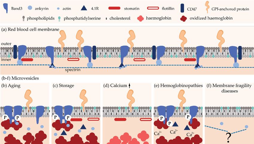
Biomolecules 2018, 8, 94 2 of 38
Biomolecules 2018, 8, x FOR PEER REVIEW 2 of 36
[8]. Yet, other
evidenced by studies
imaginghave evidenced
the budding by imaging
from the
the PM of budding
vesicles from
with sizethe PM of
closer to vesicles with size [9–11].
that of exosomes closer
to that of exosomes [9–11]. In this review, we will focus on PM-derived
In this review, we will focus on PM-derived vesicles whatever their size. vesicles whatever their size.
Figure
Figure 1.
1. Characteristics
Characteristics of
ofthe
thethree
threemain
mainclasses
classesof
ofextracellular
extracellularvesicles.
vesicles.MVB,
MVB,multivesicular
multivesicularbody;
body;
PM,
PM, plasma
plasma membrane.
membrane.
Extracellular vesicles
Extracellular vesicles have
have been
been shown
shown to to contribute
contribute to to aa large
large variety
variety of of pathophysiological
pathophysiological
processes including red blood cell (RBC) senescence [12], coagulation
processes including red blood cell (RBC) senescence [12], coagulation [13], inflammation [13,14], [13], inflammation [13,14],
migration [15],
migration [15], tumorigenesis
tumorigenesis[16] [16]and
andinfection
infection [17]. AsAs
[17]. they are are
they found in body
found fluidsfluids
in body (e.g., (e.g.,
blood,blood,
urine,
cerebrospinal fluid, milk), they are easily accessible and might represent useful
urine, cerebrospinal fluid, milk), they are easily accessible and might represent useful diagnostic diagnostic biomarkers
and/or targets
biomarkers for therapeutic
and/or applicationsapplications
targets for therapeutic (reviewed in [18]). In this
(reviewed review,
in [18]). we review,
In this will focusweon vesicles
will focus
derived
on from
vesicles the PMfrom
derived of RBCs, platelets,
the PM immune
of RBCs, cells, nervous
platelets, immunecells cells,and tumor cells
nervous cells. and
Before providing
tumor cells.
detailed information regarding their biogenesis, we present below a short
Before providing detailed information regarding their biogenesis, we present below a short non- non-exhaustive overview of
their pathophysiological effects.
exhaustive overview of their pathophysiological effects.
Erythrocytes undergo
Erythrocytes undergo multiple
multiple changes
changes during
during their their 120-day
120-day lifespan
lifespan in in the
the circulation,
circulation,
including the decreased activity of multiple enzymes, the gradual accumulation
including the decreased activity of multiple enzymes, the gradual accumulation of oxidative damage, of oxidative damage,
the redistribution
the redistribution of of ions,
ions, the
the loss
loss ofof membrane
membrane by by vesiculation
vesiculation as as well
well asas cell
cell volume,
volume, density
density and and
deformability alterations (for reviews, see [19–21]). Microvesicle generation
deformability alterations (for reviews, see [19–21]). Microvesicle generation constitutes a central constitutes a central
mechanism in
mechanism in the
the RBC
RBC homeostasis
homeostasis and and isis responsible
responsible for for the
the loss
loss of
of ~20%
~20% of of the
the PM
PM while
while thethe
hemoglobin concentration
hemoglobin concentration increases
increases by by ~14%
~14% [22,23].
[22,23]. Microvesicles
Microvesicles have have been
been proposed
proposed to to contribute
contribute
to RBC senescence by two opposite mechanisms. On one hand, they
to RBC senescence by two opposite mechanisms. On one hand, they protect RBCs from premature protect RBCs from premature
elimination via
elimination via transport
transportofof molecules
molecules thatthat
could induce
could inducerecognition by theby
recognition reticuloendothelial
the reticuloendothelial system
such assuch
system non-functional hemoglobin,
as non-functional oxidized
hemoglobin, and aggregated
oxidized and aggregated Band3 and oxidized
Band3 and oxidizedproteins [23]
proteins
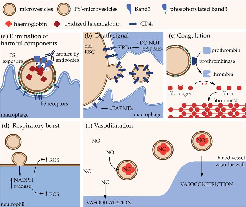
(Figure 2a). On the other hand, they appear to contain CD47, a self-marker
[23] (Figure 2a). On the other hand, they appear to contain CD47, a self-marker that prevents the that prevents the recognition
and clearance
recognition andofclearance
RBCs byofmacrophages. Elimination
RBCs by macrophages. of CD47 from
Elimination of CD47the from
RBC themembrane
RBC membranethrough
selective shedding could then promote the removal of old RBCs [24]
through selective shedding could then promote the removal of old RBCs [24] (Figure 2b). Studies (Figure 2b). Studies on miceon
suggest that MVs from RBCs are removed very fast from the
mice suggest that MVs from RBCs are removed very fast from the circulation by the circulation by the reticuloendothelial
system [25] because
reticuloendothelial they [25]
system can because
have deleterious
they can have effects on other effects
deleterious cells. on Forother
instance, MVsinstance,
cells. For bear at
MVs bear at their external leaflet phosphatidylserine (PS), which acts as an “eat me” signal but
their external leaflet phosphatidylserine (PS), which acts as an “eat me” signal for macrophages for
also promotesbut
macrophages coagulation,
also promotesas PS coagulation,
enhances prothrombinase
as PS enhances activity and other coagulation
prothrombinase activity andfactorsother
(Figure 2a,c).factors
coagulation Moreover, RBC-derived
(Figure MVs induce
2a,c). Moreover, an excessive
RBC-derived MVsproduction
induce anofexcessive
reactive oxygen
production speciesof
reactive oxygen species (ROS) in neutrophils and could be responsible for exhortation of the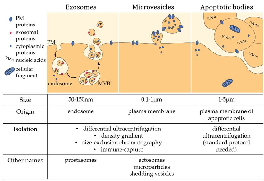
Biomolecules 2018, 8, 94 3 of 38
Biomolecules 2018, 8, x FOR PEER REVIEW 3 of 36
(ROS) in neutrophils
respiratory burst, i.e.,and
the could
rapid be responsible
release of ROSfor exhortation
necessary of the to
to answer respiratory burst,
an infection [26]i.e., the rapid
(Figure 2d).
release of ROS necessary to answer to an infection [26] (Figure 2d). Finally,
Finally, they contain hemoglobin, which allows them to bind nitric oxide modifying therebythey contain hemoglobin,
which allows them
bioavailability to latter
of the bind nitric oxide modifying
for vascular homeostasis thereby bioavailability
regulation of the
[27] (Figure 2e).latter for vascular
However, these
homeostasis regulation [27] (Figure 2e). However, these effects have been shown
effects have been shown with MVs isolated from blood storage. Although the biological content with MVs isolated
from blood
should reflectstorage. Although
the functional the biological
properties content
of circulating should
MVs, reflect
further the functional
investigations in vivo properties
are needed of
circulating
to confirm MVs,
thesefurther investigations
hypotheses. in vivothe
Nevertheless, are properties
needed to confirm
describedthese hypotheses.
above Nevertheless,
could partly explain
the properties described above could partly explain reduced post-transfusion efficacy
reduced post-transfusion efficacy and increased risk of adverse reactions in patients after transfusion and increased
risk of adverse reactions in patients after transfusion [28,29].
[28,29].
Figure 2. Pathophysiological
Figure 2. Pathophysiologicaleffects
effectsof of erythrocyte-derived
erythrocyte-derived microvesicles.
microvesicles. PS, phosphatidylserine;
PS, phosphatidylserine; RBC,
RBC, red blood
red blood cell; SIRPα,
cell; SIRPα, signal regulatory
signal regulatory proteinprotein α; NADPH;
α; NADPH; reducedreduced form of nicotinamide
form of nicotinamide adenine
adenine dinucleotide
dinucleotide phosphate; phosphate; ROS, oxygen
ROS, reactive reactivespecies;
oxygen NO,
species; NO,
nitric nitric oxide.
oxide.
Platelet is another blood cell type able to release a high quantity of EVs. Platelet EVs represent
~30% ofofthe
theblood
blood EVs
EVs [30][30]
and,and, in contrast
in contrast to RBCs,
to RBCs, these include
these include MVs asMVs well as exosomes.
well as exosomes. Platelet-
Platelet-derived
derived vesicles vesicles are essential
are essential for the regulation
for the regulation of the hemostasis,
of the hemostasis, as revealedas revealed by the
by the bleeding
bleeding disorders
disorders caused bycaused by the decreased
the decreased formationformation of platelet-derived
of platelet-derived MVs in
MVs in patients patients
with with Scott
Scott Syndrome
Syndrome
[31]. [31]. Their pro-coagulant
Their pro-coagulant activity is dueactivity
to theisexposition
due to the onexposition on their
their surface surface of pro-coagulant
of pro-coagulant molecules,
molecules,
such suchtissue
as PS and as PS factor
and tissue
(TF),factor (TF), one
one initiator ofinitiator of the coagulation
the coagulation cascade
cascade [30]. [30]. Additionally,
Additionally, platelet-
platelet-derived
derived MVs promote
MVs promote cell proliferation,
cell proliferation, survival and survival and migration,
migration, which are
which are essential foressential for
endothelial
endothelial
repair repair and
and wound wound
healing [32].healing
They are [32].also
They are also
effectors of effectors of the
the immune immunebyresponse
response by
increasing
increasing monocyte
monocyte adhesion, adhesion,
promoting promoting inflammatory
inflammatory pathways pathways and cytokine
and cytokine releaserelease in monocytes
in monocytes and
and endothelial
endothelial cellscells
(ECs)(ECs)
[33],[33], stimulating
stimulating antigen-specific
antigen-specific IgGIgGproduction
production[34]
[34] and
and upregulating
neutrophil aggregation, activation and phagocytic activity [35].
Monocytes and
Monocytes andneutrophils
neutrophils themselves
themselves are able
are also alsotoable to MVs
release release
and MVs and producing
exosomes, exosomes,
producing autocrine effects or serving as mediators to communicate with other
autocrine effects or serving as mediators to communicate with other cells (reviewed in [36,37]). They cells (reviewed
can have either
in [36,37]). They pro-inflammatory or anti-inflammatory
can have either pro-inflammatory effects [38]. They
or anti-inflammatory also [38].
effects promoteTheyblood
also
coagulation through exposure of PS and TF in monocyte MVs and through direct interaction between
neutrophil MVs and platelets, which promotes platelet activation [39] and TF expression [36].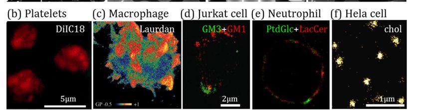
Biomolecules 2018, 8, 94                                                                            4 of 38

promote blood coagulation through exposure of PS and TF in monocyte MVs and through direct
interaction between neutrophil MVs and platelets, which promotes platelet activation [39] and TF
expression [36].
       In the central nervous system, only exosome-like EVs have been described in neurons,
oligodendrocytes and Schwann cells in physiological conditions [40]. On the other side, microglia cells
(i.e., the macrophage resident cells) and astrocytes (i.e., support cells, also involved in the blood brain
barrier) have been shown to release both exosomes and MV-like EVs [41]. The major MVs described
are released by microglia cells and astrocytes upon ATP activation of P2 X7 receptors and contain
the interleukin-1β which is released at the site of tissue damage to initiate an acute inflammatory
response [42]. Extracellular vesicles also appear to play critical role in neurodegenerative diseases.
For example, in Alzheimer disease, MVs released by microglia promote the amyloid-β pathogenesis
by increasing the solubility of the misfolded protein (i.e., the soluble form is more neurotoxic than the
aggregated one) [43]. Moreover, as microglia-derived MVs regulate the inflammatory response,
they have been shown to be increased and to play major role in multiple sclerosis, a form of
neuroinflammation [44]. Those MVs carry inflammation factors that promote the degradation of
the extracellular matrix and tight and adherens junctions, leading to the disruption of the blood brain
barrier [45].
       Cancer cell-associated vesicles were reported for the first time in 1978 in patients suffering
from Hodgkin disease. Since then, evidence has been accumulating that tumor-derived MVs
constitute important players in cancer initiation and progression through communications between
cancer cells. Microvesicles also facilitate intercellular communication between cancer cells and
microenvironmental cells (e.g., stromal, immune and vascular cells), either located directly in the
primary tumor-environment or at distance, promoting pre-metastatic niche formation. Microvesicles
are implicated in several stages of tumorigenesis and metastasis by increasing angiogenesis and
extracellular matrix remodeling, promoting escape from the immune system, inducing resistance to
therapy and triggering blood coagulation (reviewed in [16,18,46,47]). Because of the multiple roles of
MVs in cancer, they could be seen as prognostic and/or diagnostic biomarkers. Hence, they could
represent emerging targets for cancer therapy (reviewed in [18]).

2. Microvesicle Isolation and Characterization
     Several methodologies have been developed over the years to optimize EV isolation. Differential
ultracentrifugation is the most frequent one, even if protocols can considerably vary in terms of
speed and time intervals. Increasing centrifugal forces allow to separate EVs from cell debris and
intact cells thanks to their difference in size and density [48–51]. This method makes it possible
to reach a recovery of up to 80% and offers the possibility to process large volumes without the
need for chemicals that could interfere with downstream analysis [50]. Among limitations, one can
cite EV aggregation, contamination by protein aggregates and viruses as well as EV damaging
during high-speed centrifugation [48]. Moreover, since it is moderately time-consuming and
the equipment is expensive, it is not considered to be a clinically applicable isolation technique.
Another method based on centrifugation is the density gradient centrifugation, which presents
a lower recovery of 10–50% but avoids protein contamination of samples thanks to density
differences [48,52]. Nevertheless, co-isolation of lipoproteins cannot be excluded. For the same
reasons as for differential ultracentrifugation, this methodology finds no clinical application [48].
Filtration represents an alternative method that can be applied alone or in combination with
ultracentrifugation. However mechanical damage due to the pressure applied for passing EVs through
the filter can affect their properties [50]. According to the size cut-off of the column, size-exclusion
chromatography allows EV separation from non-aggregated proteins and high density lipoproteins
(HDL) but contamination with material of similar size, such as aggregated proteins and viruses,
cannot be excluded. The excellent recovery rate (up to 90%), the absence of EV damaging, the low cost
and the quickness make this methodology interesting for clinical applications. However, this method is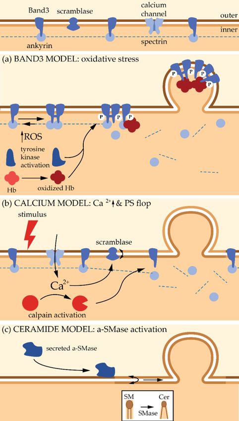
Biomolecules 2018, 8, 94                                                                           5 of 38

not suitable for large volumes and requires a pre-concentration step by ultracentrifugation [48]. Finally,
the affinity-based methods (better known as immune-capturing methods) are based on the interaction
of EV surface molecules with antibodies, lectins or lipid-binding proteins, either biotinylated or
coupled to magnetic beads. These techniques are fast and simple and contamination of purified sample
is minimal. However, it is cost-effective and not adapted to processing large-volume samples [50].
In the face of the large diversity of isolation methods, the quantity and the quality of the starting
material must guide the researcher.
      Another criterion to consider is the type of analysis downstream of EV purification, including their
structural characterization. Here also several techniques exist. Electron microscopy makes it possible
to assess EV size and morphology and to identify their cellular origin [48,53]. Since dehydration and
fixation required for traditional electron microscopy could possibly lead to EV morphology changes,
cryo-electron microscopy is widely recommended. Atomic force microscopy (AFM) also makes it
possible to determine EV structural properties via the interaction of a probing tip (cantilever) with the
surface of the sample, which generates a 3D-image of the surface topography. However, as for electron
microscopy, changes in EV morphology can occur due to the necessity of immobilizing the material [53].
Additionally, one can cite the dynamic light scattering and nanoparticle tracking, which are both based
on the same principle. Thanks to the recording of light scattering over time and its modification due
to EV Brownian motion, it is possible to determine their size and size distribution. This approach
is more reliable when the sample size is homogenous and not polydispersed [53]. For larger EVs,
flow cytometry is an alternative technique combining light scattering and fluorescence. The labeling
of EVs with fluorescent antibodies/probes allows the specific recognition of surface markers [48,53].
In addition to structural characterization, it is also possible to biochemically characterize the EVs.
This is beyond the scope of the manuscript and we invite the reader to refer to the review by
Ramirez et al. for more insights regarding techniques for biochemical and in vivo characterization [51].

3. Microvesicle Molecular Properties
      The abundance and properties of MVs appear to fluctuate depending on the cell origin,
the pathophysiological context and also the subject tested (e.g., age, gender, fasting state,
medication exposure, physical activities, pregnancy and diseases) [54–58]. Moreover, variations
in isolation techniques, culture conditions and methods to stimulate the shedding (e.g., calcium [Ca2+ ]
ionophores, lipopolysaccharide, hypoxia, tumor necrosis factor TNFα, ATP) could lead to conflicting
data [59]. Finally, most studies related to MV content are based on “MV pellets” obtained by differential
ultracentrifugation, which most of the time contain mixed populations (especially when extracted from
body fluids) [8]. It is then difficult to provide a digest of the MV content and even more to erect rigid
rules. Nevertheless, efforts have been made to collect datasets from many EV studies and put them
online (Vesiclepedia; EVpedia) [60,61] and to carefully characterize co-isolated mixed EV populations
from “traditional” isolation procedures to refine and determine new optimized protocols [8].
      Microvesicles are limited by a lipid bilayer (Section 3.3) and can carry a diversity of proteins
(Section 3.1) and nucleic acids (Section 3.2). Although they are expected to exhibit a similar content
as the PM from which they derive, accumulated evidence highlights that the MV composition is the
outcome of a regulated sorting mechanism at the PM, leading to enrichment or despoliation of some
chosen components.

3.1. Protein Content
     Proteins associated with MV biogenesis are generally found in these vesicles. For instance,
the MVs produced by the tumoral LOX cell line are positive for the small GTPase ARF6 known
to regulate their release [62]. Likewise, Rab GTPases suspected to play a role in MVs released by
neuroblastoma cells associate with these vesicles [63]. Regarding cytoskeleton proteins, actin has
been detected in neutrophil- [64] and RBC-derived MVs while spectrin, the structural basis of RBC
cytoskeleton, is lacking [65,66]. Proteins known to localize in lipids rafts appear also enriched in
Biomolecules 2018, 8, 94 6 of 38
some MVs [39,66,67], but it is not a common rule (see Section 5.3). Some MVs, in particular those
released by ECs, neutrophils and tumor cells, are charged with proteolytic enzymes, allowing tissue
microenvironment remodeling which is essential for angiogenesis, tissue repair or cancer cell invasion.
For instance, matrix metalloproteinases are found in large oncosomes from prostate cancer tissues
and cells [68,69] and in MVs from human breast carcinoma cells, neutrophils [64] and ECs [70]. For a
complete review on metalloproteinases in EVs, see [71]. Adhesion molecules are also commonly found
in MVs as they can mediate direct stimulation of the recipient cells or initiate MV internalization. Thus,
different classes of integrins are associated with MVs from monocytes/macrophages, neutrophils,
platelets, endothelial progenitor cells and tumoral cells [39,72–76]. For example, P-selectin is
found in platelet-derived MVs [73] and the P-selectin glycoprotein ligand-1 (PSGL-1) is detected in
monocyte/macrophage-derived MVs able to fusion with platelets [39]. Finally, MVs could contribute
to the propagation of oncoproteins among the tumoral cells, as it was shown for the oncogenic form of
the epidermal growth factor receptor (EGFRvIII) present in aggressive human brain tumors [77].
3.2. Nucleic Acid Content
The study of RNA in EV samples represents a growing field of research thanks to technical
advances in the detection of scarce and complex RNA samples. Using high-throughput RNA
sequencing, various mRNAs and many types of non-coding RNAs have been found in EVs [78].
For instance, MVs from endothelial progenitor cells are loaded with mRNAs associated with
the PI3K/Akt signaling pathway, which triggers angiogenesis in ECs and promotes cell survival,
proliferation and organization in capillary-like structures [76]. Another example is the transfer
of mRNA for growth factors from tumoral-derived MVs to monocytes. This enhances monocyte
survival in vitro [79]. Non-coding RNAs might also be present in MVs [80]. For instance, miRNA has
been detected in embryonic stem cell MVs [81] and human adult liver stem cell-derived MVs [82].
The presence of far more types of non-coding RNAs has been assessed in mixed EV populations or
non-defined EVs [78,80]. However, several investigations have pointed out that, when comparing
MVs with exosomes, the latter is the richest reservoir for almost all RNAs [83,84]. Even if that does
not mean the RNA transfer through MVs is inefficient, it suggests that transcellular transfer of genetic
material is less important in MVs than in exosomes. To the best of our knowledge there is no indication
that DNA is present in MVs, but it was already found in apoptotic bodies, exosomes, “exosome-like”
vesicles (i.e., unknown origin) and mixed EV populations [85]. However, it is possible to artificially
load MVs with plasmid DNA with effective transfer to the recipient [86].
3.3. Lipid Content
Lipids are the basic structural constituents of EVs but are the least-studied components and
the least-appreciated topic in dedicated reviews. Although EVs have for a long time been mainly
distinguished based on their size, origin and protein content, protein-to-lipid ratio has recently been
proposed as an alternative criterion, at least until selective markers become available. For instance,
apoptotic bodies exhibit the highest protein-to-lipid ratio, followed by MVs and then by exosomes,
as revealed by a comprehensive analysis of EV preparations from various myeloid and lymphoid
cells lines as well as blood plasma [87]. As lipids present a density of approximately ≈1 g cm−3 and
proteins of >1.3 g cm−3 , density gradients (see above Section 2) could therefore be used to separate
subpopulations of EVs with differential protein-to-lipid ratios [52].
Focusing on MV lipid content, only a few reliable data can be found in the literature,
with sometimes conflicting information (Table 1). Although ceramide (Cer) and sphingomyelin
(SM) are enriched in the MVs originating from some tumor cells (namely U87 glioblastoma and
Huh7 hepatocellular carcinoma cells) and from human bone marrow-derived mesenchymal stem
cells [88], these two sphingolipids (SLs) are less enriched in MVs from platelets and not at all
in MVs isolated from plasma or RBCs upon storage. Regarding phospholipid (PLP) content,
while PS and phosphatidylethanolamine (PE) appear to be depleted from MVs, phosphatidylcholineBiomolecules 2018, 8, 94 7 of 38
(PC) seems to present a similar content as in the PM. No reliable information can be found on
cholesterol (chol) content, except in two studies showing no spectacular enrichment. To summarize,
some studies reveal that MVs exhibit a similar lipid composition as the PM, while others show
specific enrichment/depletion, leading to the suggestion that MVs can shed from specific PM locations.
Discrepancies between studies could be related to the cell origin and the pathophysiological context,
but also to the MV isolation, purification and characterization conditions.
Table 1. Lipid content in microvesicles and enrichment as compared to the originating cells.
Data are expressed as percent of total lipid quantified and as MV/cell ratios (brackets), except when
specified. Percentages or ratios were calculated from raw data when furnished or estimated on
graphs. MS, mass spectrometry; TLC, thin layer chromatography; PM, plasma membrane; -,
not determined; SM, sphingomyelin; Cer, ceramide; PC, phosphatidylcholine; PS, phosphatidylserine;
PE, phosphatidylethanolamine; chol, cholesterol; PLPs, phospholipids.
Lipid MV Size % of Total Lipid Content (MV/Cell Ratio) Ref.
Cell Type/Body Fluid
Analysis (nm) SM Cer PC PS PE Chol/PLPs
15% 0.7% 25% 17% 9.6%
U87 glioblastoma cells MS 50–600 - [88]
(2) (2.4) (0.9) (1) (0.9)
Huh7 hepatocellular 15% 0.3% 27% 7.4% 6.8%
MS 50–600 - [88]
carcinoma cells (3.6) (1.5) (1.3) (0.6) (0.7)
Bone marrow derived 8.6% 0.5% 25% 8% 3.7%
MS 50–600 - [88]
stem cells (1.6) (1.9) (1.2) (0.7) (0.5)
Placenta MS - 37% 1% 15% 17% 2% - [89]
Plasma EVs (ratio on 21% 59% 3.6% 9.4%
TLC - - - [90]
platelets) (1) (1.8) (0.3) (0.3)
Stored RBCs (53 ± 4 days) MS - (1) - (1) (7) (0.8) - [91]
33% 26% 10% 30%
Stored RBCs (35–42 days) MS 100–300 - - [92]
(1) (1) (1) (1)
26% 27% 25%
ATP-depleted RBCs Enzymatic ~200 - - (0.9) [93]
(0.9) (0.9) (0.8)
14% 0.7% 27% 17% 37%
Platelets MS - - [94]
(1.3) (1.3) (0.7) (1) (1)
Platelets (ratio on PM 25% 31% 14% 30%
TLC - - (1.5) [95]
platelets) (1.1) (1.1) (0.8) (1)
Remodeling of membrane asymmetry in MVs appears at first glance less debated. Accordingly,
PS exposure at the outer leaflet is the most widespread tool to identify EVs. However, it should be
noted that Annexin V, a specific tool for outer PM leaflet PS, seems not able to unveil the entire EV
population. For instance, by cryo-electron microscopy, Arraud et al. revealed that a large amount of
EVs from plasma does not expose PS [30]. Whether this population refers to exosomes remains to be
determined. Moreover, Annexin V appears to bind only 20% of unstimulated platelet-derived MVs and
its binding on activated platelets depends on the agonist used for platelet activation [96]. Furthermore,
severe disruption of protein-protein interactions associated with RBC morphology changes can induce
increased MV production without increased PS exposure [97]. Finally, MVs induced upon RBC
treatment with sphingomyelinase (SMase) are much more heterogeneous in PS exposure than those
generated by spontaneous vesiculation, suggesting distinct mechanisms for biogenesis [98]. Therefore,
one should be careful not to discard relevant MV population(s) when using Annexin V as a MV
marker. Hence, these observations could suggest that different MV types can be generated by the
same cell population depending on its activation state, suggesting distinct mechanisms of biogenesis
(see Sections 4 and 5).
Lipids present in MVs could also act as messengers (for a complete review [99]). For instance,
diacylglycerol and PLPs of MVs released from platelets, RBCs, ECs and thymocytes can be hydrolyzed
by phospholipases A2 to release a polyunsaturated fatty acid that is necessary for eicosanoidsBiomolecules 2018, 8, 94 8 of 38
(i.e., lipophilic hormones) production [100]. In vivo, phospholipases A2 could trigger the incorporation
of the target MVs to the recipient cells with the help of the produced eicosanoids. For instance,
in rheumatoid arthritis, the concerted action of secreted phospholipase A2 enriched in inflamed joint
fluid and platelet-type 12-lipoxygenase present in platelet-derived MVs produces an eicosanoid
(the 12(S)-hydroxyeicosatetranoic acid) which triggers the fusion of MVs with the neutrophil
membrane [101].
4. Microvesicle Biogenesis and Shedding—General Mechanisms
In this
Biomolecules section,
2018, wePEER
8, x FOR describe in detail
REVIEW the mechanisms involved in RBC microvesiculation. We8then
of 36
provide some clues on nucleated cells.
4.1. Red Blood Cells
4.1. Red Blood Cells
4.1.1. Main Determinants of Red Blood Cell Integrity Maintenance
4.1.1. Main Determinants of Red Blood Cell Integrity Maintenance
Through its life span, alterations of one or several factors that regulate RBC deformability will
Through
rapidly its life
affect the RBCspan, alterations
integrity of one or
and therefore several
initiate MVfactors
sheddingthat regulate
from the RBC deformability
membrane. will
Four major
rapidly affect
factors the RBC
regulating theintegrity and thereforehave
RBC deformability initiate
beenMVdescribed:
shedding from thecytoskeleton
(i) the membrane. Four major
structural
factors regulating the RBC deformability have been described: (i) the cytoskeleton
properties and its vertical interactions with the membrane; (ii) the cytoplasmic viscosity; (iii) ion structural properties
and its vertical
balance interactions
and subsequent volumewithregulation;
the membrane; (ii) metabolic
and (iv) the cytoplasmic
processesviscosity; (iii) ion
controlling ATPbalance and
levels and
subsequent
redox state. volume regulation; and (iv) metabolic processes controlling ATP levels and redox state.
First, the
First, theRBCRBCcytoskeleton
cytoskeleton strengthens
strengthensthe lipid bilayerbilayer
the lipid and endows the membrane
and endows with durability
the membrane with
and flexibility
durability and toflexibility
survive in to the circulation
survive in the[102]. It is made
circulation [102].ofIta is
pseudohexagonal meshwork of
made of a pseudohexagonal
spectrins linked
meshwork to the membrane
of spectrins linked tobythe twomembrane
multiproteinby anchorage complexes:
two multiprotein the ankyrin
anchorage and the 4.1R
complexes: the
complexes (Figure 3a). Ankyrin links the spectrin tetramers to the membrane
ankyrin and the 4.1R complexes (Figure 3a). Ankyrin links the spectrin tetramers to the membrane through association with
the membrane
through channel
association withBand3. This complex
the membrane is completed
channel Band3. This by complex
association with the “marker
is completed of self”
by association
with the “marker of self” CD47, among others. 4.1R forms the second anchorage complex with (for
CD47, among others. 4.1R forms the second anchorage complex with actin and Band3, inter alia actina
complete
and Band3, review
inter see
alia[102]).
(for a The modulation
complete reviewofsee
the[102]).
interactions between cytoskeleton
The modulation and membrane
of the interactions between is
tightly regulated by protein phosphorylation [103–105], association with PLPs [106,107] and Ca 2+ [108],
cytoskeleton and membrane is tightly regulated by protein phosphorylation [103–105], association
among
with others.
PLPs [106,107] and Ca2+ [108], among others.
Figure 3. Schematic
Figure 3. Schematic representation
representation ofof lipid
lipid and
and protein
protein composition
composition of of red blood cell-derived
red blood cell-derived
microvesicles. (a) RBC plasma membrane. (b–f) RBC-derived microvesicles in (b,c)
microvesicles. (a) RBC plasma membrane. (b–f) RBC-derived microvesicles in (b,c) physiological physiological
processes
processes (senescence
(senescence in vivo and
in vivo and storage
storage at at 44 ◦°C), (d) pharmacological
C), (d) pharmacological Ca 2+ boost, and (e,f)
Ca2+ boost, and (e,f)
pathological situations (hemoglobinopathies
pathological situations (hemoglobinopathies and and membrane
membrane fragility
fragility diseases).
diseases).
Second, RBC cytoplasmic viscosity, determined by hemoglobin concentration (comprised
between 32 and 36 g/dL [109]) and state (i.e., polymerization, crystallization, degradation and
oxidation [110]), is finely regulated.
Third, RBC ion balance and subsequent volume control is regulated by ion channels, symporters,Biomolecules 2018, 8, 94 9 of 38
Second, RBC cytoplasmic viscosity, determined by hemoglobin concentration (comprised between
32 and 36 g/dL [109]) and state (i.e., polymerization, crystallization, degradation and oxidation [110]),
is finely regulated.
Third, RBC ion balance and subsequent volume control is regulated by ion channels, symporters,
antiporters and pumps. Among ion channels, one can cite Piezo1, a mechanosensitive non-selective
cation channel recently identified as the link between mechanical forces, Ca2+ influx and RBC volume
homeostasis. The Ca2+ -activated K+ channel (named Gardos), the Cl− /HCO3 − antiporter Band3
and the plasma membrane Ca2+ ATPase pump (PMCA) are also essential for the RBC homeostasis.
For additional information regarding the regulation of RBC hydration and volume, please refer
to [110,111].
Fourth, deformability of RBCs is affected by metabolic processes controlling ATP content and
redox state. Intracellular ATP represents an energy source needed for (i) ion pumps like Na+ /K+ - and
Ca2+ -ATPases, ATP-dependent glucose transporters, flippases and floppases; (ii) modulation of the
compliance of the membrane with the cytoskeleton; and (iii) de novo synthesis of glutathione that is
essential for the antioxidant system [104,112–114]. The extensive antioxidant system in RBC is designed
to neutralize the harmful ROS generated through the constant exposure to variable oxygen pressures.
Indeed, the major source of RBC oxidative stress is hemoglobin redox reactions. The reactive free
radical species generated by hemoglobin reactions and the interactions of hemoglobin with membrane
and cytoskeleton proteins both induce oxidative stresses and are involved in RBC aging. In addition,
exogenous oxidants enter the RBC and react with hemoglobin [115]. The main antioxidant protein is
the glutathione which presents two forms: the reduced GSH and oxidized GSSG. GSH scavenges ROS
and reacts with another glutathione to form the inoffensive GSSG. The GSH pool is then restored by
the action of the glutathione reductase and the reduced form of nicotinamide adenine dinucleotide
phosphate (NADPH) [19].
4.1.2. Microvesicles upon Red Blood Cell Senescence, Blood Storage and Intracellular Calcium Boost
In plasma, RBC-derived MVs are a homogeneous population of ~150 nm in diameter [116].
Regarding composition, RBC-derived MVs from the plasma of healthy individuals (i) exhibit a very
high content of Band3 and actin, contrasting with a lack of spectrin and ankyrin, (ii) are enriched in
enzymes involved in redox homeostasis and in irreversibly modified hemoglobin, (iii) present PS at
their outer lipid leaflet, and (iv) contain the glycosylphosphatidylinositol (GPI)-anchored proteins
CD55 and CD59 (Figure 3b; reviewed in [21]).
During blood storage, remodeling of the RBC membrane is associated with the oxidative
cross-linking and subsequent loss of Band3, lipid raft rearrangement and loss, as well as caspases
activation [117]. Accordingly, RBC storage-derived MVs (i) accumulate oxidized and clustered
Band3 and actin but lack spectrin, (ii) contain aggregated hemoglobin, (iii) expose PS at the surface,
and (iv) contain the GPI-anchored proteins acetylcholinesterase and CD55 as well as stomatin and
flotillins [65,66].
As the features of MVs stored in vitro are reminiscent of those of aging-released MVs, one can
suggest a similar if not identical mechanism of shedding, even though some aspects of RBC aging
in vivo may be more pronounced in blood bank RBC concentrates [118]. However, the loss of
Band3 and several raft proteins from the RBC membrane upon storage seems to occur with distinct
kinetics [117], suggesting several distinct vesiculation processes during storage. In agreement with this
hypothesis, RBC-derived MVs upon storage present size and total protein content that increase
over time. Moreover, the oxidation index of the MVs is very high before 3 weeks of storage,
then abruptly decreases. Finally, while the vesicles contain apoptosis-related signaling molecules after
day 10 of storage, the presence of CD47 is only visible from day 17 [65]. Our unpublished data also
suggest multiple vesiculation processes during RBC storage.
Two non-mutually exclusive mechanisms have been proposed in the literature to explain MV
release from RBCs:Biomolecules 2018, 8, 94 10 of 38
• Band3 Model
Accumulation of Band3 and actin, which contrasts with the absence of spectrin in MVs generated
upon RBC aging and blood storage, supports the hypothesis that partial membrane:cytoskeleton
uncoupling, due to the breakage of ankyrin:Band3 binding, could contribute to the vesiculation
process [21]. Accordingly, a simulation study highlighted that a significant reduction of the local
anchorage density is required for vesiculation [119]. Furthermore, it has been shown that cytoskeleton
stiffness and density both increase upon RBC senescence, leading to larger compressive forces on the
cell membrane. These cytoskeleton modifications have been hypothesized to result from the vesicle
detachment from the membrane and the subsequent increased membrane curvature [120–122].
However, cytoskeleton instability is probably not the primary event leading to vesiculation.
Indeed, MV enrichment in enzymes involved in redox homeostasis and in irreversibly modified
hemoglobin suggests that oxidative damage also contribute to the vesiculation process. Bosman et al.
even suggested that this constitutes the primary trigger for vesiculation [21]. The increase in
oxidative stress during RBC senescence results from a decrease in the anti-oxidative defense due to a
lower activity of superoxide dismutase, catalase, glucose-6-phosphate dehydrogenase and aspartate
aminotransferase. These latter two enzymes are involved in the formation of anti-oxidant glutathione
(GSH) and NADPH molecules [123–125]. Oxidative stress appears to lead to the clustering of Band3
thanks to two mechanisms (Figure 4a). First, ROS activate Src tyrosine kinases, which in turn induce
phosphorylation of Band3. Accordingly, hyper-phosphorylation of Band3 has been evidenced upon
RBC aging and storage [126]. This phosphorylation in turn induces Band3 detachment from membrane
skeleton, most probably by disruption from ankyrin, increasing its mobility and its clustering [126].
Second, ROS induce oxidation of hemoglobin into hemichromes, which are unable to bind O2 .
The hemichromes interact with the Band3 cytoplasmic tail, also favoring its aggregation and its
detachment from the cytoskeleton. The key role of hemoglobin in Band3 clustering is supported by
two recent observations: (i) in the early phase of RBC storage, a significant amount of hemoglobin is
associated with the lipid bilayer in MVs [127]; (ii) accumulation of oxidized hemoglobin during storage
occurs together with its enrichment into MVs [128]. It should be noted that membrane peroxidation
also seems to be required for Band3 clustering and termination of the RBC life [129]. Once Band3 is
aggregated, it will both (i) promote the binding of autologous immunoglobulin G and initiates the
removal of senescent RBCs from the bloodstream [19,130], and (ii) initiate the membrane budding and
the subsequent MV release. This vesicle release is most probably due to the membrane:cytoskeleton
anchorage destabilization. This involvement of oxidative stresses in vesicle release is supported by the
fact that a treatment with antioxidants decreases the formation of MVs from RBCs [131].termination of the RBC life [129]. Once Band3 is aggregated, it will both (i) promote the binding of
autologous immunoglobulin G and initiates the removal of senescent RBCs from the bloodstream [19,130],
and (ii) initiate the membrane budding and the subsequent MV release. This vesicle release is most
probably due to the membrane:cytoskeleton anchorage destabilization. This involvement of oxidative
stresses in2018,
Biomolecules vesicle
8, 94 release is supported by the fact that a treatment with antioxidants decreases
11 ofthe
38
formation of MVs from RBCs [131].
Figure 4. Models described in the literature for the biogenesis and shedding of red blood cell-derived
microvesicles. Hb: haemoglobin; P: phosphorylation; a-SMase: acid sphingomyelinase.
• Calcium Accumulation Model
An alternative model to Band3 aggregation relies on the increase of intracellular Ca2+
concentration. This increase is observed during RBC physiological senescence [132], which could
partly result from the decreased efficiency of Ca2+ extrusion due to the accumulation of oxidative
stresses. This increase is also seen in eryptosis (i.e., apoptosis of anucleated cells) which is triggered by
a variety of stimuli including hyperosmolarity, oxidative stress and exposure to xenobiotics [133].
To induce Ca2+ accumulation inside the RBCs, Ca2+ ionophores like A23187 are often used.
The sudden increase in Ca2+ is known to trigger biochemical and morphological changes that
finally result into the release of vesicles. Vesicles collected under this treatment (i) are free of
cytoskeleton components, (ii) contain hemoglobin, and (iii) are enriched in GPI-anchored proteins
(e.g., acetylcholinesterase and CD55) and raft lipids (Figure 3d). Two types of vesicles differing in size
have been described: on one hand MVs with a diameter of ~150 nm and on the other hand nanovesicles
(NVs) with a diameter of ~60 nm [67,134,135]. These two types of vesicles can be further distinguished
based on biochemical contents. For instance, synexin and sorcin are the most abundant proteins after
hemoglobin in NVs, while stomatin is highly enriched in MVs [67]. Two populations of Ca2+ -induced
vesicles differing in size (~200 vs. ~120 nm in diameter) have further been confirmed by anotherBiomolecules 2018, 8, 94 12 of 38
group [136]. The correspondence between the NVs described in [67] with the smallest MV population
observed in [136] remains to be determined. Anyway, all these data suggest that different types of
vesicles exist [136], which is an additional argument in favor of their formation and shedding from PM
specific regions (see Section 5).
Mechanistically, budding and release of MVs under Ca2+ increase correlate with the production
of diacylglycerol on the inner leaflet and its flop to the outer one [137]. Moreover, altered Ca2+
levels induce the recruitment and activation of Ca2+ -dependent enzymes such as scramblases with
subsequent PS externalization, which appears to be one of the main features of MVs, even if PS-negative
MVs have been reported [138]. Last but not least, increased Ca2+ level activates proteolytic enzymes
such as calpains which disrupt the membrane:cytoskeleton connection, favoring vesiculation [67,139]
(Figure 4b).
Since Ca2+ ionophore-induced MVs and those generated upon senescence and storage differ in
protein composition, Bosman et al. suggested that alteration in intracellular Ca2+ concentration is
not the primary factor in RBC MV generation in vivo nor in the blood banks [21]. We propose the
alternative hypothesis that different types of MVs are produced by the same cell, either simultaneously
or sequentially, generating MVs with differential lipid and protein compositions. In favor of this
hypothesis, Le Van Kim et al. postulated that, only from day 35 of storage, RBCs become very old and
exhibit a clustered form of Band3 and membrane microvesiculation [126], which could suggest that
another process is responsible for MV release before day 35.
4.1.3. Microvesicles in Red Blood Cell Hemoglobinopathies
The concentration of RBC-derived MVs is increased in the blood of patients with
hemoglobinopathies like thalassemia or sickle cell disease [140]. In α- and β-thalassemia, one of
the globin chains is mutated leading to an insufficient quantity of hemoglobin heterotetramers and to
the formation of hemoglobin precipitates within the erythroid precursors [141]. Blood MVs of those
patients contain high concentrations of oxidized denatured globin chains, as well as catalase and
peroxiredoxin-2, two enzymes involved in the control of the redox status (Figure 3e). Mechanistically,
hemichromes (i.e., oxidized hemoglobin) bind to Band3, inducing the formation of Band3 dimers that
are subsequently phosphorylated by tyrosine kinases. According to the Band3 vesiculation model
(see Section 4.1.2), this phosphorylation leads to the weakening of the binding between the membrane
and the cytoskeleton, as well as the clustering of Band3, finally leading to the membrane instability
and the release of MVs [142].
Sickle cell disease is associated with the formation of hemoglobin S (HbS) polymers of
deoxygenated hemoglobin. Sickle RBCs were the first pathologic cells described as a source of
MVs [140]. Oxidative stress is nowadays recognized as a key component of the chronic inflammatory
state associated with sickle cell disease. Reactive oxygen species-mediated damage to sickle RBC
membrane proteins and lipids contribute to their rigidity and fragility [143]. It leads to membrane
destabilization, poor deformability, changes of the hydration status, increase in intracellular Ca2+
and tyrosine phosphorylation of Band3 [110]. One of the consequence is the production of MVs
which (i) contain Band3, glycophorin A and protein 4.1 but lack spectrin, (ii) exhibit increased
Ca2+ level, (iii) contain, but are not enriched in, SM, PC, PS, PE nor in chol, (iv) present a similar
acetylcholinesterase activity as in the parental cell membrane, and (vi) contain heme [140,144]. Thus,
in haemoglobinopathies, oxidative damage inducing Band3 clustering appears as the initial key step
in the microvesiculation process.
4.1.4. Microvesicles in Red Blood Cell Membrane Fragility Diseases
Besides haemoglobinopathies, some membrane fragility diseases like hereditary spherocytosis are
associated with an increased vesicle release. This disease is caused by defects in proteins of the ankyrin
complexes that vertically connect the membrane to the cytoskeleton [145]. When these interactions
are compromised, membrane:cytoskeleton cohesion is lost, leading to membrane destabilization,Biomolecules 2018, 8, 94 13 of 38
decrease of the RBC surface area-to-volume ratio with the formation of spherocytes that are trapped
and destroyed in the spleen, resulting into hemolysis [146]. Elliptocytosis is another RBC membrane
fragility disease that is linked to disruptions of horizontal cytoskeleton interactions, resulting into
an alteration of the spectrin tetramer self-association. The RBCs are characterized by an elliptical
or elongated shape and by a decreased deformability [147]. Shear-stress induced vesiculation could
contribute to membrane loss in this disease but this is not supported by sound evidence.
A recent simulation study nevertheless revealed that (i) vesicles released from spherocytotic
and elliptocytotic RBC membranes are more diverse in size than those released from healthy RBCs,
and (ii) vesicles released from the elliptocytotic, but not from the spherocytotic, membrane may
contain fragments of the cytoskeleton [148] (Figure 3f). However, to the best of our knowledge,
no comprehensive analysis of the MV content from the blood of these patients is available in
the literature. Even less information is available regarding their biogenesis and shedding from
the PM. Several hypotheses have nevertheless been proposed to provide a link between cytoskeleton
alteration and vesiculation in spherocytosis. First, since proteins of the ankyrin complex are needed
for the vertical anchorage of the membrane to the cytoskeleton, their simple loss could result in
reduced mechanical strength and the subsequent vesiculation. Second, secondary loss of cytoskeleton
components may create an area of weakness in the membrane. Third, loss of Band3, the most abundant
integral membrane protein of the RBC surface, could affect RBC membrane integrity [149]. However,
the vesiculation process might differ depending on the underlying molecular defect (i.e., ankyrin,
spectrin or Band3 mutation) and thus lead to MVs with different compositions [150]. Accordingly,
Band3 has been found in MVs from spherocytotic RBCs with a defect in ankyrin or spectrin, but not
from spherocytotic RBCs with a mutation in Band3 [150,151]. A comprehensive study on vesicle
composition and shedding mechanisms in spherocytosis and elliptocytosis is therefore required.
4.2. Nucleated Cells
Most of the knowledge regarding the mechanism of MV biogenesis in nucleated cells comes
from studies on cancerous cells [152,153]. Following cell stimulation, the shedding mechanism seems
to start with the influx of Ca2+ , resulting in the activation of Ca2+ -dependent proteases, such as
calpains. This in turn disrupts the membrane cytoskeleton with formation of membrane protrusions.
At the same time, the Ca2+ -dependent scramblase is activated, leading to PS exposition to the external
leaflet [154,155].
To start outward-budding vesiculation at the PM, membrane curvature is required and can be
induced by several mechanisms (reviewed in [156]), including changes in lipid composition and
asymmetry (see Section 5) and clustering of integral membrane proteins with an inherent curvature.
However, little is known about the involvement of these processes in membrane vesiculation and
only assumptions can be made. For example, while tetraspanins (integral membrane proteins
known to gather together to form PM microdomains [157]) are often proposed as exosomal markers,
specialized tetraspanins can also induce PM curvature [158], and their presence in shedding vesicles
has been reported [83]. The establishment of a membrane bud could then participate in the sorting
of proteins into the shedding MVs. Assisting proteins could also actively help sorting other proteins
into MVs. For example, some matrix metalloproteases are delivered to nascent MVs through
the association of vesicle-associated membrane protein 3 (VAMP3) with tetraspanin CD9 [159].
Other studies suggest that proteins can be sorted through the endosomal recycling pathway regulated
by the GTPase ARF6. This idea arises from the observation that MHC class I, β1-integrin and VAMP3
are contained within MVs and known to be trafficked via ARF6 pathway [16,160].
The mechanisms underlying MV production involve multiple partners, depending on cell type
and stimulation. However, Ras superfamily GTPases are postulated to be major mediators of MV
formation. Indeed, activated RhoA promotes actin-myosin contraction that is required for MV
formation through the downstream signaling of ROCK (Rho-associated coiled-coil containing kinases)
and ERK (extracellular signal-regulated kinases) [161]. In cancerous cells in hypoxic conditions,Biomolecules 2018, 8, 94 14 of 38
the small GTPase RAB22A colocalizes with budding MVs. Moreover, MV release under hypoxic
conditions is completely abrogated upon RAB22A knockdown while it is modestly preserved under
non-hypoxic conditions, suggesting that alternative mechanisms exist depending on the hypoxia state
of the cell [162]. Muralidharan-Chari et al. showed that ARF6 is responsible for the regulation
of MV release in tumor cells. Indeed, once ARF6 is activated, it promotes the recruitment of
ERK to the PM. ERK then phosphorylates myosin light-chain kinase which in turn phosphorylates
myosin light-chain. This allows the contraction of actomyosin at the necks of MVs and thus MV
release [62]. Another pathway implies the endosomal sorting complexes required for transport
(ESCRT). This complex was initially thought to only play critical role into exosome biogenesis from the
endosomal membrane, but it was later described that some proteins from the ESCRT (named TSG101
and VPS4 ATPase) can be relocated from the endosomal membrane to the PM where they mediate the
release of MVs [11]. Accordingly, Booth et al. visualized the budding of domains enriched in proteins
from the ESCRT at the lymphocyte PM [9].
5. Microvesicle Biogenesis and Shedding—Role of Plasma Membrane Composition and
Biophysical Properties
As highlighted above, data available for RBC microvesiculation upon aging in vivo and in vitro
and upon Ca2+ intracellular boost are rapidly increasing. Accordingly, two models for MV shedding
have been proposed (Figure 4a,b). On the other hand, it is not known whether there are different types
of EVs that are simultaneously or sequentially released by cells and that could have different roles.
Last but not least, the above models do not include the contribution of PM lipids in the vesiculation
process. For instance, it is not known whether the budding of MVs could occur from specific regions
of the PM and if some specific lipid domains could represent the starting point of the vesiculation
process. Although only limited data are available in this respect, most of them appear to converge to
the idea that (modulation of) PM lipid content (Section 5.1), transversal asymmetry (Section 5.2) and
lateral heterogeneity (Section 5.3) may play a significant role in the vesiculation process.
5.1. Plasma Membrane Lipid Composition
As highlighted in Section 3.3, MVs and the PM from which they derive can differ in terms of
lipid composition, suggesting a selective sorting into MVs. In this section, we summarize current
knowledge of the role of specific SLs and chol in the formation and release of MVs. Comparison to
exosome formation is sometimes provided. However, for extended information regarding the role of
SLs and chol in the biogenesis of exosomes, please refer to [163,164], respectively.
5.1.1. Sphingolipids and Sphingomyelinases
SLs exhibit both structural and signaling roles. Among SLs, Cer not only serve of structural roles
in biomembranes through its conical shape (see below), but also have a variety of effects on signal
transduction and the regulation of cell function, particularly the potentiation of signaling pathway
leading to cell death. On the other hand, sphingosine 1-phosphate is an important signaling lipid,
controlling cell growth, adhesion, migration, survival and inflammatory response, highlighting the
importance to maintain an adequate Cer/sphingosine 1-phosphate balance [165]. Among other ways,
Cer can be generated upon hydrolysis of SM through the action of SMases. Neutral SMases (n-SMases)
are found in the Golgi and the endoplasmic reticulum or in the Golgi and the nucleus but also at the
PM [166]. Although n-SMases have been shown to facilitate exosome biogenesis (reviewed in [163]),
their role in MV biogenesis has been less explored. One recent study shows that n-SMase inhibition
increases the basal release of MVs from epithelial cells while decreasing secretion of exosomes,
suggesting that n-SMase differentially controls the release of exosomes and MVs in these cells [167].
This contrasts with the observation by Bianco et al. that n-SMase activity is not required for MV
shedding from the cell surface of primary microglia under stimulation with ATP, a very efficient
way to promote EV release [168]. Besides n-SMase, acid SMase (a-SMase) is able to generate Cer inYou can also read