The Functional Crosstalk between Myeloid-Derived Suppressor Cells and Regulatory T Cells within the Immunosuppressive Tumor Microenvironment
←
→
Page content transcription
If your browser does not render page correctly, please read the page content below
cancers
Review
The Functional Crosstalk between Myeloid-Derived Suppressor
Cells and Regulatory T Cells within the Immunosuppressive
Tumor Microenvironment
Maximilian Haist *, Henner Stege, Stephan Grabbe                          and Matthias Bros

                                           Department of Dermatology, University Medical Center of the Johannes Gutenberg University,
                                           55131 Mainz, Germany; Henner.Stege@unimedizin-mainz.de (H.S.); stephan.grabbe@unimedizin-mainz.de (S.G.);
                                           mbros@uni-mainz.de (M.B.)
                                           * Correspondence: Maximilian.Haist@unimedizin-mainz.de; Tel.: +49-6131-17-8793

                                           Simple Summary: Immunotherapy improved the therapeutic landscape for patients with advanced
                                           cancer diseases. However, many patients do not benefit from immunotherapy. The bidirectional
                                           crosstalk between myeloid-derived suppressor cells (MDSC) and regulatory T cells (Treg) contributes
                                           to immune evasion, limiting the success of immunotherapy by checkpoint inhibitors. This review
                                           aims to outline the current knowledge of the role and the immunosuppressive properties of MDSC
                                           and Treg within the tumor microenvironment (TME). Furthermore, we will discuss the importance
                                           of the functional crosstalk between MDSC and Treg for immunosuppression, issuing particularly
                                           the role of cell adhesion molecules. Lastly, we will depict the impact of this interaction for cancer
                                           research and discuss several strategies aimed to target these pathways for tumor therapy.

                                           Abstract: Immune checkpoint inhibitors (ICI) have led to profound and durable tumor regression in
                                           some patients with metastatic cancer diseases. However, many patients still do not derive benefit

                                    from immunotherapy. Here, the accumulation of immunosuppressive cell populations within
Citation: Haist, M.; Stege, H.;
                                           the tumor microenvironment (TME), such as myeloid-derived suppressor cells (MDSC), tumor-
Grabbe, S.; Bros, M. The Functional        associated macrophages (TAM), and regulatory T cells (Treg), contributes to the development of
Crosstalk between Myeloid-Derived          immune resistance. MDSC and Treg expand systematically in tumor patients and inhibit T cell
Suppressor Cells and Regulatory T          activation and T effector cell function. Numerous studies have shown that the immunosuppressive
Cells within the Immunosuppressive         mechanisms exerted by those inhibitory cell populations comprise soluble immunomodulatory
Tumor Microenvironment. Cancers            mediators and receptor interactions. The latter are also required for the crosstalk of MDSC and Treg,
2021, 13, 210. https://doi.org/            raising questions about the relevance of cell–cell contacts for the establishment of their inhibitory
10.3390/cancers13020210
                                           properties. This review aims to outline the current knowledge on the crosstalk between these two
                                           cell populations, issuing particularly the potential role of cell adhesion molecules. In this regard, we
Received: 28 October 2020
                                           further discuss the relevance of β2 integrins, which are essential for the differentiation and function
Accepted: 6 January 2021
                                           of leukocytes as well as for MDSC–Treg interaction. Lastly, we aim to describe the impact of such
Published: 8 January 2021
                                           bidirectional crosstalk for basic and applied cancer research and discuss how the targeting of these
Publisher’s Note: MDPI stays neu-          pathways might pave the way for future approaches in immunotherapy.
tral with regard to jurisdictional clai-
ms in published maps and institutio-       Keywords: myeloid-derived suppressor cells; regulatory T cells; crosstalk; tumor microenvironment;
nal affiliations.                          tumor immune evasion; immunotherapy; cell–cell contact; β2 integrins; CD18; CD11

Copyright: © 2021 by the authors. Li-
                                           1. Introduction
censee MDPI, Basel, Switzerland.
This article is an open access article
                                                Immunotherapy with immune checkpoint inhibitors (ICI) has emerged as a promising
distributed under the terms and con-
                                           treatment for many different types of cancer [1], since it has demonstrated stable and
ditions of the Creative Commons At-        impressive tumor regressions even at an advanced stage of disease [1]. However, a large
tribution (CC BY) license (https://        number of cancer patients do not derive benefit from ICI therapy, which is presumably
creativecommons.org/licenses/by/           due to an intrinsic or acquired resistance [2]. Increasing evidence suggests that an im-
4.0/).                                     munologically active TME is an important predictor for the therapeutic responsiveness

Cancers 2021, 13, 210. https://doi.org/10.3390/cancers13020210                                             https://www.mdpi.com/journal/cancers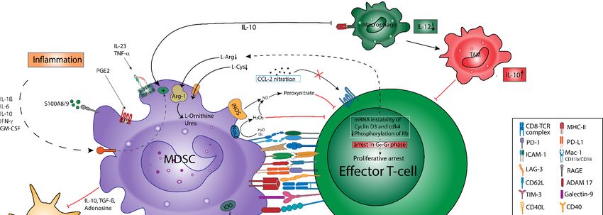
Cancers 2021, 13, 210                                                                                             2 of 34

                        toward ICI. Here, it has been demonstrated that both tumor-related factors, e.g., a high
                        mutational load of the tumor cells [3,4], the presence of neoantigens [5,6], microsatellite in-
                        stability [7,8], and host factors, i.e., the frequency, composition, and activation status [9,10]
                        of tumor-infiltrating lymphocytes (TIL), are predictive for the responsiveness toward ICI
                        treatment. Particularly referring to the activation status of TIL, it has been documented
                        that the extent of programmed cell death 1 ligand 1 (PD-L1) expression on tumor cells
                        correlates with objective response rates in melanoma and non-small cell lung cancer [11–13].
                        Hence, PD-L1 expression levels are also applied in the clinical setting to optimize patient
                        stratification prior to the introduction of ICI therapy. However, both the extent of cytotoxic
                        T-lymphocyte (CTL) infiltration into the tumor and PD-L1 expression on tumor cells do
                        not always correlate with clinical benefit [14]. So far, the various immunosuppressive
                        mechanisms, being present both locally within the tumor microenvironment (TME) and in
                        lymphoid organs, have been identified as major factors mediating immune resistance [15].
                        Next to the immunosuppressive effects conferred by soluble mediators and leukocyte
                        receptor interactions, the extensive infiltration of the tumor by immunosuppressive cell
                        populations, such as tumor-associated macrophages (TAM) [16,17], myeloid-derived sup-
                        pressor cells (MDSC) [18], and regulatory T cells (Treg) [19,20], has been identified as a
                        major driver of the pro-tumorigenic transformation in the TME. The presence of these
                        immunosuppressive cells hampers effector T cell induction and recruitment as well as
                        the capability of both natural killer (NK) cells and antigen-presenting cells (APC) to ex-
                        ert effective tumor surveillance, consequently leading to a profound inhibition of the
                        anti-tumor immune response [21]. Thus, the understanding of this immunosuppressive
                        network mediating tumor immune evasion, via cell–cell interactions and by the secretion of
                        soluble immunomodulatory mediators, is essential for the development of novel strategies
                        overcoming immune resistance in cancer treatment.
                              Recent observations in different cancer models suggest a crosstalk between MDSC
                        and Treg, but its character remains incompletely defined [21–23]. As the crosstalk between
                        MDSC and Treg has recently been proposed to be a powerful barrier counter-acting anti-
                        tumoral immune responses, this review is dedicated to give insights into the potential role
                        of cell–cell contacts as a prerequisite for the immunosuppressive mechanisms in the TME,
                        leading to tumor immune evasion. This in consequence facilitates cancer progression and
                        the development of metastases [24].
                              Furthermore, we aim to delineate the relevance of metabolic pathways and soluble
                        mediators for the functional interaction of MDSC and Treg, according to the current state of
                        scientific research. Since β2 integrins are known to be key regulators of cell adhesion and
                        cell signaling, they are essential for immune cell functions [25]. Accordingly, β2 integrins
                        may constitute potential mediators of the crosstalk between MDSC and Treg [26]. Hence,
                        this review additionally aims to outline the potential role of β2 integrins in this critical
                        cell–cell interaction within an immunosuppressive TME.

                        2. The Immunosuppressive TME
                              The TME is a complex milieu being composed of a heterogeneous assemblage of
                        distinct tumor—and host cell types such as cancer-associated fibroblasts (CAF), endothe-
                        lial cells, pericytes, and immune cells that constitute the tumor parenchyma and tumor
                        stroma [14,27]. These various cell types exhibit an extensive crosstalk that dynamically
                        regulates the phenotype and function of the individual cells within the TME. This allows
                        the establishment of a chronic pro-inflammatory state that favors the establishment of a
                        tumor-supportive microenvironment [14,28–30]. Thus, it is increasingly evident that the
                        crosstalk between cancer cells and cells of the neoplastic stroma in the TME enables tumor
                        cells to evade host immunosurveillance and thereby supports tumor growth, progression,
                        and metastasis [14,31]. Moreover, the regulatory signaling conveyed by soluble mediators
                        and cell–cell interactions is considered essential in controlling the individual cell function
                        and orchestrating the collective activity within the tumor network [14].
Cancers 2021, 13, 210                                                                                            3 of 34

                        2.1. Immunomodulatory Mediators Shape the TME
                              In the context of immunotherapy, the mutual interactions of tumor-infiltrating immune
                        cells have become an increasingly important area of research, as these cells shape the
                        unique properties of the TME [2]. The tumor-infiltrating immune cells include both tumor-
                        promoting as well as tumor-killing subclasses [14]. Here, it has been shown that tumor
                        infiltration by T cells (mainly CTL) and NK cells correlates with overall prognosis and
                        with the response to ICI treatment [32]. However, in the course of tumor development, a
                        chronic inflammatory state is frequently being induced, which includes the elevation of pro-
                        inflammatory mediators, the infiltration of regulatory immune cells, and the recruitment
                        of endothelial cells and fibroblasts [30,33]. The accumulation of both pro-inflammatory
                        mediators, including cytokines (e.g., interleukin (IL-1, IL-6); tumor-necrosis-factor-alpha,
                        (TNF-α)), chemokines (CC-chemokine ligand 2 (CCL2), and C-X-C motif ligand 2; (CXCL-
                        2)), prostaglandines (prostaglandine E2 (PGE2)) and growth factors (e.g., transforming
                        growth factor-β (TGF-β), granulocyte-macrophage colony-stimulating factor (GM-CSF)),
                        orchestrate the crosstalk between the various cells within the TME. In concert with these
                        soluble mediators, cell–cell-based interactions such as the programmed death protein (PD)-
                        1/PD-ligand (L)-1 axis contribute to the intense crosstalk between the immunosuppressive
                        cell populations, subsequently enhancing the tumor-supporting capacity of the TME, which
                        tips the scale toward immunosuppression and tumor angiogenesis [30]. Altogether, these
                        mechanisms antagonize the cancer-directed immune responses and effectively impair the
                        lytic machinery of TIL in the TME [24,34].

                        2.2. Cellular Composition of the TME
                              Notably, MDSC, TAM, and Treg are the major cellular components of the immunosup-
                        pressive TME. It has been demonstrated that the release of pro-inflammatory cytokines
                        within the TME promotes the immunosuppressive potential of regulatory myeloid cells,
                        such as tumor-associated neutrophils (TAN) [35–37], TAM [27,33,38–40], MDSC [28,41,42]
                        and regulatory dendritic cells (DC) [43–45]. Consequently, a strong tumor infiltration by
                        myeloid cells—being the most abundant cell types within the TME [46]—correlates with
                        rapid tumor growth and a poor prognosis [32]. Here, TAM primarily serves to promote
                        tumor growth and progression via the generation of angiogenetic factors such as vascular
                        endothelial growth factor (VEGF), and the secretion of immunomodulatory cytokines
                        (e.g., IL-6, IL8 and IL-10) [38]. These cytokines generated by TAM and tumor cells pro-
                        mote an aberrant activation of myelopoiesis, resulting in a defective differentiation of
                        myeloid progenitor cells toward MDSC, which exert a strong pro-tumor activity [46,47].
                        In particular, it has been shown that MDSC suppress both CTL and NK cell activity via
                        immunomodulatory mediators, including IL-1, IL-6, reactive oxygen species (ROS), and
                        nitric oxide (NO) [14,48–50]. Hence, the proliferation, activation, and retention of highly im-
                        munosuppressive MDSC are not only induced by the chronic inflammatory state within the
                        TME, but it further enhances these conditions, thus creating a positive feedback loop [34,51].
                        In this context, recent studies revealed that MDSC can modulate the de novo induction,
                        development, and activation of Treg, thus further amplifying the immunosuppressive
                        character in the TME [24]. CD4+ CD25hi Forkhead-Box-Protein P3 (FoxP3)+ Treg cells are
                        frequently found in the course of tumor progression and counteract APC activity, T cell
                        activation, and anti-tumor functions of effector T cells (Teff) [24,52]. Therefore, similar to
                        MDSC, clinical reports confirmed a negative correlation between the frequency of Treg,
                        the patient’s individual prognosis, and the response to ICI treatment [24].
                              Next to their direct immunosuppressive effects, MDSC and Treg implicitly contribute
                        to the establishment of a TME being characterized by hypoxia, the accumulation of
                        lactic acid, and adenosine (ADO). These factors prevent APC maturation, impair Teff
                        functions, and thus counteract the tumoricidal functions of activated immune effector
                        cells [14,24,46,53]. Since MDSC and Treg systemically expand in the course of tumor de-
                        velopment and strongly impair T cell driven anti-tumor immune responses, a detailed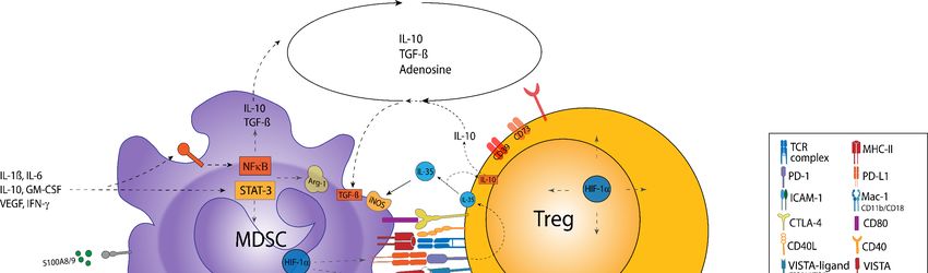
Cancers 2021, 13, 210                                                                                         4 of 34

                        characterization of the phenotype and immunosuppressive functions of these cells will be
                        provided in the following section.

                        3. Myeloid-Derived Suppressor Cells
                             Immature myeloid cells, which are generated in the bone marrow (BM) of healthy
                        individuals in response to acute infection, stress, or trauma, regularly differentiate into
                        mature myeloid cells, such as polymorphonuclear neutrophils (PMN) and monocytes,
                        without exerting immunosuppression [49]. In contrast, neoplastic cells, tumor-associated
                        stroma cells, and a frequently observed inflammation within the TME favor the aberrant
                        activation of myelopoiesis that results in the expansion and recruitment of immature
                        myeloid cells to the tumor site [50]. Indeed, a prominent effect of this “tumor-driven
                        macroenvironment” is the accumulation of highly suppressive, immature myeloid cells in
                        the tumor. Owing to their common myeloid origin and suppressive properties, these cells
                        were termed MDSC.

                        3.1. MDSC Subsets and Their Immunophenotypes
                              MDSC have first been identified in tumor-bearing mice as immature myeloid cells
                        characterized by the co-expression of CD11b and Gr-1, comprising the lineage markers
                        Ly6G and Ly6C [49,54]. Unlike other myeloid cells, MDSC exhibit a larger diversity of
                        phenotypes, which complicates their identification and characterization [24]. This hetero-
                        geneity is in part due to the unique inflammatory milieu present within different tumor
                        entities [41]. Further contributing to the high plasticity of MDSC phenotypes are the tempo-
                        ral variations in the context of tumor-immune editing, as the TME is subject to permanent
                        modulations in the course of the malignant transformation [39,55].
                              There are currently two main subsets of MDSC to be distinguished: granulocytic
                        (G)-MDSC and monocytic (M)-MDSC [56]. G-MDSC represents the predominant subset of
                        MDSC in the majority of tumor patients and tumor animal models (approximately 75%)
                        compared with M-MDSC (approximately 25%) [57,58]. However, G-MDSC are considered
                        to be less suppressive than M-MDSC when evaluated on a per-cell basis [49,58,59]. This ob-
                        servation was confirmed in human studies, demonstrating a tight correlation between
                        M-MDSC numbers and the inhibition of T cell activation [60].
                              Murine G-MDSC are generally characterized as CD11b+ , Ly-6G+ , Ly-6Clow (collec-
                        tively termed as Gr-1high ), and CD49− , whereas murine M-MDSC are defined as CD11b+ ,
                        Ly6G− , Ly-6Chigh (Gr-1high ), and CD49+ , while expressing F4/80, CD115, or CCR2 at
                        varying extents [49]. Due to the polymorphonuclear morphology of G-MDSC and the
                        expression of CD11b and Ly6G, their relationship to PMN is an ongoing issue [24,61].
                        However, as compared to PMN, G-MDSC show a diminished phagocytic activity, produce
                        higher levels of ROS, and suppress T cell activation upon activation (Table 1) [61]. There-
                        fore, the assessment of these distinctive immunosuppressive properties is important for a
                        definite characterization of G-MDSC, since no distinctive G-MDSC marker set has been
                        established yet [24,62].
                              Leaving alone the vast heterogeneity in murine MDSC phenotypes, the definition of
                        specific markers for MDSC in humans remains another important issue. Human MDSC
                        are commonly found to be CD11b+ , CD33+ , and HLA-DR− [63,64], and the majority
                        of G-MDSC is CD15+ , whereas CD14 expression is predominantly confined to M-MDSC
                        (Table 1) [24,65]. However, MDSC subsets in humans have yet not been defined consistently
                        with respect to surface marker expression [60].
                              Despite conflicting reports about MDSC surface marker expression, the clinical value
                        of MDSC has readily been demonstrated: Recent reports highlighted that the frequency
                        of MDSC per se may reflect the tumor burden of cancer patients, thus showing a strong
                        correlation between a high MDSC frequency and a poor prognosis [64,66,67]. On the other
                        hand, the tumor itself may influence the composition of the MDSC compartment. In gen-
                        eral, G-MDSC have been found to be the main MDSC subset in patients with renal cell
                        carcinoma [68], whereas M-MDSC constitute the dominant immunosuppressive MDSC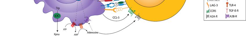
Cancers 2021, 13, 210                                                                                                                             5 of 34

                                      subpopulation in melanomas or head–neck cancer [60,65]. However, since none of the
                                      aforementioned individual markers are unique to a distinct MDSC subset, the defini-
                                      tive identification of human MDSC subsets requires the assessment of their employed
                                      suppressive mechanisms [41,55].

      Table 1. Phenotypic definitions and functional characteristics of different myeloid cell types present within solid tumors.

      Characteristics                  PMN                    TAN                  G-MDSC                   M-MDSC                       TAM
                                     CD11b+                 CD11b+                  CD11b+                   CD11b+                    CD11b+
                                     CD11c−                 Ly6Clow                  Gr-1+                    Gr-1+                    F4/80+
                                     Ly6Clow                 Ly6G+                  Ly6G+                   Ly6Chigh                   CD206+
      Murine marker                   Ly6G+                 PD-L1+                 Ly6Clow                   Ly6G−                     CD163+
         subsets                     CD101+                CD170high               CD115low                  CD49d+                     CD36+
                                     F4/80−                                         CD49−                   CD115high                 MHC-IIlow
                                     CD115−                                                                                            IL-10R+
                                                                                                                                       CD124+
                                     CD11b+                 CD45+                    CD33+                    CD33+                     CD14+
                                     CD66b+                 CD33+                   CD11b+                   CD11b+                     CD68+
                                      CD15+                 CD11b+                 HLA-DR−                  HLA-DR-                    CD205+
                                     CD14−                 HLA-DR−                   CD15+                    CD14+                    CD163+
     Human marker
                                      CD16+                 CD66b+                 STAT-3high               STAT-3high                  CD36+
       subsets
                                     CD62L+                 PD-L1+                  CD66b+                   CD124+                  HLA-DRlow
                                     CXCR1+                CD170high                CD244+                   S100A9+                   IL-10R+
                                                            LOX-1+                  S100A9+                                            PD-L1+
                                                                                    LOX-1+                                            STAT-3low
                                 predominantly           predominantly
     Maturation stage                                                              Immature                  Immature                   Mature
                                    mature                  mature
                                                                                                                                          IL-4
                                                                                     G-CSF                    M-CSF
     Potent inductors                GM-CSF                  TGF-β                                                                       IL-10
                                                                                      IL-6                     IL-6
                                                                                                                                        TGF-β
                                                                                                                                       Hypoxia
    Inhibition of T cell
                                         Ø                       ↑                      ↑                        ↑↑                        ↑
       proliferation
            ROS                          ↑                       ↑                     ↑↑                         ↓                        Ø
           MPO                           ↑                       ↑                     ↑↑                        Ø                         Ø
        Arginase-1                       Ø                       ↑                     ↑↑                         ↑                        ↑
            NO                           Ø                       ↓                      ↓                        ↑↑                        ↑
          NETosis                        ↑                       ↑                      Ø                        Ø                         Ø
            IL-8                         ↑                       ↑                      Ø                        Ø                         ↑↑
       Immune cell                                                                                                                   Functional
      polarization in            TAN, G-MDSC,                                                                                     polarization (M1
                                                              PMN               TAN, PMN (?)                 TAM, DC
        response to              APC-like-PMN                                                                                         and M2
        stimulation                                                                                                                 phenotype)
      Ø: no significant effect; ↓: lower expression/activity compared to the other listed cell types; ↑: higher expression/activity compared to the
      other cell types; ↑↑: strongest expression/activity among the listed cell types; MPO = myeloperoxidase; NET = neutrophil extracellular traps.

                                      3.2. Myeloid Cell Plasticity within Tumors
                                           Of note, MDSC entering the TME may have the plasticity to interconvert between
                                      different phenotypes. In particular, it has been shown that MDSC might convert into
                                      TAM, DC, or TAN depending on the conditions present in the TME (Figure 1) [36,49,50].
                                      For example, the culture of tumor-derived MDSC in the absence of tumor-derived fac-
                                      tors was repetitively shown to result in the generation of mature macrophages, PMN,
                                      and DC [50,69–71], whereas the presence of tumor-derived factors or the adoptive transfer
                                      of MDSC into tumor-bearing hosts promoted their differentiation into immunosuppressive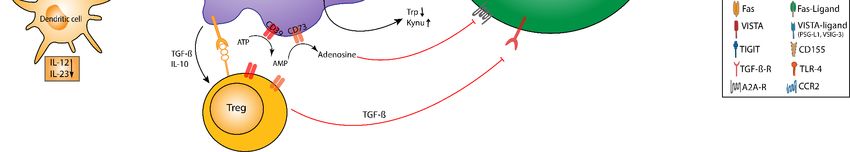
Of note, MDSC entering the TME may have the plasticity to interconvert between
different phenotypes. In particular, it has been shown that MDSC might convert into
TAM, DC, or TAN depending on the conditions present in the TME (Figure 1) [36,49,50].
For example, the culture of tumor-derived MDSC in the absence of tumor-derived factors
Cancers 2021, 13, 210 was repetitively shown to result in the generation of mature macrophages, PMN,6 and of 34 DC
[50,69–71], whereas the presence of tumor-derived factors or the adoptive transfer of
MDSC into tumor-bearing hosts promoted their differentiation into immunosuppressive
macrophages
macrophages [49,69].
[49,69].Hence,
Hence,TAN
TAN and TAMmay
and TAM mayconstitute
constitute differentiated
differentiated MDSC
MDSC or rep-
or rep-
resent a pro-tumorigenic subset of mature PMN and macrophages polarized by
resent a pro-tumorigenic subset of mature PMN and macrophages polarized by soluble soluble
mediators
mediators[24,49,53].
[24,49,53].
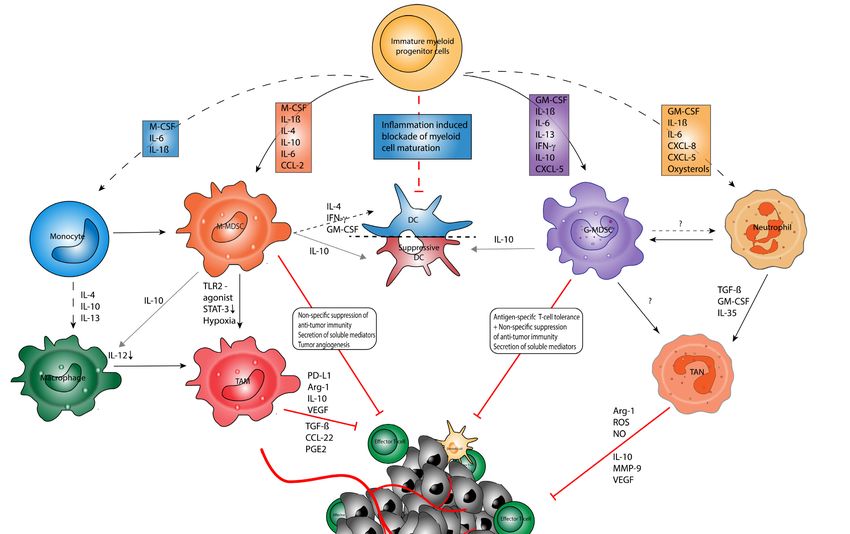
Figure 1. Myeloid
Figure cellcell
1. Myeloid plasticity in in
plasticity cancer.
cancer.Myeloid
Myeloidcell
cell types originatefrom
types originate fromhematopoietic
hematopoietic stem
stem cells
cells andand multipotent
multipotent
immature myeloid
immature progenitor
myeloid progenitor cells
cellsininthe
thebone marrow(BM).
bone marrow (BM).TheThe differentiation
differentiation toward
toward the matured
the matured cell linecell line
(i.e., (i.e., poly-
polymor-
morphonuclear neutrophils (PMN)) is promoted by soluble mediators and chemokines. In cancer
phonuclear neutrophils (PMN)) is promoted by soluble mediators and chemokines. In cancer patients, the differentiationpatients, the differenti-
ationpathways
pathwaysare are strongly
strongly affected
affected by factors
by factors produced
produced in theintumor
the tumor microenvironment
microenvironment (TME) by (TME) by stromal
stromal cells, immunecells, cells,
immune
cells,and
andtumor
tumorcells
cells (e.g.,
(e.g., granulocyte-macrophage
granulocyte-macrophage colony-stimulating
colony-stimulating factor (GM-CSF),
factor (GM-CSF), interleukin
interleukin (IL)-1β,
(IL)-1β, IL-6, IL-6,
IL-10, IL-10,
IL-23,
IL-23,interferon-gamma
interferon-gamma (IFN-γ)).
(IFN-γ)). In particular,
In particular, the promotes
the TME TME promotes the polarization
the polarization of macrophages
of macrophages toward immunosup-
toward immunosuppressive
pressive tumor-associated
tumor-associated macrophages
macrophages (TAM),(TAM), whichthe
which confer confer the inhibition
inhibition of effectorofTeffector T cells
cells (Teff) (Teff)
within within
the TME viathe TME via
various
various mechanisms [72]. PMN within the TME frequently show a polarization toward immunosuppressive
mechanisms [72]. PMN within the TME frequently show a polarization toward immunosuppressive TAN, which is driven TAN, which
is driven by soluble factors such as transforming growth factor-β (TGF-β). Tumor-associated neutrophils
by soluble factors such as transforming growth factor-β (TGF-β). Tumor-associated neutrophils (TAN) confer immunosup- (TAN) confer
pression via multiple mechanisms. The most prominent effect of the aberrant differentiation includes the accumulation of
granulocytic (G-MDSC) and monocytic (M-MDSC) myeloid-derived suppressor cells. Myeloid cells may act as an integrated
system in the context of tumor immunity [49]. Depending on the structural composition of the TME, myeloid cells polarize
from MDSC toward TAM or TAN or promote the tolerization of DC in the context of a nutrient-depleted, hypoxic, inflamed
TME. Under normoxic conditions, IFN-γ and TNF-α have been found to reverse this polarization and promote MDSC
differentiation toward immunogenic DC and inflammatory M1 macrophages. It remains questioned if MDSC and TAN
undergo an irreversible polarization or can polarize to anti-tumor PMN [24].Cancers 2021, 13, 210 7 of 34
Despite the phenotypical similarities of the various (immunosuppressive) myeloid
cell types, recent reports highlighted, that these can be discriminated by transcriptomic and
multi-omics approaches: Referring to the granulocytic cell line in particular, Fridlender et al.
revealed cell-specific transcriptome signatures of PMN, G-MDSC, and TAN, confirming
the existence of three distinct phenotypes [62]. Moreover, G-MDSC have shown a higher
immunosuppressive activity, expressed higher levels of CD115 and CD244, and lower levels
of CXCR1 than PMN [61,73]. G-MDSC exerted less phagocytic activity, show a smaller
chemotactic response, expressed higher levels of Arginase (Arg)-1 and myeloperoxidase
(MPO), and showed a higher production of ROS [49,73].
Likewise, M-MDSC, despite their similarity in morphology and phenotype with
other monocytic cell populations, are a functionally distinct population. Particularly, they
showed a strong expression of inducible NO-synthase (iNOS) and Arg-1, which explains
their highly immunosuppressive character [49,74]. In parallel, it has been reported that
hypoxia and hypoxia inducible factor 1α (HIF-1α) within the TME might be key drivers for
the upregulation of immunosuppressive Arg-1 and iNOS in M-MDSC and may promote
the differentiation of CD11b+ Ly6C+ M-MDSC into immunosuppressive TAM [53,75]. Since
the polarization toward a macrophage M2 phenotype is more likely in MDSC at the tumor
site compared to spleen-derived MDSC, it remains an issue to clarify whether the origin of
MDSC within the TME might determine the modulation of their phenotype [24,53].
Collectively, these findings provide a mechanistic link between different myeloid cells
and indicate that MDSC have the plasticity to interconvert between different phenotypes
depending on the specific conditions present within the TME (see Figure 1) [24,53]. How-
ever, knowledge of the factors that govern the interconversion of the various granulocytic
and monocytic (immunosuppressive) cell types is still far from being complete. There-
fore, in vivo strategies and multi-omics approaches are vital to elucidate (combinations
of) TME-derived factors that may induce the differentiation, expansion, activation, and
interconversion of MDSC populations [24,76].
3.3. Mechanisms of Tumor-Induced MDSC Accumulation
Evidence suggests that the release of tumor-derived soluble mediators, such as GM-
CSF, VEGF, or IL-6 impairs the myeloid compartment and thus contributes to defective
myeloid cell maturation. Moreover, it has been proposed that the relative amounts of G-CSF
and M-CSF present within the bone marrow may account for the different shares of the
aforementioned MDSC subsets [59]. Here, Waight et al. reported that G-CSF facilitates the
accumulation of G-MDSC in the TME, subsequently promoting tumor growth. Moreover,
tumor-derived CCL2, CCL12, CXCL5, S100A8, and S100A9 promote the recruitment of
immature myeloid cells to the tumor stroma, facilitating the enrichment of both MDSC
subpopulations within the TME [49,77–79]. Tumor-derived TGF-β has also been found to
regulate MDSC accumulation and the polarization of other myeloid cell populations, such
as tumor-infiltrating PMN toward an immunosuppressive phenotype [80]. Furthermore,
soluble factors such as IL-1β, IL-6, and S100A9 [81,82], and T cell-derived cytokines such as
IFN-γ, IL-4, IL10, and IL-13 have been reported to promote immunosuppressive MDSC [83].
The regulation of the integrated myeloid cell network via tumor-derived soluble medi-
ators is controlled on multiple levels via the activation of various transcription factors. Here,
the Toll-like receptor (TLR) family, namely TLR-4, which is triggered by S100A8 and S100A9
proteins, contributes to myeloid cell development via the downstream induction of nuclear
factor-kB (NFκB), thus supporting the mobilization of myeloid cells to sites of inflammation
and their inflammation-driven suppressive potency [49,84]. Other suppressive properties
of MDSC are controlled by signal transducer and activator of transcription (STAT)-1 and
STAT-6, which regulate myeloid cells by inducing iNOS and Arg-1 expression [69,83].
Further, STAT-3 has been identified as a crucial regulator of MDSC expansion that
conveys the recruitment of MDSC to the tumor site by upregulating the pro-inflammatory
S100A8 and S100A9 proteins [85]. Hence, S100A9 protein has been proposed as a potential
marker characterizing human CD14+ HLA-DR− M-MDSC. STAT-3 has also been reportedCancers 2021, 13, 210 8 of 34
to induce the upregulation of NADPH oxidase (Nox) components, thereby adding up to
the immunosuppressive features of MDSC, such as ROS production [49,86]. However,
an unsolved question remains: How do these molecular markers relate to the suppressive
function of MDSC? Hence, the most definitive characterization of MDSC remains their
immunosuppressive function, which will be addressed in the following section.
4. Immunosuppressive Properties of MDSC
MDSC are considered key regulators of immune responses in many pathophysi-
ological conditions, including anti-tumor immune responses. G-MDSC and M-MDSC
apply antigen-specific and antigen-non-specific mechanisms to regulate immune responses
and thus inhibit Teff via a plethora of mechanisms [24]: In peripheral lymphoid organs,
the MDSC-mediated suppression of CTL usually requires antigen presentation by MDSC
and direct MDSC/T cell contact [87,88]. Otherwise, at the tumor site [53,89] and in the
periphery [90], MDSC can suppress nearby T cells in an antigen-independent manner.
Although none of these mechanisms are exclusively used by either MDSC subpopulation,
it has been demonstrated that ROS generation is characteristic for G-MDSC, whereas Arg-1
expression and the generation of NO has primarily been found in M-MDSC [50,58,75].
4.1. Depletion of Nutrients
MDSC confer immunosuppression by various mechanisms (Figure 2), such as the
depletion of nutrients. This involves the Arg-1-dependent consumption of L-arginine and
deprivation of L-cysteine via its consumption and sequestration in MDSC [91], which
causes the proliferative arrest of antigen-activated T cells due to the downregulation
of the TCR (T cell receptor) complex and a cell cycle arrest in the G0-G1 phase [49,68].
This phenomenon could be reversed by the replenishment of L-arginine in vitro, but more
importantly, in vivo studies reported that the depletion of G-MDSC re-established T cell
growth, emphasizing their role in cancer immunosuppression [92]. The inhibition of T cell
activation is further enhanced via the consumption of L-tryptophan by MDSC-derived
indoleamine-2,3-dioxygenase (IDO) and the subsequent accumulation of kynurenines [93].
Additionally, it was shown that the ADO-generating ectoenzymes CD39 and CD73 are
upregulated by MDSC upon HIF-1α induction [94]. ADO impedes Teff function via A2A-
receptor (A2AR) and promotes TAM and MDSC suppressive functions via A2BR [95,96].
Whereas the depletion of nutrients and oxygen within the TME comprises T cell func-
tion [97,98], tumor hypoxia and lactate accumulation drive HIF-1α stabilization in MDSC,
thus upregulating PD-L1 expression and promoting a metabolic switch to fatty acid oxi-
dation (FAO). FAO further induces Arg-1 expression, NO, and peroxynitrite generation,
resulting in Teff impairment [99].
4.2. Oxidative Stress
Another suppressive mechanisms is the generation of oxidative stress via ROS and
reactive nitrogen species [49]. The production of ROS is mediated by Nox-2. Here, studies
conducted by Corzo et al. found an upregulation of ROS in G-MDSC isolated from seven
different murine tumor models and in tumor-derived G-MDSC obtained from patients
with head neck cancer [86]. Interestingly, in the absence of ROS production, G-MDSC
did not only lose their ability to confer T cell hyporesponsiveness in vivo, they also dif-
ferentiated into mature DC [86]. On the other hand, MDSC themselves are protected
from the cytotoxic ROS effects by induction of the antioxidant nuclear factor erythroid
related factor 2 (Nrf2) and the accumulation of the ROS scavenger phosphoenolpyruvate
(PEP) [100]. Peroxynitrite is produced by the cooperative activities of Nox-2, Arg-1, and
iNOS [24,101]. Peroxynitrites can cause the nitration of several proteins in tumor and
immune cells including the TCR, leading to subsequent TCR desensitization and T cell
apoptosis [49]. Moreover, nitration mediates several molecular blocks in T cells, including
conformational changes in the TCR–CD8 complex, which renders CTL unresponsive to
antigen-specific stimulation [87]. Furthermore, it was found that peroxynitrite interferesCancers 2021, 13, 210 9 of 34
with IL-2 receptor signaling [102] and leads to the nitration of CCL-2 chemokines. Con-
sequently, antigen-specific CTLs do not infiltrate into the tumor but instead remain in
the tumor-surrounding stroma [79]. Notably, iNOS-driven NO generation may further
induce cyclooxygenase-2 (COX-2) activity, resulting in an enhanced PGE2 production,
which serves as a potent inductor of IDO, Arg-1, IL-10, and VEGF secretion by MDSC [98].
Lastly, it has been documented that MDSC counteract the upregulation of CD44 and CD162
Cancers 2021, 13, x 9 of 35
by T cells in an NO-dependent manner, thus impairing T cell extravasation and tissue
infiltration [103,104].
Figure 2. MDSC-mediated
Figure 2. MDSC-mediated inhibition
inhibitionofofTTcell
cell activation andproliferation.
activation and proliferation. Direct
Direct inhibition
inhibition of Teffof Teff involves
involves cell–cell cell–cell
contacts con-
tacts (e.g., via checkpoint molecules), which induce proliferative arrest apoptosis, a reduced migratory
(e.g., via checkpoint molecules), which induce proliferative arrest apoptosis, a reduced migratory activity, and attenuated activity, and atten-
uatedTTcell cellrecirculation.
recirculation.
T cell activation is further inhibited via soluble mediators and metabolic pathways: MDSC contribute to con-
T cell activation is further inhibited via soluble mediators and metabolic pathways: MDSC
tribute to L-arginine and
L-arginine and L-cysteine L-cysteine depletion
depletion in the TME, in the TME,
which which
causes causes proliferative
proliferative arrest via mRNA arrest via mRNA
instability instability of cyclin-
of cyclin-dependent
dependent
kinase kinase
4 (cdk4),4reduced
(cdk4),phosphorylation
reduced phosphorylation of retinoblastoma
of retinoblastoma protein (Rb), andprotein
the loss(Rb),
of theand
T cellthe loss of
receptor the ζ-chain
(TCR) T cell receptor
on
(TCR)Teff.
ζ-chain
G-MDSC on Teff. G-MDSC
express express
high levels of NADPHhigh levels
oxidaseof(Nox)-2,
NADPH oxidaseROS-dependent
mediating (Nox)-2, mediating ROS-dependent
inhibition inhibition of
of Teff. The cooperative
Teff. The cooperative
activities of Nox-2,activities
Arginaseof(Arg)-1,
Nox-2, andArginase (Arg)-1,
inducible nitricand inducible
oxide synthasenitric
(iNOS) oxide synthase
generate (iNOS) generate
peroxynitrite, peroxyni-
which drives
trite, which drives protein nitration resulting in desensitization of the TCR and the interference with
protein nitration resulting in desensitization of the TCR and the interference with IL-2 receptor signaling. The consumption IL-2 receptor signal-
ing. The
of L-tryptophan and the accumulation of kynurenines in the TME add up to the inhibition of Teff and regulatory T cellsof Teff
consumption of L-tryptophan and the accumulation of kynurenines in the TME add up to the inhibition
and regulatory T cellsCD39
(Treg) induction. (Treg)andinduction. CD39extracellular
CD73 degrade and CD73 degrade extracellular
ATP to adenosine, ATP
which to adenosine,
enhances which enhances
T cell inhibition and Treg T cell
inhibition and Treg
induction. induction.
Indirect mechanismsIndirect mechanisms ofimmunosuppression
of MDSC-mediated MDSC-mediated immunosuppression
include the induction and include the induction
expansion of Treg and
expansion of Treg both via cell–cell contact-dependent mechanisms and soluble mediators
both via cell–cell contact-dependent mechanisms and soluble mediators (e.g., TGF-β, IL-10, prostaglandine-E2; PGE2;(e.g., TGF-β, IL-10, prostaglan-
and
dine-E2; PGE2; and A2A-receptor mediated signaling). Likewise, MDSC imprint a tolerogenic
A2A-receptor mediated signaling). Likewise, MDSC imprint a tolerogenic function in DC via IL-10, TGF-β, and adenosine. function in DC via IL-10,
TGF-β, Bothand theadenosine.
accumulationBothof the
Tregaccumulation
and TAM addof upTreg and
to Teff TAM add
inhibition up to
within theTeff
TME. inhibition
Macrophages withinarethe TME.toward
skewed Macrophages
an
are skewed toward an M2 phenotype via IL-10, thus impairing IL-12 production. Tumor-derived
M2 phenotype via IL-10, thus impairing IL-12 production. Tumor-derived soluble factors contribute to STAT3-mediated soluble factors contribute
to STAT3-mediated upregulation
upregulation of proteins of proteins
including Nox-2, cell including Nox-2, cell
survival proteins survival
(Cyclin D1), orproteins
S100A8/9, (Cyclin D1), MDSC
promoting or S100A8/9, promoting
accumulation
MDSC accumulation
(via (via S100A8/9
S100A8/9 ligation to RAGE), ligation
survival,to andRAGE), survival, and immunosuppression.
immunosuppression.
4.2. Oxidative Stress
4.3. Receptor-Mediated Inhibition
Another suppressive mechanisms is the generation of oxidative stress via ROS and
The interference with lymphocyte trafficking and viability is another immunosup-
reactive nitrogen species [49]. The production of ROS is mediated by Nox-2. Here, studies
pressive mechanism exerted by MDSC: Here, the expression of membrane-bound ADAM-
conducted by Corzo
metallopeptidase et al. 17
domain found an upregulation
(ADAM17) of ROS in
on MDSC decreased G-MDSC
CD62 isolated expres-
ligand (CD62L) from seven
different murine tumor models and in tumor-derived G-MDSC obtained from patients
with head neck cancer [86]. Interestingly, in the absence of ROS production, G-MDSC did
not only lose their ability to confer T cell hyporesponsiveness in vivo, they also differen-
tiated into mature DC [86]. On the other hand, MDSC themselves are protected from the
cytotoxic ROS effects by induction of the antioxidant nuclear factor erythroid related fac-Cancers 2021, 13, 210 10 of 34
sion on CD4+ and CD8+ T cells, thereby limiting the recirculation into lymph nodes [105].
Furthermore, several checkpoint molecules were shown to be critically involved in MDSC-
mediated immunosuppression: Among these, PD-L1 and CTLA-4 are prominent negative
regulators of T cell functions [106]. PD-L1 exerts its effects via ligation of PD-1 on T cells,
resulting in T cell anergy and apoptosis [104], promoting the induction and function of
Treg [107] and thus contributing to tumor immune evasion. Treg express CTLA-4, which
mainly interacts with CD80/CD86 as expressed by APC-like DC. This interaction causes
an impairment of APC-dependent T cell activation [108], enhances the immunosuppres-
sive properties of Treg, and augments peripheral tolerance [109]. Blocking checkpoint
molecules via monoclonal antibodies has in fact proven to restore effective anti-tumor
immune responses in many patients with advanced malignancies. This effect has been
attributed in part to the blockade of MDSC-mediated immunosuppression of Teff [104].
Youn and coworkers additionally suggested that PD-L2 might add up to MDSC-
induced T cell inhibition, since PD-L2-/PD-1 interaction skewed T cells toward T-helper
cells type 2 (Th2) [61,110,111].
Notably, more recent observations revealed the pivotal role of additional checkpoint
molecules, such as the V domain-containing immunoglobulin suppressor of T-cell activa-
tion (VISTA), Galectin-9 (Gal-9), and CD155 for MDSC-mediated immunosuppression [112].
In particular, VISTA has been reported to enhance the inhibition of T cell [113,114] and B
cell responses [115] by MDSC, whereas a blockade of VISTA allowed for the restoration
of a protective anti-tumor response [116,117]. Next, it has been documented that Gal-9-
expression on MDSC induced T cell apoptosis via ligation to the checkpoint protein T cell
immunoglobulin and mucin domain-containing protein (TIM)-3 [118]. Gal-9 has been also
been reported to promote a suppressive TME by enhancing the degradation of stimulator
of interferon genes (STING) [119]. As suggested by Dardalhon et al., the interaction of
TIM-3+ , IFN-γ-secreting T cells with Gal-9+ MDSC might add up to both MDSC expansion
and immunosuppressive functions [120]. Last, recent observations indicated that CD155
might also be involved in MDSC-mediated T cell inhibition, since it may serve as a ligand
for T-cell Ig and ITIM domain (TIGIT), which is found on T and NK cells promoting the
immunosuppressive functions of Treg [121,122]. Despite conflicting reports about the
role of Fas-(L)igand-Fas signaling for MDSC homeostasis and function [90,123], it is well
documented that MDSC are able to induce T cell apoptosis via FasL [124]. Next to the T cell-
specific inhibition, MDSC also interfere with NK cell cytotoxicity via receptor-mediated
mechanisms, e.g., the interaction of membrane-bound TGF-β with the NK cell receptor
NKp30 [49,125,126].
4.4. Induction of Protolerogenic APC
Additionally, MDSC promote immunosuppression indirectly by the interaction with
other cells of the myeloid cell lineage, such as the inhibition of conventional DC and
macrophages. This observation further complicates the understanding of the myeloid
cell network within tumors, since myeloid cells engage with each other but also have
the plasticity to transdifferentiate between different phenotypes. The interdependency of
cells in the myeloid linage can be exemplified by the IL-10 and cell–cell contact-mediated
mechanisms by which MDSC decrease macrophage IL-12 production, tipping them toward
an M2-like phenotype [127]. This initiates a positive feedback loop, as macrophages
themselves promote IL-10 synthesis in MDSC, further enhancing the shift toward an
M2-like phenotype [49]. An inflamed TME enhances the infiltration of MDSC into the
tumor, promotes TLR-4 signaling, the expression of CD14 on MDSC, and their activation.
Thus, inflammation is considered a key driver of MDSC and macrophage crosstalk within
the TME [128]. Next to the interaction between MDSC and macrophages, MDSC impair
DC function via the production of IL-10, which inhibits IL-12 production in DC and the
subsequent DC-mediated activation of T cells [49,129]. Adding up more recently to the
wide array of immunosuppressive features, it has been observed that MDSC significantly
enhance their immunosuppressive potential via the activation and expansion of TregCancers 2021, 13, 210 11 of 34
populations [49]. The character of this interaction is discussed in the following after a brief
presentation of Treg characteristics.
5. Regulatory T Cells
It has been shown that regulatory T cells play a crucial role in regulating the home-
ostasis of the immune system and maintaining tolerance [130]. Moreover, Treg have been
found to limit the anti-tumor immune response. In accordance, the number of Treg cir-
culating in the blood of cancer patients and the infiltration of Treg into the tumor have
been documented to be closely related to the progression and prognosis of multiple cancer
entities [20]. More interestingly, the extent of Treg infiltration into human tumors has
been proposed to show an inverse correlation with the response to ICI therapy [131,132].
Not least, this observation emphasizes the importance of Treg in the understanding of the
anti-tumor immunity and thus the development of novel therapeutic approaches.
5.1. Characteristics and Classification of Treg
Treg are defined as a T helper cell subpopulation characterized by the co-expression
of CD4, CD25, and in large parts of FoxP3, which inhibit the activation and differentia-
tion of CD4+ and CD8+ T cells, subsequently impairing reactivity against autologous and
tumor-expressed antigens [130,133,134]. According to their biological properties, Treg are
generally divided into two groups: natural (n) regulatory T cells and induced (i) regulatory
T cells, which commonly express FoxP3 [135]. Whereas nTreg develop in the thymus and
exert their inhibitory activity for maintaining immune tolerance largely through intercellu-
lar contact, iTreg are derived from peripheral naïve tumor antigen-specific T cells, which are
induced by TME-derived cytokines and other soluble mediators [130]. However, both types
of Treg act in a tumor-antigen specific manner [136]. In contrast to Th cells and CTL, which
rely largely on glycolysis, glucose transporter (GLUT)-1 expression, and on mammalian
target of rapamycin (mTOR) signaling, to sustain their metabolic activity, Treg express low
levels of GLUT-1, are negatively regulated by mTOR, and depend largely on oxidative
phosphorylation and FAO to sustain their metabolic and suppressive activity [98,137,138].
5.2. Immunosuppressive Properties of Treg
Treg use several mechanisms to inhibit the anti-tumor immune activity of Teff, NK cells,
and DC, thus driving tumor progression. First, it has been shown that Treg-derived soluble
mediators, such as IL-10, TGF-β, and IL-35, suppress antigen presentation by DC, promote
T cell exhaustion and CTL dysfunction [139,140]. Next, it has been reported that Treg largely
interfere with the cell metabolism both within the TME and in secondary lymphatic organs,
inhibiting the proliferation of Teff by the competitive consumption of IL-2 [136]. Addition-
ally, the expression of the ectonucleotidases CD39 and CD73 enables Treg to hydrolyze
extracellular adenosine triphosphate (ATP) into adenosine monophosphate (AMP) and
subsequently to immunosuppressive ADO, which inhibits Teff via engagement with the
A2AR [141]. Moreover, the intercellular transfer of cyclic AMP (cAMP) to Teff via gap junc-
tions is considered another metabolic mechanism of Treg to inhibit an effective anti-tumor
immune response [130]. Similar to TAM and MDSC, Treg contribute to Arg-1 mediated
arginine depletion within the TME [142]. In contrast to Teff, Treg are largely unaffected by
limitations of either glutamine or leucine within the TME [143]. Treg counterbalance the
high ROS levels within the TME via antioxidants such as glutathione. In agreement, the
removal of this ROS-inactivating mechanism in Treg significantly impaired their inhibitory
activity [144]. Lastly, Treg hampered Teff and NK cell function and activity via immunosup-
pressive receptor interactions and the application of cytotoxic enzymes [145]. In particular,
Treg are capable of killing effector cells using granzymes or perforins and orchestrate the
quiescence of memory T cells by inhibiting effector programs via checkpoint molecules
such as cytotoxic T-lymphocyte-associated protein 4 (CTLA-4) [130,146]. Furthermore,
Treg hamper anti-tumor immunity via the interaction of CTLA-4 with the co-stimulatory
receptors CD80 and CD86, which are expressed by APC-like DC, resulting in the inhibitionCancers 2021, 13, 210 12 of 34
of their T cell stimulatory capacity [130,147]. In the course of this interaction, it has been
found that Treg might enhance immunosuppression via the upregulation of IDO and Arg-1
on APC, which impaired the induction of Teff and in turn also inhibited mTOR signaling
in Treg [148,149].
Recent reports indicate that the interaction of Treg with MDSC might further contribute
to the immunosuppressive activity and potential of Treg, forming a positive feedback
loop that facilitates the enforcement of their suppressive activity [130], as described in
the following.
6. Functional Crosstalk between MDSC and Treg
The interactions of MDSC and Treg in different cancer models have been proposed to
play a critical role in shaping the TME (Table 2) [21]. Although a strong influx of MDSC and
Treg has been described for many different tumor entities, there is only little evidence yet
for a direct mechanistic link between these major immunoregulatory cell populations. Here,
different modes of interactions have been proposed, namely those conferred by soluble
mediators, metabolic cooperations, or cell–cell contacts (Figure 3) [21]. Furthermore, it has
been suggested that MDSC promote both the conversion of naïve CD4+ T cells toward
iTreg and the expansion of nTreg [150–152].
Table 2. A selection of important mediators in the functional crosstalk between MDSC and Treg.
Receptors/Soluble Disease Model,
Cell Type Species Observations Reference
Mediators Immune State
Treg-derived TGF-β enhanced Arg-1, PD-L1, and
iNOS expression on MDSC, thus promoting their
TGF-β Treg and MDSC mouse Murine colitis immunosuppressive properties [153]
MDSC themselves showed a stronger induction of
Treg after TGF-β stimulation
Depletion of Treg downregulated PD-L1 expression
PD-1/ on MDSC and inhibited IL-10 production
Treg, MDSC and Diminished PD-L1 expression on MDSC led to a
PD-L1, mouse Ret-melanoma [154]
CD4+ T cells reduced inhibition of CD4+ T cells
IL-10
iNOS expression was not affected by Treg depletion
MDSC mediated Treg induction via IL-10
IL-10, and TGF-β
MDSC and Treg mouse Metastatic colon cancer Treg induction was independent of NO-mediated [151]
TGF-β
immunosuppression by MDSC
Physical interactions between MDSC and Treg
Cell-cell contacts (video-microscopic analysis)
Pancreatic ductal +
MDSC mediated Treg induction and
(receptors not MDSC and Treg mouse [21]
Adeno-Carcinoma immunosuppression via cell–cell contacts
specified)
(transwell system)
CD40-deficient MDSC failed to induce Treg (after
adoptive transfer)
CD40/CD40L MDSC and Treg mouse B16-OVA Melanoma anti-CD40 antibody treatment promoted the [155]
differentiation of MDSC toward DC
and macrophages
MDSC enhanced the immunosuppressive properties
of Treg via the engagement of CTLA-4 with CD80
CD80/CTLA-4 MDSC and Treg mouse Ovarian carcinoma CD80 depletion led to a significant reduction in [156]
tumor growth
Mac-1 and ROS production were required for the
Acute systemic inhibition of T cell function by a suppressive subset
Mac-1 MDSC and T cells human [157]
inflammation of human PMN MDSC enhanced the immunosuppressive prop-
erties of Treg via the engagement of CTLA-4
CD80/CTLA-4 MDSC and Treg mouse Ovarian carcinoma with CD80 [156]
CD80 depletion led to a significant reduction in
tumor growth
Cancers 2021, 13, 210 Mac-1 and ROS production were required for the 13 of 34
Acute systemic in-
Mac-1 MDSC and T cells human inhibition of T cell function by a suppressive sub- [157]
flammation
set of human PMN
Figure
Figure3.3.Crosstalk
CrosstalkofofMDSC
MDSCand andTreg.
Treg. MDSC
MDSC andand Treg interactions are
Treg interactions are enhanced
enhancedby bysoluble
solublemediators,
mediators,aacloseclosemetabolic
metabolic
cooperation,
cooperation,andand cell–cell interactions. Particularly,
cell–cell interactions. Particularly,MDSC-derived
MDSC-derived IL-10
IL-10 and
and TGF-β
TGF-β promote
promote TregTreg induction,
induction, prolifera-
proliferation,
tion, and activation. The secretion of TGF-β and IL-10 by Treg enhances the generation of these cytokines
and activation. The secretion of TGF-β and IL-10 by Treg enhances the generation of these cytokines in MDSC, establishing in MDSC, estab-
lishing a positive feedback loop. IL-10 and TGF-β promote the expression of immunosuppressive
a positive feedback loop. IL-10 and TGF-β promote the expression of immunosuppressive receptors (e.g., PD-L1) and receptors (e.g., PD-L1)
and enzymes (e.g., Arg-1, iNOS, and CD73) on MDSC. Autocrine IL-35 secretion by Treg, which is promoted via the PD-
enzymes (e.g., Arg-1, iNOS, and CD73) on MDSC. Autocrine IL-35 secretion by Treg, which is promoted via the PD-L1-PD-1
L1-PD-1 pathway, contributes to enhanced IL-10 secretion. The cooperative generation of adenosine (ADO) via the
pathway, contributes to enhanced IL-10 secretion. The cooperative generation of adenosine (ADO) via the CD39/73 axis
CD39/73 axis and the IDO-mediated accumulation of kynurenines (Kynu) further serve as important mechanisms of the
and the IDO-mediated accumulation of kynurenines (Kynu) further serve as important mechanisms of the bidirectional
bidirectional crosstalk. First, ADO prevents the maturation of MDSC via A2B-receptor (A2BR) stimulation. A2A-receptor
crosstalk.
(A2AR) First, ADO
stimulation prevents
augments thethe maturationand
proliferation of MDSC via A2B-receptor
immunosuppressive (A2BR)ofstimulation.
potential A2A-receptor (A2AR)
Treg. Indoleamine-2,3-dioxygenase
stimulation augments
(IDO)-mediated the of
depletion proliferation
tryptophanand immunosuppressive
(Trp) potential ofinTreg.
and the Kynu accumulation Indoleamine-2,3-dioxygenase
the TME add up to the induction (IDO)- of Treg
mediated
and depletionofofMDSC
the recruitment tryptophan (Trp) and
to the tumor site.the Kynu accumulation
Checkpoint in the TME
molecules contribute addcrosstalk
to the up to thebetween
inductionMDSC of Treg
and and
Treg
thePD-L1/PD-1,
via recruitment of MDSC to the MHC-II/LAG-3,
CD80/CTLA-4, tumor site. Checkpoint molecules contribute
V domain-containing to the crosstalk
immunoglobulin betweenofMDSC
suppressor T-cell and Treg
activation
(VISTA)-Ligand/VISTA,
via PD-L1/PD-1, CD80/CTLA-4, Gal-9/TIM-3 (not shown), V
MHC-II/LAG-3, ordomain-containing
CD155/TIGIT (not immunoglobulin
shown) interaction, promoting
suppressor the suppressive
of T-cell activation
activities of MDSC and Treg. Notably, CD80 expression is upregulated after direct MDSC–Treg
(VISTA)-Ligand/VISTA, Gal-9/TIM-3 (not shown), or CD155/TIGIT (not shown) interaction, promoting the suppressive interaction. In addition,
CD40–CD40L interaction is involved in MDSC-mediated immunosuppression and Treg expansion
activities of MDSC and Treg. Notably, CD80 expression is upregulated after direct MDSC–Treg interaction. In addition, at the tumor site.
Lastly, the interaction of CD11b/CD18 on MDSC with intercellular adhesion molecule (ICAM)-1
CD40–CD40L interaction is involved in MDSC-mediated immunosuppression and Treg expansion at the tumor site. Lastly, expressed by Treg might
enhance MDSC-derived ROS generation. Here, it seems plausible that the engagement of other β2 integrins might also be
the interaction of CD11b/CD18 on MDSC with intercellular adhesion molecule (ICAM)-1 expressed by Treg might enhance
involved in the crosstalk between MDSC and Treg, e.g., lymphocyte function-associated antigen-1 (LFA-1) on Treg with
MDSC-derived ROS generation. Here, it seems plausible that the engagement of other β2 integrins might also be involved
ICAM-1 on MDSC. The inflammatory and hypoxic TME further enhances MDSC/Treg interaction via mediators, such as
in the crosstalk between MDSC and Treg, e.g., lymphocyte function-associated antigen-1 (LFA-1) on Treg with ICAM-1 on
IL-1β, IL-6, IL-10, IFN-γ, GM-CSF, or VEGF, which enhance the secretion of IL-10 and TGF-β or promote STAT-3 signaling,
MDSC. The to
contributing inflammatory and hypoxic
the upregulation TME further
of cell surface enhances
molecules (e.g.,MDSC/Treg
PD-L1, CD80, interaction
Mac-1) andvia enzymes
mediators, such as
(CD39, IL-1β,
Nox-2 orIL-6,
Arg-
IL-10, IFN-γ, GM-CSF, or VEGF, which enhance
1) involved in the bidirectional positive feedback loops. the secretion of IL-10 and TGF-β or promote STAT-3 signaling, contributing
to the upregulation of cell surface molecules (e.g., PD-L1, CD80, Mac-1) and enzymes (CD39, Nox-2 or Arg-1) involved in
the bidirectional positive feedback loops.
6.1. Functional Interactions Based on Soluble Mediators
Soluble mediators in the TME are considered vital for orchestrating the regulatory
tumor immune network. It has been shown as early as 2005 that MDSC promote Treg
proliferation in vivo in a TGF-β-dependent manner [158]. Subsequent reports further
revealed that IFN-γ and IL-10 are required for the production of both TGF-β and IL-
10 by MDSC in tumor-bearing mice [125,151,159]. Additionally, it has been found that
IFN-γ and IL-10 upregulated ligands for several co-stimulatory molecules on MDSC
(e.g., CD86 and PD-L1). In concert with the aforementioned molecules, the production of
soluble mediators (IL-10 and TGF-β) may provide signals for the induction of Treg [151].
Therefore, the authors concluded that MDSC mediate Treg development and subsequent
immunosuppression within the TME through a combination of pathways dependent on
TGF-β and/or IL-10, which may also involve cell–cell contacts. In the same study, theYou can also read


