World Journal of Stem Cells - World J Stem Cells 2021 January 26; 13(1): 1-138 - NET
←
→
Page content transcription
If your browser does not render page correctly, please read the page content below
ISSN 1948-0210 (online)

World Journal of
Stem Cells
World J Stem Cells 2021 January 26; 13(1): 1-138

                                                   Published by Baishideng Publishing Group Inc
World Journal of
WJ S C                                                    Stem Cells
Contents                                                         Monthly Volume 13 Number 1 January 26, 2021

         REVIEW
  1      Perspectives of pluripotent stem cells in livestock
         Kumar D, Talluri TR, Selokar NL, Hyder I, Kues WA

  30     Adipose-derived stem cells: Pathophysiologic implications vs therapeutic potential in systemic sclerosis
         Rosa I, Romano E, Fioretto BS, Matucci-Cerinic M, Manetti M

  49     Mesenchymal stem cell-derived small extracellular vesicles in the treatment of human diseases: Progress
         and prospect
         Shi J, Zhao YC, Niu ZF, Fan HJ, Hou SK, Guo XQ, Sang L, Lv Q

         MINIREVIEWS
  64     Pancreatic β cell regeneration induced by clinical and preclinical agents
         Wang KL, Tao M, Wei TJ, Wei R

         ORIGINAL ARTICLE
         Basic Study
  78     Stem cell transplantation and/or adenoviral glial cell line-derived neurotrophic factor promote functional
         recovery in hemiparkinsonian rats
         Tsai MJ, Hung SC, Weng CF, Fan SF, Liou DY, Huang WC, Liu KD, Cheng H

  91     Vascularization and osteogenesis in ectopically implanted bone tissue-engineered constructs with
         endothelial and osteogenic differentiated adipose-derived stem cells
         Najdanović JG, Cvetković VJ, Stojanović ST, Vukelić-Nikolić MĐ, Živković JM, Najman SJ

 115     Proliferation and tenogenic differentiation of bone marrow mesenchymal stem cells in a porous collagen
         sponge scaffold
         Zhang BY, Xu P, Luo Q, Song GB

 128     Transcriptomic alterations underline aging of osteogenic bone marrow stromal cells
         Cheng YH, Liu SF, Dong JC, Bian Q

       WJSC    https://www.wjgnet.com                        I                    January 26, 2021   Volume 13   Issue 1
World Journal of Stem Cells
Contents
Monthly Volume 13 Number 1 January 26, 2021
ABOUT COVER
Editorial Board Member of World Journal of Stem Cells, Dr. Yong-Can Huang received his Bachelor’s and Master’s
degrees from Sichuan University (China) in 2007 and 2010, respectively; in 2015, he obtained his PhD from the
University of Hong Kong. Currently, he is Associate Professor at Peking University Shenzhen Hospital in
Shenzhen (China). His major research interests include bone marrow- and placenta-derived stem cells for
musculoskeletal regeneration, and intervertebral disc transplantation. Dr. Huang is member of the International
Society of Orthopaedic Surgery and Traumatology, the International Chinese Musculoskeletal Research Society,
and the Tissue Engineering and Regenerative Medicine International Society. (L-Editor: Filipodia)
AIMS AND SCOPE
The primary aim of World Journal of Stem Cells (WJSC, World J Stem Cells) is to provide scholars and readers from
various fields of stem cells with a platform to publish high-quality basic and clinical research articles and
communicate their research findings online. WJSC publishes articles reporting research results obtained in the field
of stem cell biology and regenerative medicine, related to the wide range of stem cells including embryonic stem
cells, germline stem cells, tissue-specific stem cells, adult stem cells, mesenchymal stromal cells, induced
pluripotent stem cells, embryonal carcinoma stem cells, hemangioblasts, lymphoid progenitor cells, etc.
INDEXING/ABSTRACTING
The WJSC is now indexed in Science Citation Index Expanded (also known as SciSearch ®), Journal Citation
Reports/Science Edition, Biological Abstracts, BIOSIS Previews, Scopus, PubMed, and PubMed Central. The 2020
Edition of Journal Citation Reports® cites the 2019 impact factor (IF) for WJSC as 3.231; IF without journal self cites:
3.128; Ranking: 18 among 29 journals in cell and tissue engineering; Quartile category: Q3; Ranking: 113 among 195
journals in cell biology; and Quartile category: Q3. The WJSC’s CiteScore for 2019 is 4.9 and Scopus CiteScore rank
2019: Histology is 15/60; Genetics is 124/324; Genetics (clinical) is 35/90; Molecular Biology is 177/381; Cell
Biology is 143/274.
RESPONSIBLE EDITORS FOR THIS ISSUE
Production Editor: Yan-Xia Xing; Production Department Director: Yun-Xiaojian Wu; Editorial Office Director: Ze-Mao Gong.
NAME OF JOURNAL INSTRUCTIONS TO AUTHORS
World Journal of Stem Cells https://www.wjgnet.com/bpg/gerinfo/204
ISSN GUIDELINES FOR ETHICS DOCUMENTS
ISSN 1948-0210 (online) https://www.wjgnet.com/bpg/GerInfo/287
LAUNCH DATE GUIDELINES FOR NON-NATIVE SPEAKERS OF ENGLISH
December 31, 2009 https://www.wjgnet.com/bpg/gerinfo/240
FREQUENCY PUBLICATION ETHICS
Monthly https://www.wjgnet.com/bpg/GerInfo/288
EDITORS-IN-CHIEF PUBLICATION MISCONDUCT
Shengwen Calvin Li, PhD, MPhil., FRSM, Tong Cao, Carlo Ventura https://www.wjgnet.com/bpg/gerinfo/208
EDITORIAL BOARD MEMBERS ARTICLE PROCESSING CHARGE
https://www.wjgnet.com/1948-0210/editorialboard.htm https://www.wjgnet.com/bpg/gerinfo/242
PUBLICATION DATE STEPS FOR SUBMITTING MANUSCRIPTS
January 26, 2021 https://www.wjgnet.com/bpg/GerInfo/239
COPYRIGHT ONLINE SUBMISSION
© 2021 Baishideng Publishing Group Inc https://www.f6publishing.com
© 2021 Baishideng Publishing Group Inc. All rights reserved. 7041 Koll Center Parkway, Suite 160, Pleasanton, CA 94566, USA
E-mail: bpgoffice@wjgnet.com https://www.wjgnet.com
WJSC https://www.wjgnet.com II January 26, 2021 Volume 13 Issue 1World Journal of
WJ S C Stem Cells
Submit a Manuscript: https://www.f6publishing.com World J Stem Cells 2021 January 26; 13(1): 64-77
DOI: 10.4252/wjsc.v13.i1.64 ISSN 1948-0210 (online)
MINIREVIEWS
Pancreatic β cell regeneration induced by clinical and preclinical
agents
Kang-Li Wang, Ming Tao, Tian-Jiao Wei, Rui Wei
ORCID number: Kang-Li Wang Kang-Li Wang, Tian-Jiao Wei, Rui Wei, Department of Endocrinology and Metabolism, Peking
0000-0002-6209-5936; Ming Tao University Third Hospital, Beijing 100191, China
0000-0002-8647-6178; Tian-Jiao Wei
0000-0003-1546-4257; Rui Wei 0000- Ming Tao, Department of General Surgery, Peking University Third Hospital, Beijing 100191,
0002-8838-703X. China
Author contributions: Wang KL Corresponding author: Rui Wei, MD, PhD, Department of Endocrinology and Metabolism,
and Tao M contributed equally to Peking University Third Hospital, No. 49 North Garden Road, Haidian District, Beijing
this work; Wang KL, Tao M, and 100191, China. weirui@bjmu.edu.cn
Wei TJ collected the supportive
literature and drafted the
manuscript; Wei R conceived the Abstract
manuscript, reviewed the
Diabetes, one of the most common chronic diseases in the modern world, has
literature, and revised and pancreatic β cell deficiency as a major part of its pathophysiological mechanism.
proofread the manuscript; all Pancreatic regeneration is a potential therapeutic strategy for the recovery of β cell
authors have read and approved loss. However, endocrine islets have limited regenerative capacity, especially in
the final manuscript. adult humans. Almost all hypoglycemic drugs can protect β cells by inhibiting β
cell apoptosis and dedifferentiation via correction of hyperglycemia and
Supported by the National Key
amelioration of the consequent inflammation and oxidative stress. Several agents,
Research and Development
including glucagon-like peptide-1 and γ-aminobutyric acid, have been shown to
Program of China, No.
promote β cell proliferation, which is considered the main source of the
2016YFA0100501; the National
regenerated β cells in adult rodents, but with less clarity in humans. Pancreatic
Natural Science Foundation of
progenitor cells might exist and be activated under particular circumstances.
China, No. 81770768 and No.
Artemisinins and γ-aminobutyric acid can induce α-to-β cell conversion, although
81970671; and the Natural Science
some disputes exist. Intestinal endocrine progenitors can transdeterminate into
Foundation of Beijing, No. 7192225.
insulin-producing cells in the gut after FoxO1 deletion, and pharmacological
Conflict-of-interest statement: The research into FoxO1 inhibition is ongoing. Other cells, including pancreatic acinar
author declares no conflict of cells, can transdifferentiate into β cells, and clinical and preclinical strategies are
interest for this article. currently underway. In this review, we summarize the clinical and preclinical
agents used in different approaches for β cell regeneration and make some
Open-Access: This article is an suggestions regarding future perspectives for clinical application.
open-access article that was
selected by an in-house editor and Key Words: β cell regeneration; β cell dedifferentiation; Cell proliferation; Pancreatic
fully peer-reviewed by external
progenitors; α-to-β cell transdifferentiation; Enteroendocrine progenitor cells
reviewers. It is distributed in
accordance with the Creative
©The Author(s) 2021. Published by Baishideng Publishing Group Inc. All rights reserved.
Commons Attribution
NonCommercial (CC BY-NC 4.0)
license, which permits others to
WJSC https://www.wjgnet.com 64 January 26, 2021 Volume 13 Issue 1Wang KL et al. β cell regeneration
distribute, remix, adapt, build
upon this work non-commercially, Core Tip: Pancreatic regeneration is a potential therapeutic strategy for β cell recovery
and license their derivative works in diabetes. Previous studies have focused on the various cell types and different
on different terms, provided the strategies for islet regeneration. However, how far this is from clinical application has
original work is properly cited and not been fully elucidated. In this review, we focus on the clinical and preclinical agents
the use is non-commercial. See: htt used in different approaches. The clinical potential and disadvantages of the various
p://creativecommons.org/License approaches are discussed, and some suggestions are made for future perspectives.
s/by-nc/4.0/
Manuscript source: Invited Citation: Wang KL, Tao M, Wei TJ, Wei R. Pancreatic β cell regeneration induced by clinical
manuscript and preclinical agents. World J Stem Cells 2021; 13(1): 64-77
URL: https://www.wjgnet.com/1948-0210/full/v13/i1/64.htm
Specialty type: Endocrinology and
DOI: https://dx.doi.org/10.4252/wjsc.v13.i1.64
metabolism
Country/Territory of origin: China
Peer-review report’s scientific
quality classification INTRODUCTION
Grade A (Excellent): 0 Diabetes is a critical global health concern due to its high prevalence[1], as well as
Grade B (Very good): B related disability and mortality[2]. Pancreatic β cell deficiency is a major component of
Grade C (Good): C the pathophysiological mechanism[3]. Substantial β cell loss results in permanent
Grade D (Fair): 0 endocrine deficiency and irreversible diabetes. Pancreatic regeneration is a potential
Grade E (Poor): 0 therapeutic strategy for β cell recovery. However, the endocrine pancreas (islet) has
limited regenerative capacity, especially in adults[4]. Therefore, strategies for
Received: June 29, 2020 promoting β cell regeneration have profound implications for the treatment of
Peer-review started: June 29, 2020 diabetes, especially for type 1 diabetes (T1D) and late-stage type 2 diabetes (T2D) with
First decision: October 21, 2020 substantial β cell loss.
There are two ways to regenerate pancreatic β cells. The first is to avoid β cell loss,
Revised: November 16, 2020
including inhibiting β cell apoptosis/necrosis and dedifferentiation. The second is to
Accepted: November 28, 2020
promote newborn, including endogenous regeneration (stimulating β cell
Article in press: November 28, 2020 proliferation, inducing transdifferentiation and transdetermination of other cells to β
Published online: January 26, 2021 cells, reactivating pancreatic endocrine progenitors, and facilitating differentiation into
β cells in vivo) and exogenous supplementation (promoting the differentiation of stem
P-Reviewer: Abdelalim EM, Khan I cells into β cells, inducing the reprogramming of mature cells into β cells in vitro, and
S-Editor: Zhang L then transplanting the obtained cells to patients with diabetes). There is a long history
L-Editor: Wang TQ of investigations regarding pancreatic regeneration that dates back to nearly a century.
P-Editor: Zhang YL Under some physiological conditions (e.g., during pregnancy, in obesity, and under
insulin resistance conditions), islet adaption and increased β cell mass occur in both
animal models and humans[5-8]. Recently, the emerging technologies have supplied
more evidence for β cell regeneration. Single-cell RNA sequencing data have showed
that human islets contain four distinct subtypes of β cells[9], as well as potentially
intermediate stages[10], suggesting that β cells can adapt, transdifferentiate, or undergo
neogenesis. Investigations regarding physiological regeneration may provide
information for drug development. Several strategies have been used to promote β cell
regeneration, including pancreatectomy, partial duct ligation, and chemical-induced
massive β cell loss[11-15]. The molecular pathways that drive increases in β cell mass
have been widely studied. Thousands of materials have been investigated, and
hundreds have been proved to be functional in the process of β cell regeneration, but
only a small proportion are clinical, pre-clinical, or clinical potential drugs.
CORRECTION OF METABOLIC DISORDER AND INHIBITION OF
APOPTOSIS
The main clinical manifestation of diabetes is hyperglycemia, usually accompanied by
dyslipidemia. Glucotoxicity, lipotoxicity, and the subsequent inflammation and
oxidative stress result in β cell dysfunction and death, which further exacerbate
hyperglycemia and create a vicious circle. Therefore, antidiabetic drugs can lower
glycaemia and disturb the circle, and thus, inhibit functional β cell loss[16-18]. However,
the gradual loss of β cell function cannot be reversed using recently developed
therapies. In some cases, earlier and longer use of the hypoglycemic drug has better
protective effects on β cells[19]. Considering the wide existence of autoimmune damage
in T1D and islet microenvironment inflammation in T2D, some immunomodulation
WJSC https://www.wjgnet.com 65 January 26, 2021 Volume 13 Issue 1Wang KL et al. β cell regeneration
therapies can protect β cells[20]. For instance, the activation of the nuclear receptor LRH-
1/NR5A2 induces immune self-tolerance and increases β cell survival[21]. CD3
monoclonal antibody (mAb), which suppresses the immune system, has significant
albeit transient preservation capabilities of β cell function, resulting in a decrease in
insulin requirements for at least 24 mo in patients with recent-onset T1D, and delays
clinical symptoms by approximately 2 years in high-risk relatives of patients with
T1D[22]. γ-aminobutyric acid (GABA) acts as an immunosuppressive regulator in T1D
by mediating cytokine secretion from human peripheral blood mononuclear cells and
CD4+ T cells[23]. Notably, the antidiabetic drug glucagon-like peptide-1 (GLP-1)
analogue directly modulates innate immunity-mediated inflammation in patients with
T2D[24]. In summary, the hypoglycemic drugs and immunomodulation therapies can
protect β cells, via delay or prevention of β cell loss, by correcting metabolic disorders
and improving the microenvironment.
Except for the indirect effects, several antidiabetic drugs have direct protective
effects on β cells by inhibiting stress-induced cell death. The most remarkable drug is
GLP-1. Native GLP-1 and GLP-1 analogs (e.g., liraglutide, exenatide, and lixisenatide)
exert antiapoptotic effects on human and rodent β cells through AMPK/mTOR and
PI3k/Akt signaling pathways, as previously summarized[25]. In addition, GLP-1
prevents pancreatic β cell death by increasing autophagic flux and restoring lysosomal
function[26]. The classic hypoglycemic drug metformin decreases human islet apoptosis,
increases insulin content, and increases the number and density of mature insulin
granules, which is mediated by a reduction in oxidative stress[27]. Other clinical drugs,
such as angiotensin-converting enzyme inhibitors, protect human islets from
glucotoxicity by inhibiting oxidative stress[28]. In summary, the direct antiapoptotic
effect, together with indirect metabolic improvement and amelioration of
inflammation, attenuates β cell loss.
INHIBITION OF β CELL DEDIFFERENTIATION
Cell dedifferentiation is considered a mechanism of diabetic β cell failure[29]. Following
physiological or pathological stress, β cells lose fully mature healthy characteristics
and revert to progenitor-like cells, with changes in gene expression patterns
(upregulation of genes that are typically expressed in embryonic endocrine
progenitors, and downregulation of genes that are expressed in mature β cells) and in
structural and functional elements (loss of mature secretory granule). The Accili group
and other groups extend this concept to humans and observe chromogranin
A/synaptophysin+ and hormone- endocrine cells in patients with T2D and T1D[30-32].
The Accili group concludes that an approximately 30% deficit of β cells in T2D is not
due to death but instead to dedifferentiation or transdifferentiation of β cells to other
islet types[30]. In contrast, the Butler group finds that the chromogranin+ hormone- islet
cells accounts for no more than a 2% deficit of β cells in T2D[33]. Single-cell
transcriptomics in islets from young children reveal that multiple α cell signature
genes are preferentially observed in juvenile β cells, suggesting the immature state.
This pattern is also observed in T2D donors, indicating a dedifferentiation process[34].
Nevertheless, there is no consistent conclusion regarding cell dedifferentiation in
humans due to the incapability of lineage tracing.
This concept of dedifferentiation is of great significance for clinical treatments. In
contrast to cell death, β cells do not disappear; they exist but lose their characteristics,
implying that the dedifferentiated cells can quickly restore the functional β cell mass.
For instance, dysfunctional β cells can recover in patients with T2D with proper
management, such as diet, exercise, or intensive insulin therapy[35,36]. Except for the
antidiabetic drugs and the diabetes management, other drugs also own the ability of
inhibition of β cell dedifferentiation. Salsalate, an anti-inflammatory drug with anti-
diabetic properties, diminishes β cell dedifferentiation by inhibiting the Notch1
pathway[37]. Renin–angiotensin system inhibitors, either angiotensin II type 1 receptor
blockers or angiotensin-converting enzyme inhibitors, efficiently reverse the
dedifferentiated status of β cells via inhibition of NF-κb signaling[38]. It should be noted
that continued intervention may be required to alter the progressive loss of β cell
function. As is shown by a recent report, liraglutide plus metformin improves β cell
function during the treatment period, but the effects disappears after the treatment is
stopped, in adults with impaired glucose tolerance or newly-diagnosed T2D[39].
WJSC https://www.wjgnet.com 66 January 26, 2021 Volume 13 Issue 1Wang KL et al. β cell regeneration
STIMULATION OF β CELL PROLIFERATION
Cell proliferation is considered the main source of regenerated β cells in adult
rodents[40]. The proliferative rate of β cells is notably high in fetal and neonatal rodents
but declines rapidly with age[41]. A large number of growth factors and mitogenic
agents have been shown to promote β cell proliferation in animal models. These
include hepatocyte growth factor, GLP-1, insulin-like growth factors, epidermal
growth factors, and others[42]. However, these agents have generally failed to promote
the significant proliferation of human β cells, possibly due to molecular, structural,
and functional differences, as well as developmental disparities between mouse and
human islets[43-45]. In fact, the normal turnover of human β cells is considerably lower
than that of mice[4,46], and β cells adapt to stressors (such as pregnancy or obesity) in
completely different ways[47]. Therefore, further consideration should be given to the
validity of the mouse model to draw conclusions in humans. Thanks to high-
throughput screening technology and the availability of human islets, an increasing
amount of information regarding human β cells is becoming available. Tyrosine-
regulated kinases Dyrk1a and Nfat manipulate human β cell proliferation[48].
Hepatocyte-derived secretory SerpinB1 and its partial mimic GW311616A enhance β
cell proliferation by inhibiting elastase activity and activating key proteins in the
growth factor signaling pathway[49]. GABA promotes β cell replication in grafted
human islets by activating a calcium-dependent signaling pathway and the
downstream PI3K/Akt and CREB-IRS2 signaling pathways[50]. The antidiabetic drug
GLP-1 analog exendin-4 stimulates human β cell proliferation in juvenile, but not
adult, islets and requires calcineurin/Nfat signaling[51]. However, human β cells
appear to resist proliferation, and their proliferation response to mitogenic stimuli is
limited at an approximately 0.3%-0.5% increase compared to the basal proliferation
index (approximately 0.1%-0.2%)[46]. Notably, combining of the Dyrk1a inhibitor with
the GLP-1 receptor agonist[52], or combined inhibition of Dyrk1a, SMAD, and Trithorax
pathways[53] induces a synergistic increase in human β cell replication (5%-8%).
Nevertheless, the approach of stimulating β cell self-replication is not applicable for
treating patients with complete or near-complete absence of β cells.
PROMOTION OF STEM CELL DIFFERENTIATION
Stem cells, possessing abilities of self-renewal and multilineage differentiation, are the
ideal source for cell replacement therapy. There are two methods of stem cell-based β
cell regeneration. One is to promote stem cell differentiation into β cells in vitro. The
other is to activate pancreatic progenitors and promote them to differentiate into β
cells in situ. Pluripotent stem cells, including embryonic stem cells (ESCs) and induced
pluripotent stem cells, which have unlimited self-renewal abilities, are appealing seed
cells for β cell regeneration. A stepwise protocol was established to guide cell
differentiation through four successive stages (definitive endoderm, pancreatic
epithelium, endocrine progenitors, and β-like cells)[54]. The stepwise protocol replicates
the signaling events that control β cell formation during human pancreas
development. At present, most differentiation protocols are based on this protocol.
After decades of work, optimized cocktails of cytokines and chemicals have yielded
cells with remarkable transcriptional, morphological, and functional resemblance to
bona fide β cells[55,56]. More recent studies have defined conditions that greatly improve
the functional maturation of β cells, achieving first- and second-phase insulin
secretion[57,58]. Notably, diabetic patient-specific β cells can be derived from induced
pluripotent stem cells or nuclear transfer ESCs, which can avoid immune rejection[59,60].
Except for the stem cell-derived β cells, stem cell-derived pancreatic endoderm cells or
pancreatic progenitors can also be the source of cell replacement for diabetes
treatment[61]. Transplantation of these progenitors has led to further functional
maturation in vivo and rescue of experimental diabetes in mice[62]. Other stem cells,
including mesenchymal stem cells (MSCs), can also differentiate into β cells via the
stepwise induction protocol[63]. Interestingly, MSCs (unlike MSC-derived β-like cells)
protect β cells and promote islet regeneration by secreting numerous immuno-
modulatory and tissue regenerative factors[64], thereby inhibiting inflammation in the
islet microenvironment and promoting microcirculation. At present, ESC-derived
pancreatic progenitors and MSCs are already undergoing clinical trials. During in vitro
stepwise differentiation, different protocols are used. In each step, many cytokines and
chemicals are used to induce stem cell differentiation toward β cells. It is worth
mentioning that the antidiabetic drug GLP-1 promotes ESCs (including mouse and
WJSC https://www.wjgnet.com 67 January 26, 2021 Volume 13 Issue 1Wang KL et al. β cell regeneration
human) and other stem cells (e.g., pancreatic progenitors) to differentiate into β
cells[65-68]. Other clinical agents, including ascorbic acid, zinc sulfate, and N-acetyl
cysteine, are also used for β cell differentiation[56,58]. However, the clinical agents cannot
work alone and must interact with other agents.
There is a long-standing hypothesis that pancreatic stem or progenitor cells exist in
the adult animal or even human pancreas. In most circumstances, the pancreatic duct
serves as a pool for progenitors of both endocrine and exocrine cells after birth and
into adulthood[69]. The early studies have showed that after pancreatectomy or
pancreatic duct ligation, a rare population of Ngn3+ endocrine progenitors appears in
ductal structures in mouse models[14,15]. A recent study also shows that Ngn3+ cells are
around islets and ducts in experimental models of α-to-β cell transdifferentiation[70].
The notion of pancreatic duct-derived neogenesis has been confirmed by using
lineage-tracing experiments in mice. However, lineage tracing cannot be conducted in
the human pancreas, and there are some conflicting findings, but more evidence
supports this notion than not. In pregnant, obese, insulin-resistant, and diabetic
humans, the number of single/small clusters of insulin+ cells and bihormone-
expressing cells, as well as the proportion of insulin+ cells, within ducts has been
observed to increase, suggesting that neogenesis may be the main mechanism in adult
humans[7,8,71]. A very recent study conducts single-cell RNA sequencing and confirms
the existence of multipotent progenitor-like cells within the pancreatic ducts of the
human pancreas in patients with T1D and T2D, regardless of the duration of the
disease[72]. Nevertheless, investigations into the specific markers that characterize the
progenitors are still in process. Carbonic anhydrase II[73], Hnf1β, Sox9[74], stage-specific
embryonic antigen 4[75], and activin-like kinase 3[72] are suggested as progenitor
markers but the results are inconsistent[76]. Notably, except for the ductal tree, the
progenitors might also exist in intraislets. The recent identification of a “virgin β cell
subpopulation”, a urocortin 3-, MafA-, and insulin+ subpopulation in the periphery of
the islet, adds to the potential list of progenitors that can contribute toward a
functional β cell pool[77]. A very recent study finds that the adult mouse islets contain a
population of protein C receptor-positive endocrine progenitors, which undergo clonal
expansion and generate all four endocrine cell types during adult homeostasis[78]. For
clinical drugs, GLP-1/exendin-4 has been reported to facilitate β cell neogenesis from
duct cells in streptozocin-induced T1D rats and in cultured human ducts[79]. The
dipeptidyl peptidase 4 (DPP4) inhibitor (which inhibits GLP-1 degradation)
vildagliptin promotes β cell neogenesis in streptozotocin-induced diabetic rodents[80,81].
Interestingly, a novel diet therapy with a 4-d fasting-mimicking diet induces a
stepwise expression of Sox17 and Pdx1 (resembling that observed during pancreatic
development), followed by the Ngn3-driven neogenesis of β cells, and restores insulin
secretion and glucose homeostasis in both T1D and T2D mice. In human T1D
pancreatic islets, fasting conditions also activate pancreatic progenitors and promote
insulin production[82].
INDUCTION OF CELL TRANSDIFFERENTIATION AND TRANSDETERMI-
NATION
Cell transdifferentiation, also known as lineage reprogramming, can also be used for β
cell regeneration. Distantly related cells (e.g., fibroblasts and keratinocytes) and
developmentally related cells (e.g., liver, gastrointestinal, and pancreatic exocrine cells)
can be converted into functional β cells (summarized in our previous review[83]).
Endogenous α and δ cells are attractive sources for β cell reprogramming due to their
same developmental transcriptional mechanisms, similar epigenetic landscape, and
distinctive location. Under a condition of more than 99% β cell loss, slow but
significant recovery of β cell mass occurs in mice. Lineage-tracing studies suggest that
the new insulin-producing cells arise from the conversion of pancreatic α cells (in
adults) or δ-cells (before puberty)[12,13]. The molecular mechanism of this conversion
between islet cell phenotypes is currently unknown. The genetic deletion of Arx (a
master regulator of α cell development, and a key transcription factor for maintenance
of the α cell identity) or forced expression of Pax4 (a master regulator of β cell
development, and a key transcription factor for maintenance of the β cell identity)
converts α cells to β cells in mouse models[84,85]. Forced expression of Pdx1 and MafA
induces the transdifferentiation of human α cells to β cells[86]. The ability of cell
transdifferentiation is confirmed by lineage-tracing in mice, and preclinical strategies
are underway. GABA is an inducer of α-to-β cell conversion in mice and human islets,
which is useful information for clinical trials[87]. Artemisinins, which have already been
WJSC https://www.wjgnet.com 68 January 26, 2021 Volume 13 Issue 1Wang KL et al. β cell regeneration
used for malaria treatment clinically, improve glucose-stimulated insulin release,
change gene profiles of human islets, and induce α-to-β transdifferentiation[88]. These
data provide a possible unprecedented β cell regeneration strategy using a known and
approved therapeutic agent. However some paradoxical effects of artemisinin on β cell
regeneration warrant further verification[89]. The widely used hypoglycemic drug GLP-
1 has showed some interesting results. An early report has found that 9-wk treatment
of liraglutide increases β cell number and insulin content and secretion, while it
decreases α cell number in ZDF rats[90]. A recent study by Lee et al[91] finds that
recombinant adenovirus expressing GLP-1 can promote α-to-β cell conversion in
streptozotocin-induced T1D mice, which is proved by using lineage-tracing
technology[91]. The determination of whether the currently marketed GLP-1 drugs
(including GLP-1 analogs and DPP4 inhibitor) and other drugs that can increase GLP-1
levels can induce phenotype conversion from α cells to β cells is of considerable
interest. In fact, we prove that glucagon receptor mAb, which increases GLP-1
secretion in the islets and intestines[92,93], can induce α-to-β cell conversion in T1D
mice[93]. Our most recent study reveals that dapagliflozin, a sodium-glucose co-
transporter type 2 inhibitor, also induces α-to-β cell conversion in T2D mice[94]. Lee’s
study concludes that fibroblast growth factor 21 (FGF21) participates in GLP-1-
induced α-to-β cell conversion[91], suggesting that FGF21, which is undergoing clinical
trials[95], might also possess this ability. In contrast to α-to-β cell conversion, δ cell-
derived β cell regeneration is faster and more efficient, always leading to diabetes
recovery, but only occurs before puberty[13]. Further study shows that instead of direct
conversion, δ-cells dedifferentiate to a progenitor stage, reenter the cell cycle, and
recapitulate embryonic development to become insulin producers[13]. This juvenile
adaptability relies, at least in part, upon the combined action of FoxO1 and
downstream effectors. The juvenile mechanism can be somewhat mimicked with the
pharmacological inhibition of FoxO1 after injury, which promotes the δ-to-β
conversion in adulthood[13].
Acinar cells comprise the most abundant pancreatic cell type, and therefore,
constitute an attractive source for β cell reprogramming. A combination of Ngn3,
Pdx1, and MafA (three developmental regulators of β cells) efficiently converts
pancreatic acinar cells into β-like cells after delivery into the adult mouse pancreas
using adenoviral vectors[96]. The lineage-reprogrammed cells achieve long-term
stability and undergo epigenetic, transcriptional, anatomical, and functional
development toward a β cell phenotype, as well as acquire the ability to reverse
diabetes[97]. A transient cytokine mixture of epidermal growth factor and ciliary
neurotrophic factor activates Stat3 signaling, leads to in vivo conversion of acinar-to-β
cells, and reverses alloxan-induced diabetes in mice[98]. Without any genetic
manipulation, bone morphogenetic protein 7 induces the conversion of adult human
nonendocrine pancreatic tissue into endocrine cell types[99]. However, the insulin-
expressing cells arise mainly from extrainsular progenitors rather than the mature
exocrine cells[99]. It appears that there is not an easy way to induce phenotype
conversion of mature acinar cells to endocrine cells via clinic-related strategies.
Enteroendocrine progenitors are ideal cell sources for insulin production.
Enteroendocrine progenitors express Ngn3, the same marker as pancreatic endocrine
progenitors, and continually arise from gut stem cells as well as contribute to the
repopulation of the high-turnover enteroendocrine population. Conditional deletion of
FoxO1 from Ngn3+ intestinal endocrine progenitors leads to the formation of insulin-
producing cells in the gut (namely, transdetermination)[100]. Pharmacological or RNA
interference-based FoxO1 inhibition in the gut might also achieve this goal. The
inhibitors of FoxO1 have been studied extensively, but to date, their application in the
management of metabolic disorders is limited[101]. Oral administration of the FoxO1
inhibitor AS1842856 to diabetic db/db mice leads to a drastic decrease in fasting plasma
glucose levels[102], which is suggestive of clinical potential. We have previously showed
that FoxO1 inhibition by an inhibitor or siRNA promotes human ESCs to differentiate
into β cells[103]. Based on these results, we infer that the FoxO1 inhibitor might promote
the production of β cells from intestinal endocrine progenitors. Another target for
intestine-to-β cell conversion is GLP-1. Intraintestinal injection of a recombinant
adenovirus constitutively expressing GLP-1 produces insulin-positive cells in the
intestines, significantly increases serum insulin, reduces blood glucose levels, and
improves glucose tolerance[104]. GLP-1 treatment induces insulin production in
developing intestinal epithelial cells, and to a lesser extent, in adult intestinal epithelial
cells both in vitro and in vivo. The insulin+ cells become responsive to a glucose
challenge in vitro and reverse insulin-dependent diabetes after implantation into
diabetic mice[105]. The GLP-1-induced conversion is mediated by the activation of Ngn3
and its downstream genes[104,105]. We recently find that glucagon receptor mAb
WJSC https://www.wjgnet.com 69 January 26, 2021 Volume 13 Issue 1Wang KL et al. β cell regeneration
promotes intestinal L cell proliferation and increases circulating and intestinal GLP-1
levels in T2D mice. In addition, GLP-1 production is upregulated in the mouse L cell
line and primary mouse and human enterocytes[92]. These results suggest that glucagon
receptor mAb might produce insulin+ cells in the intestines. Other progenitors and
mature cells, including hepatocytes and neuroendocrine cells, can also convert into β
cells[83], but no clinical or preclinical drugs have been reported.
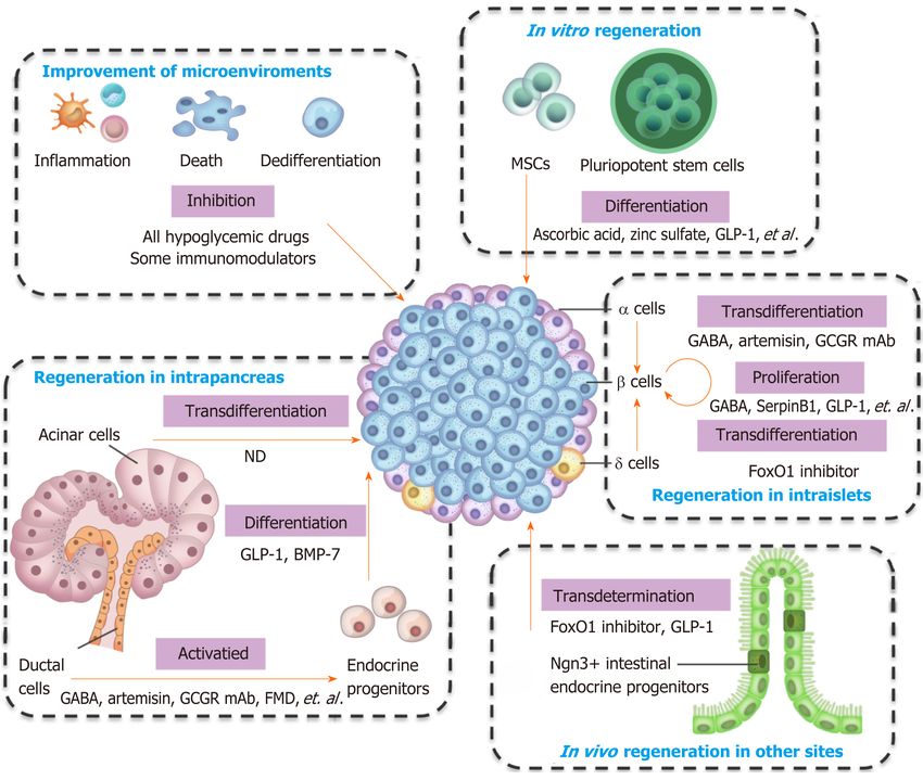
CONCLUSION
Pancreatic β cells own the ability of regeneration, and several types of cells can convert
into β cells. The types of cells that can generate β cells, and the preclinical and clinical
agents used for regeneration are summarized in Figure 1 and Table 1. Almost all
hypoglycemic drugs and some immunomodulators can protect β cells by inhibiting β
cell death and dedifferentiation through the correction of hyperglycemia and
improvement of the consequent inflammation and oxidative stress. GLP-1 and other
optimized molecules may promote stem cells to differentiate into β cells in vitro and
supplement β cell mass. Endogenous regeneration is an attractive approach.
Regeneration in intraislets (including β cell proliferation and α/δ-to-β cell conversion),
intrapancreas (including acinar-to-β cell reprogramming, reactivation of endocrine
progenitor, and differentiation into β cells), and other sites (e.g., transdetermination of
enteroendocrine progenitors to β cells) have all been investigated with some
promising results. The different regeneration paths might represent different
compensatory mechanisms for occasional β cell demise, relatively common stressors,
or widespread islet loss and/or partial organ remodeling (as summarized in a
previous review[69]). Notably, two or more regeneration paths often occur
simultaneously in one condition. For instance, β cell replication and neogenesis from
progenitors often occur together; α-to-β cell conversion generally induces progenitor
activation. Notably, one drug often protects β cells from different aspects. For instance,
GLP-1 and GABA promote the replication of β cells, enhance the conversion of α-to-β
cells, and suppress immune reactions and cell apoptosis. As a result, GLP-1 and GABA
have valuable significance for β cell regeneration and diabetes treatment. Several
studies have identified the long-term effects of GLP-1. For example, exenatide
improves β cell function for up to 3 years of treatment in patients with T2D[106,107]. The
safety and effects on the β cell function recovery of GABA in patients must be
determined.
In addition, there are other areas that require further investigation. First, the
regeneration mechanism between humans and rodents is different. Most strategies for
promoting regeneration have only been successfully applied to animals and have
failed in humans. Therefore, conclusions should be drawn cautiously from mouse
models when interpreting the results for humans; moreover, carrying out experiments
using human islets and conducting clinical trials would help. Second, the efficiency of
β cell recovery induced by current clinical drugs is low, and other strategies that have
high clinical efficiency and potential are required. Third, the win-win aim of
hypoglycemia and islet regeneration is challenging when using a single agent, and a
combination strategy is needed. A possible strategy might involve agents with good
glucose-lowering efficacy and agents that have been demonstrated potential to
preserve and regenerate β cells. On one hand, the agents with good glucose-lowering
efficacy (such as insulin), achieve glycemic goals rapidly, thereby minimizing the
exposure of β cells to glucotoxicity and lipotoxicity. On the other hand, several agents,
such as GLP-1 or GABA, have been demonstrated to be potentially able to preserve
and regenerate β cells, and may potentially contribute to the aim of recovering β cell
mass. Fourth, the promotion of proliferation and neogenesis may lead to the
development of cancer. The investigation of signals that mediate the physiological
expansion of β cell mass in obesity and insulin resistance might lead to novel β cell
regeneration reagents without significant tumorigenic risks. Besides, regulatory
mechanisms to turn on and off regenerative and oncogenic pathways require
investigations before applying regenerative approaches clinically. Additionally,
further investigation is required into how the interventions to expand β cell mass can
be specifically targeted to β cells. Taken together, in the last century, considerable
efforts have been made to achieve complete β cell regeneration, and several agents
have showed clinical potential, but there is still a long way to go.
WJSC https://www.wjgnet.com 70 January 26, 2021 Volume 13 Issue 1Wang KL et al. β cell regeneration
Table 1 Cell types, regeneration mechanisms, and clinical/preclinical agents for pancreatic β cell regeneration
Ref.
Regeneration
Cell types Clinical/preclinical agents
mechanisms
[16-18]
Correction of glucotoxicity All antidiabetic drugs
and lipotoxicity
[21-24]
Suppression of the Immunomodulation therapies, for instance, activation of the nuclear receptor LRH-
immune system 1/NR5A2, CD3 mAb, GABA, GLP-1
[25,27,28]
Inhibition of cell death Islet β cells GLP-1, metformin, angiotensin-converting enzyme inhibitors
[35,36];
Inhibition of cell Islet β cells Antidiabetic drugs and the diabetes management, such as diet, exercise, or intensive [37,38]
dedifferentiation insulin therapy; Other drugs, such as salsalate, renin–angiotensin system inhibitors
[42];
Stimulation of cell Islet β cells A large number of growth factors and mitogenic agents, including hepatocyte growth [48-51];
proliferation factor, GLP-1, insulin-like growth factors, epidermal growth factors in rodent models; [52,53]
Inhibition of Dyrk1a, SerpinB1, GABA, GLP-1, etc.; Combining of the Dyrk1a
inhibitor with the GLP-1 receptor agonist; combined inhibition of Dyrk1a, SMAD and
Trithorax pathways
[54-58];
Promotion of stem cell Pluriopotent stem cells Stepwise induction with cocktails of cytokines and chemicals; GLP-1, ascorbic acid, [56,58,65-
differentiation zinc sulfate, N-acetyl cysteine, etc. 66]
[67]
Stem cell-derived pancreatic GLP-1
endoderm cells or
progenitors
[63]
Mesenchymal stem cells Stepwise induction
[79-82]
Pancreatic stem or GLP-1, dipeptidyl peptidase 4 inhibitor, fasting-mimicking diet, etc.
progenitor cells in vivo
[87,88,90,
Induction of cell Pancreatic α cells GABA, artemisins, GLP-1, glucagon receptor mAb, sodium-glucose co-transporter 91,93,94]
transdifferentiation and type 2 inhibitor inhibitor, fibroblast growth factor 21, etc.
transdetermination
[13]
Pancreatic δ-cells FoxO1 inhibitor, etc.
[98]
Pancreatic acinar cells Cytokine mixture of epidermal growth factor and ciliary neurotrophic factor
[100,104,
Enteroendocrine progenitors FoxO1 inhibition, GLP-1, etc. 105]
Other progenitors and Not determined.
mature cells, including
hepatocytes, neuroendocrine
cells
GABA: γ-aminobutyric acid; GLP-1: Glucagon-like peptide-1; mAb: Monoclonal antibody.
WJSC https://www.wjgnet.com 71 January 26, 2021 Volume 13 Issue 1Wang KL et al. β cell regeneration
Figure 1 Integrative view of the cell types and clinical/preclinical agents for β cell regeneration. FMD: Fasting-mimicking diet; GABA: γ-
aminobutyric acid; GCGR mAb: Glucagon receptor monoclonal antibody; GLP-1: Glucagon-like peptide-1; MSCs: Mesenchymal stem cells; ND: Not determined.
REFERENCES
1 Li Y, Teng D, Shi X, Qin G, Qin Y, Quan H, Shi B, Sun H, Ba J, Chen B, Du J, He L, Lai X, Li Y,
Chi H, Liao E, Liu C, Liu L, Tang X, Tong N, Wang G, Zhang JA, Wang Y, Xue Y, Yan L, Yang J,
Yang L, Yao Y, Ye Z, Zhang Q, Zhang L, Zhu J, Zhu M, Ning G, Mu Y, Zhao J, Teng W, Shan Z.
Prevalence of diabetes recorded in mainland China using 2018 diagnostic criteria from the American
Diabetes Association: national cross sectional study. BMJ 2020; 369: m997 [PMID: 32345662 DOI:
10.1136/bmj.m997]
2 Zhou M, Wang H, Zeng X, Yin P, Zhu J, Chen W, Li X, Wang L, Wang L, Liu Y, Liu J, Zhang M,
Qi J, Yu S, Afshin A, Gakidou E, Glenn S, Krish VS, Miller-Petrie MK, Mountjoy-Venning WC,
Mullany EC, Redford SB, Liu H, Naghavi M, Hay SI, Wang L, Murray CJL, Liang X. Mortality,
morbidity, and risk factors in China and its provinces, 1990-2017: a systematic analysis for the
Global Burden of Disease Study 2017. Lancet 2019; 394: 1145-1158 [PMID: 31248666 DOI:
10.1016/S0140-6736(19)30427-1]
3 Matveyenko AV, Butler PC. Relationship between beta-cell mass and diabetes onset. Diabetes Obes
Metab 2008; 10 Suppl 4: 23-31 [PMID: 18834430 DOI: 10.1111/j.1463-1326.2008.00939.x]
4 Cnop M, Igoillo-Esteve M, Hughes SJ, Walker JN, Cnop I, Clark A. Longevity of human islet α-
and β-cells. Diabetes Obes Metab 2011; 13 Suppl 1: 39-46 [PMID: 21824255 DOI:
10.1111/j.1463-1326.2011.01443.x]
5 Rieck S, Kaestner KH. Expansion of beta-cell mass in response to pregnancy. Trends Endocrinol
Metab 2010; 21: 151-158 [PMID: 20015659 DOI: 10.1016/j.tem.2009.11.001]
6 Kahn SE, Hull RL, Utzschneider KM. Mechanisms linking obesity to insulin resistance and type 2
diabetes. Nature 2006; 444: 840-846 [PMID: 17167471 DOI: 10.1038/nature05482]
7 Butler AE, Cao-Minh L, Galasso R, Rizza RA, Corradin A, Cobelli C, Butler PC. Adaptive changes
in pancreatic beta cell fractional area and beta cell turnover in human pregnancy. Diabetologia 2010;
53: 2167-2176 [PMID: 20523966 DOI: 10.1007/s00125-010-1809-6]
8 Mezza T, Muscogiuri G, Sorice GP, Clemente G, Hu J, Pontecorvi A, Holst JJ, Giaccari A, Kulkarni
RN. Insulin resistance alters islet morphology in nondiabetic humans. Diabetes 2014; 63: 994-1007
[PMID: 24215793 DOI: 10.2337/db13-1013]
9 Dorrell C, Schug J, Canaday PS, Russ HA, Tarlow BD, Grompe MT, Horton T, Hebrok M, Streeter
PR, Kaestner KH, Grompe M. Human islets contain four distinct subtypes of β cells. Nat Commun
2016; 7: 11756 [PMID: 27399229 DOI: 10.1038/ncomms11756]
WJSC https://www.wjgnet.com 72 January 26, 2021 Volume 13 Issue 1Wang KL et al. β cell regeneration
10 Segerstolpe Å, Palasantza A, Eliasson P, Andersson EM, Andréasson AC, Sun X, Picelli S, Sabirsh
A, Clausen M, Bjursell MK, Smith DM, Kasper M, Ämmälä C, Sandberg R. Single-Cell
Transcriptome Profiling of Human Pancreatic Islets in Health and Type 2 Diabetes. Cell Metab
2016; 24: 593-607 [PMID: 27667667 DOI: 10.1016/j.cmet.2016.08.020]
11 Li WC, Rukstalis JM, Nishimura W, Tchipashvili V, Habener JF, Sharma A, Bonner-Weir S.
Activation of pancreatic-duct-derived progenitor cells during pancreas regeneration in adult rats. J
Cell Sci 2010; 123: 2792-2802 [PMID: 20663919 DOI: 10.1242/jcs.065268]
12 Thorel F, Népote V, Avril I, Kohno K, Desgraz R, Chera S, Herrera PL. Conversion of adult
pancreatic alpha-cells to beta-cells after extreme beta-cell loss. Nature 2010; 464: 1149-1154
[PMID: 20364121 DOI: 10.1038/nature08894]
13 Chera S, Baronnier D, Ghila L, Cigliola V, Jensen JN, Gu G, Furuyama K, Thorel F, Gribble FM,
Reimann F, Herrera PL. Diabetes recovery by age-dependent conversion of pancreatic δ-cells into
insulin producers. Nature 2014; 514: 503-507 [PMID: 25141178 DOI: 10.1038/nature13633]
14 Xu X, D'Hoker J, Stangé G, Bonné S, De Leu N, Xiao X, Van de Casteele M, Mellitzer G, Ling Z,
Pipeleers D, Bouwens L, Scharfmann R, Gradwohl G, Heimberg H. Beta cells can be generated from
endogenous progenitors in injured adult mouse pancreas. Cell 2008; 132: 197-207 [PMID: 18243096
DOI: 10.1016/j.cell.2007.12.015]
15 Bonner-Weir S, Baxter LA, Schuppin GT, Smith FE. A second pathway for regeneration of adult
exocrine and endocrine pancreas. A possible recapitulation of embryonic development. Diabetes
1993; 42: 1715-1720 [PMID: 8243817 DOI: 10.2337/diab.42.12.1715]
16 Bonora E. Protection of pancreatic beta-cells: is it feasible? Nutr Metab Cardiovasc Dis 2008; 18:
74-83 [PMID: 18096375 DOI: 10.1016/j.numecd.2007.05.004]
17 Del Prato S, Bianchi C, Marchetti P. beta-cell function and anti-diabetic pharmacotherapy. Diabetes
Metab Res Rev 2007; 23: 518-527 [PMID: 17883249 DOI: 10.1002/dmrr.770]
18 Boland BB, Brown C Jr, Boland ML, Cann J, Sulikowski M, Hansen G, Grønlund RV, King W,
Rondinone C, Trevaskis J, Rhodes CJ, Grimsby JS. Pancreatic β-Cell Rest Replenishes Insulin
Secretory Capacity and Attenuates Diabetes in an Extreme Model of Obese Type 2 Diabetes.
Diabetes 2019; 68: 131-140 [PMID: 30305366 DOI: 10.2337/db18-0304]
19 Kimura T, Obata A, Shimoda M, Okauchi S, Kanda-Kimura Y, Nogami Y, Moriuchi S, Hirukawa
H, Kohara K, Nakanishi S, Mune T, Kaku K, Kaneto H. Protective effects of the SGLT2 inhibitor
luseogliflozin on pancreatic β-cells in db/db mice: The earlier and longer, the better. Diabetes Obes
Metab 2018; 20: 2442-2457 [PMID: 29873444 DOI: 10.1111/dom.13400]
20 Eguchi K, Nagai R. Islet inflammation in type 2 diabetes and physiology. J Clin Invest 2017; 127:
14-23 [PMID: 28045399 DOI: 10.1172/JCI88877]
21 Cobo-Vuilleumier N, Gauthier BR. Time for a paradigm shift in treating type 1 diabetes mellitus:
coupling inflammation to islet regeneration. Metabolism 2020; 104: 154137 [PMID: 31904355 DOI:
10.1016/j.metabol.2020.154137]
22 Herold KC, Bundy BN, Long SA, Bluestone JA, DiMeglio LA, Dufort MJ, Gitelman SE, Gottlieb
PA, Krischer JP, Linsley PS, Marks JB, Moore W, Moran A, Rodriguez H, Russell WE, Schatz D,
Skyler JS, Tsalikian E, Wherrett DK, Ziegler AG, Greenbaum CJ; Type 1 Diabetes TrialNet Study
Group. An Anti-CD3 Antibody, Teplizumab, in Relatives at Risk for Type 1 Diabetes. N Engl J Med
2019; 381: 603-613 [PMID: 31180194 DOI: 10.1056/NEJMoa1902226]
23 Bhandage AK, Jin Z, Korol SV, Shen Q, Pei Y, Deng Q, Espes D, Carlsson PO, Kamali-
Moghaddam M, Birnir B. GABA Regulates Release of Inflammatory Cytokines From Peripheral
Blood Mononuclear Cells and CD4+ T Cells and Is Immunosuppressive in Type 1 Diabetes.
EBioMedicine 2018; 30: 283-294 [PMID: 29627388 DOI: 10.1016/j.ebiom.2018.03.019]
24 Hogan AE, Gaoatswe G, Lynch L, Corrigan MA, Woods C, O'Connell J, O'Shea D. Glucagon-like
peptide 1 analogue therapy directly modulates innate immune-mediated inflammation in individuals
with type 2 diabetes mellitus. Diabetologia 2014; 57: 781-784 [PMID: 24362727 DOI:
10.1007/s00125-013-3145-0]
25 Tudurí E, López M, Diéguez C, Nadal A, Nogueiras R. Glucagon-Like Peptide 1 Analogs and their
Effects on Pancreatic Islets. Trends Endocrinol Metab 2016; 27: 304-318 [PMID: 27062006 DOI:
10.1016/j.tem.2016.03.004]
26 Zummo FP, Cullen KS, Honkanen-Scott M, Shaw JAM, Lovat PE, Arden C. Glucagon-Like
Peptide 1 Protects Pancreatic β-Cells From Death by Increasing Autophagic Flux and Restoring
Lysosomal Function. Diabetes 2017; 66: 1272-1285 [PMID: 28232493 DOI: 10.2337/db16-1009]
27 Marchetti P, Del Guerra S, Marselli L, Lupi R, Masini M, Pollera M, Bugliani M, Boggi U, Vistoli
F, Mosca F, Del Prato S. Pancreatic islets from type 2 diabetic patients have functional defects and
increased apoptosis that are ameliorated by metformin. J Clin Endocrinol Metab 2004; 89: 5535-
5541 [PMID: 15531508 DOI: 10.1210/jc.2004-0150]
28 Lupi R, Del Guerra S, Bugliani M, Boggi U, Mosca F, Torri S, Del Prato S, Marchetti P. The direct
effects of the angiotensin-converting enzyme inhibitors, zofenoprilat and enalaprilat, on isolated
human pancreatic islets. Eur J Endocrinol 2006; 154: 355-361 [PMID: 16452552 DOI:
10.1530/eje.1.02086]
29 Talchai C, Xuan S, Lin HV, Sussel L, Accili D. Pancreatic β cell dedifferentiation as a mechanism
of diabetic β cell failure. Cell 2012; 150: 1223-1234 [PMID: 22980982 DOI:
10.1016/j.cell.2012.07.029]
30 Cinti F, Bouchi R, Kim-Muller JY, Ohmura Y, Sandoval PR, Masini M, Marselli L, Suleiman M,
Ratner LE, Marchetti P, Accili D. Evidence of β-Cell Dedifferentiation in Human Type 2 Diabetes. J
WJSC https://www.wjgnet.com 73 January 26, 2021 Volume 13 Issue 1Wang KL et al. β cell regeneration
Clin Endocrinol Metab 2016; 101: 1044-1054 [PMID: 26713822 DOI: 10.1210/jc.2015-2860]
31 Md Moin AS, Dhawan S, Cory M, Butler PC, Rizza RA, Butler AE. Increased Frequency of
Hormone Negative and Polyhormonal Endocrine Cells in Lean Individuals With Type 2 Diabetes. J
Clin Endocrinol Metab 2016; 101: 3628-3636 [PMID: 27472443 DOI: 10.1210/jc.2016-2496]
32 Md Moin AS, Dhawan S, Shieh C, Butler PC, Cory M, Butler AE. Increased Hormone-Negative
Endocrine Cells in the Pancreas in Type 1 Diabetes. J Clin Endocrinol Metab 2016; 101: 3487-3496
[PMID: 27300574 DOI: 10.1210/jc.2016-1350]
33 Butler AE, Dhawan S, Hoang J, Cory M, Zeng K, Fritsch H, Meier JJ, Rizza RA, Butler PC. β-Cell
Deficit in Obese Type 2 Diabetes, a Minor Role of β-Cell Dedifferentiation and Degranulation. J
Clin Endocrinol Metab 2016; 101: 523-532 [PMID: 26700560 DOI: 10.1210/jc.2015-3566]
34 Wang YJ, Schug J, Won KJ, Liu C, Naji A, Avrahami D, Golson ML, Kaestner KH. Single-Cell
Transcriptomics of the Human Endocrine Pancreas. Diabetes 2016; 65: 3028-3038 [PMID:
27364731 DOI: 10.2337/db16-0405]
35 Wang Z, York NW, Nichols CG, Remedi MS. Pancreatic β cell dedifferentiation in diabetes and
redifferentiation following insulin therapy. Cell Metab 2014; 19: 872-882 [PMID: 24746806 DOI:
10.1016/j.cmet.2014.03.010]
36 Accili D, Talchai SC, Kim-Muller JY, Cinti F, Ishida E, Ordelheide AM, Kuo T, Fan J, Son J. When
β-cells fail: lessons from dedifferentiation. Diabetes Obes Metab 2016; 18 Suppl 1: 117-122 [PMID:
27615140 DOI: 10.1111/dom.12723]
37 Han F, Li X, Yang J, Liu H, Zhang Y, Yang X, Yang S, Chang B, Chen L, Chang B. Salsalate
Prevents β-Cell Dedifferentiation in OLETF Rats with Type 2 Diabetes through Notch1 Pathway.
Aging Dis 2019; 10: 719-730 [PMID: 31440379 DOI: 10.14336/AD.2018.1221]
38 Chen H, Zhou W, Ruan Y, Yang L, Xu N, Chen R, Yang R, Sun J, Zhang Z. Reversal of
angiotensin ll-induced β-cell dedifferentiation via inhibition of NF-κb signaling. Mol Med 2018; 24:
43 [PMID: 30134927 DOI: 10.1186/s10020-018-0044-3]
39 RISE Consortium. Lack of Durable Improvements in β-Cell Function Following Withdrawal of
Pharmacological Interventions in Adults With Impaired Glucose Tolerance or Recently Diagnosed
Type 2 Diabetes. Diabetes Care 2019; 42: 1742-1751 [PMID: 31178434 DOI: 10.2337/dc19-0556]
40 Dor Y, Brown J, Martinez OI, Melton DA. Adult pancreatic beta-cells are formed by self-
duplication rather than stem-cell differentiation. Nature 2004; 429: 41-46 [PMID: 15129273 DOI:
10.1038/nature02520]
41 Teta M, Long SY, Wartschow LM, Rankin MM, Kushner JA. Very slow turnover of beta-cells in
aged adult mice. Diabetes 2005; 54: 2557-2567 [PMID: 16123343 DOI:
10.2337/diabetes.54.9.2557]
42 Saunders D, Powers AC. Replicative capacity of β-cells and type 1 diabetes. J Autoimmun 2016; 71:
59-68 [PMID: 27133598 DOI: 10.1016/j.jaut.2016.03.014]
43 Dolenšek J, Rupnik MS, Stožer A. Structural similarities and differences between the human and the
mouse pancreas. Islets 2015; 7: e1024405 [PMID: 26030186 DOI:
10.1080/19382014.2015.1024405]
44 Nair G, Hebrok M. Islet formation in mice and men: lessons for the generation of functional insulin-
producing β-cells from human pluripotent stem cells. Curr Opin Genet Dev 2015; 32: 171-180
[PMID: 25909383 DOI: 10.1016/j.gde.2015.03.004]
45 Levetan CS, Pierce SM. Distinctions between the islets of mice and men: implications for new
therapies for type 1 and 2 diabetes. Endocr Pract 2013; 19: 301-312 [PMID: 23186955 DOI:
10.4158/EP12138.RA]
46 Wang P, Fiaschi-Taesch NM, Vasavada RC, Scott DK, García-Ocaña A, Stewart AF. Diabetes
mellitus--advances and challenges in human β-cell proliferation. Nat Rev Endocrinol 2015; 11: 201-
212 [PMID: 25687999 DOI: 10.1038/nrendo.2015.9]
47 Cigliola V, Thorel F, Chera S, Herrera PL. Stress-induced adaptive islet cell identity changes.
Diabetes Obes Metab 2016; 18 Suppl 1: 87-96 [PMID: 27615136 DOI: 10.1111/dom.12726]
48 Wang P, Alvarez-Perez JC, Felsenfeld DP, Liu H, Sivendran S, Bender A, Kumar A, Sanchez R,
Scott DK, Garcia-Ocaña A, Stewart AF. A high-throughput chemical screen reveals that harmine-
mediated inhibition of DYRK1A increases human pancreatic beta cell replication. Nat Med 2015;
21: 383-388 [PMID: 25751815 DOI: 10.1038/nm.3820]
49 El Ouaamari A, Dirice E, Gedeon N, Hu J, Zhou JY, Shirakawa J, Hou L, Goodman J, Karampelias
C, Qiang G, Boucher J, Martinez R, Gritsenko MA, De Jesus DF, Kahraman S, Bhatt S, Smith RD,
Beer HD, Jungtrakoon P, Gong Y, Goldfine AB, Liew CW, Doria A, Andersson O, Qian WJ,
Remold-O'Donnell E, Kulkarni RN. SerpinB1 Promotes Pancreatic β Cell Proliferation. Cell Metab
2016; 23: 194-205 [PMID: 26701651 DOI: 10.1016/j.cmet.2015.12.001]
50 Purwana I, Zheng J, Li X, Deurloo M, Son DO, Zhang Z, Liang C, Shen E, Tadkase A, Feng ZP, Li
Y, Hasilo C, Paraskevas S, Bortell R, Greiner DL, Atkinson M, Prud'homme GJ, Wang Q. GABA
promotes human β-cell proliferation and modulates glucose homeostasis. Diabetes 2014; 63: 4197-
4205 [PMID: 25008178 DOI: 10.2337/db14-0153]
51 Dai C, Hang Y, Shostak A, Poffenberger G, Hart N, Prasad N, Phillips N, Levy SE, Greiner DL,
Shultz LD, Bottino R, Kim SK, Powers AC. Age-dependent human β cell proliferation induced by
glucagon-like peptide 1 and calcineurin signaling. J Clin Invest 2017; 127: 3835-3844 [PMID:
28920919 DOI: 10.1172/JCI91761]
52 Ackeifi C, Wang P, Karakose E, Manning Fox JE, González BJ, Liu H, Wilson J, Swartz E,
Berrouet C, Li Y, Kumar K, MacDonald PE, Sanchez R, Thorens B, DeVita R, Homann D, Egli D,
WJSC https://www.wjgnet.com 74 January 26, 2021 Volume 13 Issue 1You can also read