Brain Activation Time-Locked to Sleep Spindles Associated With Human Cognitive Abilities - Owen Lab
←
→
Page content transcription
If your browser does not render page correctly, please read the page content below
ORIGINAL RESEARCH
                                                                                                                                          published: 06 February 2019
                                                                                                                                        doi: 10.3389/fnins.2019.00046

                                            Brain Activation Time-Locked to
                                            Sleep Spindles Associated With
                                            Human Cognitive Abilities
                                            Zhuo Fang 1,2 , Laura B. Ray 1,3 , Adrian M. Owen 1,4 and Stuart M. Fogel 1,2,3,4,5*
                                            1
                                              Brain and Mind Institute, Western University, London, ON, Canada, 2 School of Psychology, University of Ottawa, Ottawa,
                                            ON, Canada, 3 Sleep Unit, The Royal’s Institute of Mental Health Research, University of Ottawa, Ottawa, ON, Canada,
                                            4
                                              Department of Psychology, Western University, London, ON, Canada, 5 University of Ottawa Brain and Mind Research
                                            Institute, Ottawa, ON, Canada

                                            Simultaneous electroencephalography and functional magnetic resonance imaging
                                            (EEG–fMRI) studies have revealed brain activations time-locked to spindles. Yet, the
                                            functional significance of these spindle-related brain activations is not understood.
                                            EEG studies have shown that inter-individual differences in the electrophysiological
                                            characteristics of spindles (e.g., density, amplitude, duration) are highly correlated with
                                            “Reasoning” abilities (i.e., “fluid intelligence”; problem solving skills, the ability to employ
                                            logic, identify complex patterns), but not short-term memory (STM) or verbal abilities.
                                            Spindle-dependent reactivation of brain areas recruited during new learning suggests
                             Edited by:     night-to-night variations reflect offline memory processing. However, the functional
                         Michele Bellesi,   significance of stable, trait-like inter-individual differences in brain activations recruited
 University of Bristol, United Kingdom
                                            during spindle events is unknown. Using EEG–fMRI sleep recordings, we found that a
                        Reviewed by:
                        Fabio Ferrarelli,   subset of brain activations time-locked to spindles were specifically related to Reasoning
      University of Pittsburgh School       abilities but were unrelated to STM or verbal abilities. Thus, suggesting that individuals
           of Medicine, United States
          Sofia Isabel Ribeiro Pereira,
                                            with higher fluid intelligence have greater activation of brain regions recruited during
   Cardiff University, United Kingdom       spontaneous spindle events. This may serve as a first step to further understand
                   *Correspondence:         the function of sleep spindles and the brain activity which supports the capacity
                       Stuart M. Fogel      for Reasoning.
                    sfogel@uottawa.ca
                                            Keywords: sleep, spindles, cognitive abilities, simultaneous EEG–fMRI, NREM
                    Specialty section:
          This article was submitted to
        Sleep and Circadian Rhythms,        INTRODUCTION
                a section of the journal
             Frontiers in Neuroscience
                                            Sleep spindles are one of the defining features of non-rapid eye movement (NREM) sleep. Spindles
         Received: 22 October 2018          are traditionally defined as bursts of waxing and waning neural oscillations between 11 and 16 Hz
         Accepted: 17 January 2019
                                            (Iber et al., 2007), which stand out from the ongoing, background electroencephalographic (EEG)
        Published: 06 February 2019
                                            activity (Rechtschaffen and Kales, 1968). The brain regions activated during spontaneous spindle
                              Citation:     events have been identified (Laufs et al., 2007; Schabus et al., 2007; Tyvaert et al., 2008; Andrade
      Fang Z, Ray LB, Owen AM and
                                            et al., 2011; Caporro et al., 2012). However, the functional significance of these brain activations
     Fogel SM (2019) Brain Activation
       Time-Locked to Sleep Spindles
                                            has yet to be elucidated, thereby limiting our understanding of the function of sleep spindles.
    Associated With Human Cognitive         Spindles are remarkably stable from night-to-night, but vary considerably from one individual
      Abilities. Front. Neurosci. 13:46.    to another, and because of the trait-like nature of spindles (Silverstein and Levy, 1976), they
       doi: 10.3389/fnins.2019.00046        have even been suggested to be an “electrophysiological fingerprint” (De Gennaro et al., 2005).

Frontiers in Neuroscience | www.frontiersin.org                                   1                                              February 2019 | Volume 13 | Article 46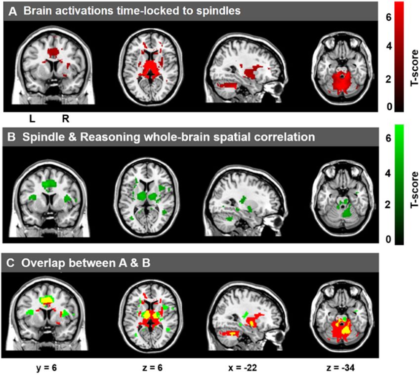
Fang et al.                                                                                                       Sleep Spindles and Cognitive Abilities

Recent work by our group and others (Nader and Smith,                           Notably, the sleep spindle is the only known spon-
2001, 2003; Bódizs et al., 2005, 2008; Schabus et al., 2006;                taneous neural oscillation that has been identified as an
Fogel et al., 2007; Ujma et al., 2014, 2015; Fang et al.,                   electrophysiological marker of cognitive abilities and aptitudes,
2017) suggests that spindles are electrophysiological markers               that are typically assessed by intelligence quotient (IQ) tests (for
of specific cognitive abilities, and in particular, Reasoning               review, see Fogel and Smith, 2011). The association between
abilities (i.e., “fluid intelligence”; problem solving skills, the          sleep spindles and individual differences in cognitive abilities has
ability to employ logic, identify complex patterns). However,               been well documented. More specifically, previous studies have
the neural basis of this relationship is not known; there is no             revealed that interindividual differences in spindle characteristics
direct evidence investigating the relationship between trait-like           are related to the capacity for Reasoning (i.e., the ability to
cognitive abilities and interindividual differences in spindle-             identify complex patterns and relationships, the use of logic,
dependent brain activations. Thus, as a first step, we sought               existing knowledge, skills, and experience to solve novel problems
to explore what brain activations time-locked to spontaneous                (Nader and Smith, 2001, 2003; Bódizs et al., 2005, 2008; Schabus
spindle events are correlated with various domains of human                 et al., 2006; Fogel et al., 2007; Ujma et al., 2014, 2015; Fang et al.,
intellectual abilities. This might shed light on the functional             2017). For example, Nader and Smith (2001, 2003) found that
significance of brain activations time-locked to sleep spindles,            both the number of sleep spindles and sigma power (12–14 Hz)
and the neural functional correlates which explain individual               was correlated with Performance IQ scores. Further studies
differences in trait-like cognitive strengths and weaknesses.               revealed this relationship to be specific to these Reasoning-
   In an effort to identify the functional brain areas recruited            related abilities, over-and-above (i.e., controlling for) Verbal
during spontaneous spindle events, a handful of studies have                IQ (Fogel et al., 2007; Fang et al., 2017). Consistently, several
employed simultaneous electroencephalography and functional                 studies (Bódizs et al., 2005; Schabus et al., 2006) found that
magnetic resonance imaging (EEG–fMRI) to explore brain                      fast spindles were positively correlated with similar metrics
activations time-locked to spindles (Laufs et al., 2007; Schabus            of Reasoning abilities (i.e., “fluid intelligence”), measured by
et al., 2007; Tyvaert et al., 2008; Andrade et al., 2011; Caporro           the Raven’s Progressive Matrices (Raven et al., 1976). Similar
et al., 2012). Spindle-related activations have been consistently           studies identified a positive correlation between right-parietal fast
found in the thalamus and the temporal lobe, for both fast                  spindles and visuospatial abilities assessed by the Rey–Osterrieth
spindles and slow spindles (Laufs et al., 2007; Schabus et al.,             complex figure test (Bódizs et al., 2008). They identified a positive
2007; Tyvaert et al., 2008; Andrade et al., 2011; Caporro                   correlation between spindles and intellectual abilities measured
et al., 2012), as well as activation of the cingulate cortex                by the Cattell Culture Fair Intelligence test, specifically in woman
and motor areas (Andrade et al., 2011; Caporro et al., 2012).               but not in men (Ujma et al., 2014). Although a relationship in
Interestingly, activation of the putamen has also been found                men was subsequently identified by the same group in daytime
to be associated with spindle events (Tyvaert et al., 2008;                 sleep (Ujma et al., 2015). Taken together, these studies support
Caporro et al., 2012) and Andrade et al. (2011) found a                     the notion that sleep spindles are an electrophysiological marker
strong interaction between spindle occurrence and hippocampal               of cognitive abilities, and specifically, the ability to solve problems
formation functional connectivity, suggesting that spindles may             using logic and Reasoning. These studies have provided insight
be related to memory and cognitive functioning. However, they               into the electrophysiological correlates of Reasoning abilities,
did not report any relationship between these activations and               insofar as to indirectly suggest that efficient functioning of the
cognitive abilities. In addition, by directly comparing fast spindles       neural substrates that support spindle generation and those
vs. slow spindles, Schabus et al. (2007) observed increased                 that are recruited during spontaneous spindle events may be
activations associated with slow spindles in the superior temporal          related to the capacity for these cognitive skills. Two recent
gyrus while fast spindles recruited activation in sensorimotor              studies employing simultaneous EEG–fMRI have investigated the
areas, mesial frontal cortex, hippocampus, and cerebellum;                  association between sleep spindle-related brain activation and
important memory centers of the brain. However, in the                      memory consolidation, including declarative (Bergmann et al.,
absence of any direct relationship between spindle-dependent                2012) and procedural memory (Fogel et al., 2017a). However,
activation and cognitive performance, the functional significance           both of these studies were interested in investigating memory
of these spindle-related activations could only be speculated               trace reactivation following new learning (i.e., acute effects of
based alone on the brain regions recruited. Taken together, the             learning on subsequent sleep), and did not investigate the stable,
extant literature, not-surprisingly, suggests that brain activations        trait-like interindividual differences in cognitive abilities and how
associated with the action of sleep spindles involve well-known             they relate to inter-individual differences in spindle-dependent
spindle-generating regions (e.g., thalamic and cortical regions),           brain activation. Thus, the functional significance of spontaneous
as well as, more intriguingly, regions which subserve executive             spindle-related brain activations, and whether these activations
functioning [prefrontal cortex (PFC)], declarative memory                   related to specific domains of cognitive abilities [e.g., Reasoning,
(hippocampus), motor skills (motor cortex and cerebellum), and              Verbal, Short-Term Memory (STM)] in healthy individuals
procedural memory (the striatum). However, these studies are                remains to be investigated, which is the principle aim of the
limited in that they can only tell us what brain regions are                current study.
recruited during spindle events. The functional significance of                 Therefore, here, using simultaneous EEG–fMRI recordings
these activations with respect to the trait-like nature of spindles         during sleep, we sought to identify, for the first time, the
remains to be elucidated.                                                   neuroanatomical function correlates of the well-established

Frontiers in Neuroscience | www.frontiersin.org                         2                                        February 2019 | Volume 13 | Article 46
Fang et al.                                                                                                             Sleep Spindles and Cognitive Abilities

relationship between sleep spindles and specific cognitive                TABLE 1 | Sleep architecture and sleep spindle parameters for spindles during
                                                                          NREM sleep from EEG–fMRI recording sessions.
abilities. We hypothesized that the neural activation patterns,
time-locked to spindles would be related to distinct cognitive                                                   M                             SD
abilities whereby, consistent with previous cognitive and EEG                                                                Sleep architecture
studies, spindle-related brain activations would be correlated to
a greater extent with Reasoning, but not STM or Verbal abilities,         Wake (min) (N = 26)                  26.87                            20.25
and include brain regions known to be involved in spindle                 NREM1 (min) (N = 26)                   5.84                             4.38
generation, and also known to support Reasoning abilities (e.g.,          NREM2 (min) (N = 29)                 23.87                            14.50

thalamus, PFC, striatum, cerebellum). This will provide insight           SWS (min) (N = 20)                   14.77                            17.17
                                                                          NREM (min)                           39.29                            19.33
into the functional significance of sleep spindles.
                                                                          REM (N = 8)                          17.80                            10.76
                                                                          Total sleep                          44.20                            23.84
                                                                          Sleep latency                          8.16                           10.11
MATERIALS AND METHODS
                                                                                                              Total bandwidth (11–16 Hz) spindles at Cz

Participants                                                              Number                              334.74                          212.29
To be included in the study, all participants were non-shift              Duration (s)                           0.49                             0.05
workers and medication-free; had no history of head injury                Amplitude (µV)                       27.21                              6.43
or seizures; had a normal body mass index (
Fang et al. Sleep Spindles and Cognitive Abilities
All 12 subtests are based on classic paradigms from cognitive TABLE 2 | Descriptive statistics of the three CBS subscales (Reasoning, STM,
and Verbal abilities).
psychology. For example, the Reasoning factor is best described
in terms of performance on five tests adapted from the cognitive IQ measures Range Mean ± SD Median
literature, including deductive reasoning (Cattell, 1940), spatial
rotation (Silverman et al., 2000), feature match (Treisman and Reasoning 78.84–108.17 95.65 ± 7.20 96.46
Gelade, 1980), spatial planning (Shallice, 1982), and polygons STM 84.38–115.33 101.60 ± 6.77 102.30
(Folstein et al., 1975). STM is best described in terms of Verbal 88.51–110.92 99.62 ± 5.12 99.52
four tests, including visuospatial working memory (Inoue and
Matsuzawa, 2007), spatial span (Corsi, 1972), paired associates
by completing the Sleep Disorder Questionnaires (Douglass
(Gould et al., 2006), and self-ordered search (Collins et al., 1998).
et al., 1994), BDI (Beck et al., 1974), BAI (Beck et al., 1988)
Finally, verbal ability is best captured by performance on three
scales, Horne–Ostberg Morningness–Eveningness Questionnaire
tests, including verbal reasoning (Baddeley, 1968), color-word
(Horne and Ostberg, 1976), and the MRI safety screening
remapping (Stroop, 1935), and digit span (Wechsler, 1981). More
questionnaire to screen for signs of sleep disorders, unusual sleep
detailed information of the 12 subtests can be found in the
habits, depression, or anxiety and MRI compatibility. Participants
Supplementary Material.
who met all these criteria were included in the study and visited
During the orientation session, participants were informed
the sleep lab for an orientation session at least 1 week prior
about the study requirements and procedures, and given detailed
to the EEG–fMRI sleep recording night. During the orientation
instructions of the online CBS tests. All participants were
session, participants were informed about the study requirements
required to register and complete the CBS tests online at home
and procedures, and given detailed instructions of the online
after the orientation session. Participants were required to use
CBS tests. All participants were required to complete the CBS
a computer mouse to perform the task. Completion of the 12
tests online at home after the orientation session, during which
tests takes between 30 and 60 min in total. The order of the tasks
participants were not allowed to consume caffeinated, alcoholic,
was randomized across participants. Prior to each test, specific
or nicotine products. Participants were required to keep a regular
instructions on how to perform the respective test were displayed
sleep-wake cycle (bed-time between 2200 and 2400 h, wake-
onscreen.
time between 0700 and 0900 h), to abstain from taking daytime
CBS Scores Calculation naps at least 7 days prior to and throughout participation in
the study. Compliance with this schedule was assessed using
Consistent with the previous literature (Hampshire et al., 2012),
both sleep diaries and wrist actigraphy (Actiwatch 2, Philips
the raw scores from each of the 12 subtests were normalized
Respironics, Andover, MA, United States) worn on the non-
using the mean and standard deviation obtained from a large,
dominant wrist for 1 week prior to the EEG–fMRI sleep recording
young population (N = 44,600; age 20–35 years) of subjects who
night. Participants who met these requirements were scheduled
completed the CBS Trials (Hampshire et al., 2012) (Equation 1).
for the EEG–fMRI sleep recording session. The experimental
Each subtest was then weighted according to the factor loadings
sleep session started between 21h00 and 24h00, during which
from Hampshire et al. (2012) (Equation 2). Finally, the respective
time simultaneous EEG–fMRI was recorded while participants
sub-tests were averaged to create the Reasoning, STM, and
slept in the scanner. Specifically, the scan procedure normally
Verbal sub-scales and transformed to standard scores (Equation
started at 21h00, at which point, the EEG equipment was installed
3), so that test scores were readily comparable to results
and tested. This was followed by localizer scans, a T1 MPRAGE
from similar studies that employed test batteries tapping into
structural scan, and an awake resting scan. These procedures took
Reasoning and Verbal abilities, such as the Multidimensional
more than 30 min to complete. The sleep recording (“lights out”)
Aptitude Battery – II (Fogel and Smith, 2006; Fogel et al.,
normally started after 22h00, within the range of the subject’s
2007) and other commonly used batteries of cognitive abilities
habitual bedtime. The average sleep latency was 8.16 ± 10.11 min
(e.g., Wechsler Adult Intelligence Scale (Wechsler, 1981). The
(Table 1) and the average sleep onset time, when participants
descriptive statistics of each subtest are shown in Table 2.
fell asleep in the scanner was 22h22 (±25 min). Following the
Z-score = (X raw - M norm )/SDnorm (1) EEG–fMRI sleep session, participants were allowed to sleep in the
nearby sleep laboratory for the remainder of the night.
Weighted Score = Z-score ∗ factor loadings (2)
Standard score = 100 + Weighted Score ∗ 15 (3) Polysomnographic Recording and
Analysis
where X raw , raw score of each test item in the current study; M norm , Recording Parameters
mean score of each test item from the literature; SDnorm , standard Simultaneous polysomnography was comprised of 64-channel
deviation of each test item from the literature; factor loading, MR-compatible EEG cap which included one electrocardiogram
weight of each test item from the literature. (ECG) lead (Braincap MR, Easycap, Herrsching, Germany) and
two MR-compatible 32-channel amplifiers (Brainamp MR plus,
Experimental Procedure Brain Products GmbH, Gilching, Germany). EEG recordings
An overview of the study procedure is shown in Figure 1. were taken referenced to FCz. Skin resistance was reduced
Participants underwent an initial screening prior to the study below 5 KOhm using high-chloride abrasive electrode paste
Frontiers in Neuroscience | www.frontiersin.org 4 February 2019 | Volume 13 | Article 46Fang et al. Sleep Spindles and Cognitive Abilities
FIGURE 1 | Experimental procedure of the study. Participants underwent initial screening to exclude any signs of sleep disorders, unusual sleep habits, or other
health-related ineligibility and MRI compatibility. Eligible participants visited the sleep lab for the orientation session at least a week prior to the EEG–fMRI sleep
recording night, in which participants were given detailed instructions about the study procedure, CBS tests, sleep diary, and actiwatch. All participants completed
the CBS tests online and kept a regular sleep-wake cycle for at least 1 week prior to the sleep recording. Compliance with this schedule was assessed using both
sleep diaries and wrist actigraphy. Participants who met these requirements were scheduled for the EEG–fMRI sleep recording session. MRI scanning started at
21h00, and lights out was from 22h00 to as late as 24h00.
(Abralyt 2000 HiCL; Easycap, Herrsching, Germany). The single of the pump noise is >80 Hz, which is outside EEG frequencies
drop-down ECG electrode from the EEG cap can have less of interest in our study (e.g., 11–16 Hz for spindles), and would
than optimal visualization of the R-peak of the QRS complex. not impact slower frequencies needed for accurate visual sleep
For this reason, we used additional bipolar electrodes for three scoring. In the second step, the R-peaks in the ECG were
ECG derivations to obtain high-quality recordings in order to semi-automatically detected, visually verified, manually adjusted
accurately identify R-peaks, using an MR-compatible 16-channel when necessary, to correct both false positives and false negative
bipolar amplifier (Brainamp ExG MR, Brain Products GmbH, R-peak detections. Then, adaptive template subtraction (Allen
Gilching, Germany). This was done in order to increase the et al., 1998) was used to remove BCG artifacts time-locked to
chances of acquiring at least one ECG channel with high quality the R-peak of the QRS complex of the cardiac rhythm. After
R-peaks, necessary for effective ballistocardiographic (BCG) these two steps, we visually verified the quality of the data and
correction. In addition, as recommended by Mullinger et al. inspected the amplitude of the residual artifacts time-locked
(2011), we repositioned subjects in the MRI scanner so that the to the R-peaks (Supplementary Figure S1). An independent
subjects were shifted away from iso-center of the magnetic field component analyses (ICAs)-based approach (Srivastava et al.,
by 40 mm. At this position, the MRI images are not impacted, but 2005; Mantini et al., 2007) was applied to remove any remaining
the BCG artifact has been reported to be reduced by up to 40%, BCG residual artifact if the peak of the maximum amplitude
thereby improving EEG quality after BCG correction. EEG data of the residual artifact exceeded 3 µV during the QRS complex
were transferred via fiber optic cables to a personal laptop where (e.g., 0–600 ms). Finally, a low-pass filter (60 Hz) was applied
Brain Products Recorder Software, Version 1.x (Brain Products, to the EEG data, which were then re-referenced to averaged
Gilching, Germany) was synchronized to the scanner clock. Data mastoids. A sample of the EEG traces after correction is shown
were digitized with a resolution of 500-nv/bit at 5 kHZ and were in Supplementary Figure S2.
analog filtered by a band-limiter low pass filter at 500 Hz and a Following the artifact correction, sleep stages were scored in
high pass filter with a 10-s time constant corresponding to a high accordance with standard criteria (Iber et al., 2007) using the
pass frequency of 0.0159 Hz. “VisEd Marks” toolbox2 for eeglab (Delorme and Makeig, 2004).
Automatic spindle detection was carried out using a previously
EEG Data Processing published and validated (Ray et al., 2015) method (n.b., against
Electroencephalographic data were first corrected for gradient- both multiple expert scorers and using crowd-sourcing from
induced and cardioballistic artifacts in two separate steps: In a large sample of non-experts) employing EEGlab-compatible
the first step, MRI gradient artifacts were removed using an (Delorme and Makeig, 2004) software3 written for MATLAB
adaptive average template subtraction method (Allen et al., 2000) R2014a (The MathWorks Inc., Natick, MA, United States). The
implemented in Brain Products Analyzer, and down-sampled detailed processing steps and procedures and validation are
to 250 Hz. Sleep-fMRI parameters were chosen to ensure that reported elsewhere (Ray et al., 2015) and are thus presented only
the lowest residual gradient artifacts (18.52 Hz) would not briefly here. The spindle data were extracted from movement
compromise the sleep spindle frequency (11–16 Hz). Due to artifact-free, NREM sleep epochs. The detection method
safety and data quality concerns, the Helium pump was not
allowed to be switched off during the EEG–fMRI acquisition. 2
https://github.com/jadesjardins/vised_marks
However, through the pilot tests, we confirmed that the frequency 3
github.com/stuartfogel/detect_spindles
Frontiers in Neuroscience | www.frontiersin.org 5 February 2019 | Volume 13 | Article 46Fang et al. Sleep Spindles and Cognitive Abilities
(Ray et al., 2015) used a complex demodulation transformation repetition time to 2160 ms, such that it matched a common
of the EEG signal with a bandwidth of 5 Hz centered about multiple of the EEG sample time (0.2 ms), the product of
a carrier frequency of 13.5 Hz (i.e., 11–16 Hz) (Iber et al., the scanner clock precision (0.1 µs), and the number of
2007). Spindle detection was visually verified by an expert slices (40 slices) used (Mulert and Lemieux, 2009). Among
following automated detection. The variables of interest extracted all participants, up to 2 h of sleep EEG–fMRI data was
from this method include spindle amplitude, duration, and acquired. An expert, registered polysomnographic technologist
density (number of spindles per minute of NREM sleep) for scored the EEG data acquired during the simultaneous
each participant and at each derivation (Fz, Cz, and Pz). EEG–fMRI sleep recordings according to standard criteria
However, given the limited amount of sleep in the current (Iber et al., 2007).
study, there was an insufficient number of spindle events
to further subdivide spindles into slow (e.g., 11–13.5 Hz at Image Preprocessing
Fz) and fast (e.g., 13.6–16 Hz at Pz) spindle types without Functional images were preprocessed and analyzed using SPM84
excluding subjects due to missing data or insufficient number of (Welcome Department of Imaging Neuroscience, London,
spindle onsets. Thus, only results from full bandwidth spindles United Kingdom) implemented in MATLAB (ver. 8.5 R2015a)
at Cz in NREM sleep were reported and included in the for Windows (Microsoft, Inc. Redmond, WA, United States).
final analyses. For each subject, functional images were corrected for slice
acquisition time differences and realigned to correct head motion
Relationship Between Sleep Spindle EEG using rigid body transformation. A mean realigned image was
then created from the resulting images. The structural T1-image
Characteristics and Cognitive Abilities was coregistered to this mean volume of functional images.
Linear regression analyses were used to examine the effects Using DARTEL in SPM8, the coregistered structural images were
of sleep spindles on cognitive abilities (Reasoning, STM, segmented into gray matter, white matter, and cerebrospinal
and Verbal) assessed by the CBS test battery. Sleep spindle fluid, and an average subject-based template was created. All
duration, amplitude, and density were entered into each functional and anatomical images were then spatially normalized
model as dependent variables separately; Reasoning, STM, using the resulting template, which was generated from the
and Verbal subscale factors were entered into the models structural scans. Finally, spatial smoothing was applied on all
as independent variables. Inspection of the resulting partial functional images (Gaussian kernel, 8 mm full-width at half-
correlation coefficients was conducted to identify which subscale maximum (FWHM).
(e.g., Reasoning, Verbal, or STM) accounted for the greatest
proportion of unique interindividual variability for each spindle First-Level Individual GLM
characteristic. The regression models included gender and The onset time and duration of each spindle were identified
whole brain volume as covariates of non-interest. These were from the EEG data. Given that the EEG and fMRI recordings
included given that the relationship between spindles and were recorded simultaneously and precisely synchronized, the
Reasoning ability was reportedly different in men and women brain activations time-locked to each spindle could be estimated
(Ujma et al., 2014), and brain volume might influence NREM using the onset of each spindle (converted to TR) and duration
slow wave oscillations (Saletin et al., 2013), sleep quality of each spindle in a fixed effects GLM using an even-related
(Branger et al., 2016), or be related to cognitive abilities fMRI design. The BOLD time series data were modeled using a
(Casey et al., 2005). canonical hemodynamic response function (HRF). In total, 27
nuisance variables were entered in the model to be removed,
MRI Imaging Acquisition and Analysis including the Friston-24 movement parameters (Friston et al.,
Recording Parameters 1996), the mean white matter intensity, and the mean cerebral
Functional magnetic resonance imaging was performed at spinal fluid intensity for each participant. In addition, the spectral
a 3.0T Magnetom Prisma MR imaging system (Siemens, power (µV2 ) in the delta band (0.5–4 Hz) for each TR window
Erlangen, Germany) using a 64-channel head coil. High- (2160 ms) was also entered as a nuisance variable, given that
resolution anatomic images were acquired using a standard 3D slow wave activity is a defining characteristic of NREM sleep
Multislice MPRAGE sequence (TR = 2300 ms, TE = 2.98 ms, (Iber et al., 2007), and is related to spindle generation (Siapas
TI = 900 ms, FA = 9◦ , 176 slices, FoV = 256 × 256 mm2 , matrix and Wilson, 1998; Mölle et al., 2011). High-pass filtering was
size = 256 × 256 × 176, voxel size = 1 × 1 × 1 mm3 ). During implemented in the design using a cut-off at 128 s to remove low
the sleep session, T2∗ -weighted fMRI images were acquired frequency drifts from the time series. These analyses generated
with a gradient echo-planar imaging (EPI) sequence using axial spindle-related contrast maps of the t-statistic [SPM(t)] for all
slice orientation (TR = 2160 ms, TE = 30 ms, FA = 90◦ , spindle events.
40 transverse slices, 3 mm slice thickness, 10% inter-slice gap,
FoV = 220 × 220 mm2 , matrix size = 64 × 64 × 40, voxel Second-Level Group GLM
size = 3.44 × 3.44 × 3 mm3 ). Sleep-fMRI parameters were The SPM(t) maps generated from the first-level GLM were
chosen to ensure that the lowest residual gradient artifacts entered into second-level GLMs for group-level analyses. One
(18.52 Hz) would not compromise the sleep spindle frequency
(11–16 Hz). This was achieved by setting the MR scan 4
http://www.fil.ion.ucl.ac.uk/spm/software/spm8/
Frontiers in Neuroscience | www.frontiersin.org 6 February 2019 | Volume 13 | Article 46Fang et al. Sleep Spindles and Cognitive Abilities
sample t-tests were conducted for the brain activations time- included in the models as dependent variables and Reasoning,
locked to spindles. In addition, to investigate the relationship Verbal, and STM scores were entered as independent variables.
between the magnitude of the spindle-dependent activations and Gender and whole brain volume were included as covariates of
the cognitive abilities assessed by the CBS subscales, whole-brain non-interest.
spatial multiple regression analyses were conducted at the group
level using the SPM8 toolbox. Spindle-related contrast maps Overlap Between Spindle-Related Maps and
of the t-statistic [SPM(t)] from first-level GLM were selected, Reasoning-Spindle Correlation Maps
and cognitive test scores for each subtest (e.g., Reasoning, To illustrate the overlap of activations between the spindle-
Verbal, and STM) were entered as covariates of interest in the related activation maps and Reasoning-spindle correlation maps,
described GLMs, separately. Gender and whole brain volume the conjunction was taken as the minimum t-statistic using the
were included in the models as variables of non-interest. Multiple conjunction null hypothesis (Friston et al., 2005; Nichols et al.,
regression analyses were conducted at the whole-brain level voxel 2005) over:
by voxel [i.e., no region of interest (ROI) mask was applied]
to explore which brain regions recruited during spindle events (1) a t-map testing for the main effect of the spindle events
were correlated to each CBS subtest. Statistical inferences were during the sleep session and
performed at a threshold of p < 0.001 (uncorrected) at the whole- (2) a t-map testing for the main effect of the correlation
brail level and p < 0.05, family wise error (FWE) corrected at the between the Reasoning ability and spindle events.
cluster level. These two statistical maps were thresholded at p < 0.001
(uncorrected) at the whole-brain level and p < 0.05, FWE
Statistical Comparisons of Brain–Behavior
corrected at the cluster level.
Correlations Among Three Cambridge
Brain Sciences (CBS) Subtests
Reasoning ability was highly inter-correlated with Verbal ability
RESULTS
(r = 0.596; p = 0.001), and marginally correlated with STM
ability (r = 0.357; p = 0.058). To further test whether the
relationship between Reasoning ability and the spindle-related
Sleep Architecture
An overview of the study procedure is shown in Figure 1. Of the
activations could be accounted for by Verbal or STM abilities,
N = 35 participants recruited in the study, only 5 participants
we directly compared the correlations between spindle-related
did not meet the 5-min consolidated NREM sleep criteria for
activations and three CBS subtests (Reasoning, Verbal, and STM).
the sleep session, and one participant did not complete the CBS
Due to high multicollinearity, we could not include all subtests
online test. Therefore, N = 29 participants were included in the
scores into one design matrix model in SPM. Therefore, we
final analyses. As shown in Table 1, among these 29 participants,
employed a ROI approach for this comparison. We selected three
N = 26 participants experienced NREM1 sleep, all N = 29
main regions for further analyses, including the thalamus, the
participants experienced NREM2 sleep, N = 20 had SWS sleep,
anterior cingulate cortex/middle cingulate cortex (ACC/MCC),
and N = 8 had rapid eye movement (REM) sleep. The (N = 29)
and the bilateral putamen, because in the current study, we
participants had at least 14.67 min of sleep, and on average, a
found the brain activation in these three regions were time-
total of 44.20 (SD = 23.84) min of sleep (including REM) during
locked to spindle events and also correlated with Reasoning
the sleep recording session in the MRI scanner. The average
abilities. In addition, these three regions have been consistently
sleep latency of these 29 participants was 8.16 ± 10.11 min and
reported to be recruited during spindle events in the extant
the average sleep onset time when participants fell asleep in the
literature (Schabus et al., 2007; Tyvaert et al., 2008). In order
scanner was 22h22 (±25 min). Given the focus of the current
to employ an independent ROI analysis to avoid circularity
investigation on NREM spindles, we analyzed only the NREM
(Kriegeskorte et al., 2009), the three ROIs were defined based
data of the 29 subjects. The average duration of NREM sleep
on the coordinates reported in the previous literature which also
was 39.29 (SD = 19.33) min and participants had on average
used EEG–fMRI approach to investigate the spindle-related brain
334.74 (SD = 212.29, min = 63, max = 1035) sleep spindles during
activation, using the MarsBaR toolbox5 . The thalamus ROI was
NREM sleep.
derived from Tyvaert et al. (2008) (L: −14, −10, 6; R: 19, −10,
5, radiums: 6 mm); the ACC/MCC was derived from Schabus
et al. (2007) (ACC, L: −6, 34, 14; R: 6, 38, 12; MCC, −2, 26, Relationship Between Sleep Spindle
24, radiums: 6 mm); the bilateral putamen was derived from Characteristics and Cognitive Abilities
Tyvaert et al. (2008) (L: −29, −3, 7; R: 28, −7, 13, radiums: Standard multiple linear regression analyses revealed that, taken
6 mm). Using the MarsBaR toolbox, we calculated parametric together, Reasoning, STM, and Verbal abilities assessed by the
estimate activation values in response to spindle events in CBS trials (Table 2) significantly accounted for variability in
the three independent ROIs and conducted standard multiple spindle amplitude [F(5,23) = 3.424, R2 = 0.427, p = 0.019],
linear regressions. Brain activation parametric estimate values in but not duration [F(5,23) = 0.366, R2 = 0.074, p = 0.867),
the thalamus, the ACC/MCC, and the bilateral putamen were or density [F(5,23) = 1.489, R2 = 0.245, p = 0.232] during
NREM sleep (Table 3). Similar to previous studies (Fogel
5
http://marsbar.sourceforge.net/ et al., 2007; Fang et al., 2017), follow-up inspection of the
Frontiers in Neuroscience | www.frontiersin.org 7 February 2019 | Volume 13 | Article 46Fang et al. Sleep Spindles and Cognitive Abilities
TABLE 3 | Multiple regression analyses of the relationship between CBS trials and analysis revealed that deductive reasoning, spatial planning,
spindles (Figure 2).
and polygons were all significantly correlated with spindle
Overall regression effect
amplitude (p < 0.05), while spatial rotation and spatial planning
were marginally correlated (p < 0.10) with spindle amplitude
Sleep spindle parameter Unadjusted R2 F(5,23) p (Supplementary Table S1).
Amplitude 0.427 3.424 0.019∗ Activation of Brain Regions Time-Locked
Duration 0.074 0.366 0.867
to Spindles During NREM Sleep
Density 0.245 1.489 0.232
As expected, and consistent with previous studies (Laufs
Follow-up analyses for amplitude et al., 2007; Schabus et al., 2007; Tyvaert et al., 2008;
Andrade et al., 2011; Caporro et al., 2012), as shown in
CBS measures Semipartial r t(23) p Figure 3A, activations time-locked to spindles were observed in
the thalamus/midbrain, the bilateral striatum (putamen/globus
Reasoning 0.435 2.314 0.030∗
pallidus and caudate), the medial frontal cortex, the cerebellum,
Verbal 0.133 0.642 0.527
and the brain stem (statistical inferences were performed
STM 0.082 0.394 0.697
at a threshold of p < 0.001 uncorrected at the whole-
Statistically significant results indicated by an asterisk (∗ ) at p < 0.05; STM, short- brain level and p < 0.05, FWE corrected at the cluster
term memory. level, Table 4).
Correlation Between Brain Activations
Time-Locked to Spindles and
Cognitive Abilities
To examine the correlation between the level of activation
of neural substrates recruited during spindles events and
cognitive abilities, we conducted whole-brain spatial correlation
analyses between brain activation maps time-locked to spindles
and the scores on the three distinct cognitive subscales
(Reasoning, STM, and Verbal abilities) assessed by the
FIGURE 2 | Correlation between spindle amplitude and Reasoning ability. The
unique relationship (i.e., partial correlation, r = 0.435, p = 0.030) between
Reasoning ability, controlling for STM and Verbal abilities with spindle
amplitude during NREM sleep. Gender and the whole brain volume were
included in the model as covariates of no interest. Values reported in the
scatterplot are residuals (showing the partial correlation) and shown in
standardized arbitrary units.
partial coefficients revealed that Reasoning ability, controlling
for STM and Verbal abilities [t(23) = 2.314, r = 0.435,
p = 0.030] accounted for variability in spindle amplitude
(Figure 2). By contrast, neither STM, controlling for Reasoning
and Verbal abilities [t(23) = 0.394, r = 0.082, p = 0.697),
nor Verbal abilities, controlling for STM and Reasoning
FIGURE 3 | Cerebral activations time-locked to sleep spindles and correlation
abilities [t(23) = 0.642, r = 0.133, p = 0.527) accounted
between spindle-related activation and Reasoning abilities. (A) Activations
for variability in sleep spindle amplitude. Thus, Reasoning time-locked to sleep spindles during NREM sleep. (B) Spatial correlation
abilities (but not STM or Verbal abilities) were uniquely related maps between activations time-locked to sleep spindles and Reasoning
to spindle characteristics. To further explore which subtest abilities. (C) Overlap between A (red) and B (green), with the conjunction of A
score (i.e., deductive reasoning, spatial rotation, feature match, and B shown in yellow. Statistical inferences were performed at a threshold of
p < 0.001 (uncorrected) at the whole-brail level and p < 0.05, FWE corrected
spatial planning, and polygons) from the Reasoning subscale
at the cluster level.
was correlated with spindle amplitude, partial correlation
Frontiers in Neuroscience | www.frontiersin.org 8 February 2019 | Volume 13 | Article 46Fang et al. Sleep Spindles and Cognitive Abilities
TABLE 4 | Statistically significant activations time-locked to sleep spindles
(Figure 3A).
Hemisphere Region MNI Peak FWE-
coordinate z-score corrected
p-value
X Y Z
Right Thalamus 10 −22 10 6.14 < 0.001
Left Thalamus −12 −24 18 6.03 < 0.001
Left Caudate −14 12 12 6.07 < 0.001
Left Putamen/pallidum −18 −2 −4 4.22 0.001
Right Putamen/pallidum 18 −4 −4 5.32 < 0.05
Bilateral Cerebellum 2 −62 −10 5.88 < 0.001
Left Anterior cingulate −16 32 18 5.16 0.001
Right Anterior cingulate 12 22 28 3.94 0.001
Middle Middle cingulate 0 24 32 4.53 0.001
Significant brain responses after FWE correction p < 0.05 at the cluster level.
TABLE 5 | Whole brain correlations between Reasoning ability and spindle-related
activations (Figure 3B).
Hemisphere Region MNI Peak Cluster-
coordinate z-score level FWE
corrected
X Y Z p-value
Left Paracentral lobule −12 −32 58 4.83 < 0.001
Middle Anterior cingulate −6 12 26 4.19 < 0.001
Middle Middle cingulate −6 10 42 4.30 < 0.001
Left Precuneus −14 −58 32 4.83 < 0.001
Left Putamen/pallidum −16 −6 −2 4.34 < 0.001
Left Thalamus −12 −10 6 3.99 < 0.001
Right Thalamus 16 −10 8 3.88 < 0.001
Right Brain stem 14 −32 −32 4.58 < 0.001
Left Brain stem −8 −30 −26 4.09 < 0.001
Right Cerebellum 14 −64 −34 3.94 < 0.001
Left Temporal lobe −42 −58 −2 4.01 < 0.05
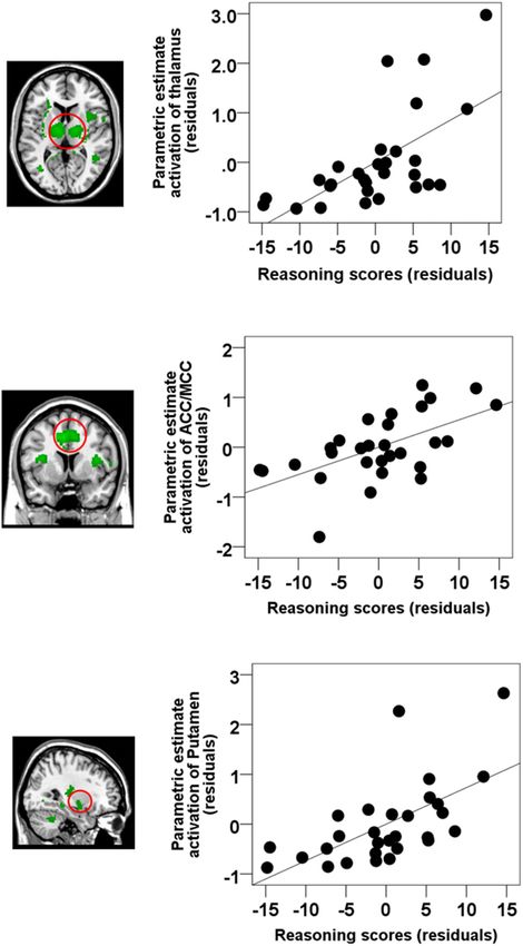
Right Temporal lobe 48 −52 −4 4.14 < 0.005 FIGURE 4 | Semi-partial correlations between brain activations time-locked to
spindles and Reasoning scores. ROI analyses revealed that Reasoning
Significant brain responses after FWE correction p < 0.05 at the cluster level. abilities were correlated with activations in the (A) the thalamus (partial
correlation, r = 0.628, p < 0.001), (B) ACC/MCC (partial correlation,
r = 0.585, p = 0.001), and (C) the bilateral putamen (partial correlation,
CBS trials. As shown in Figure 3B, Reasoning ability was r = 0.616, p = 0.001). Gender and the whole brain volume were included in
significantly correlated with activations time-locked to spindle the model as covariates of no interests. Values are standardized residuals
events in the thalamus, bilateral putamen, brainstem/pons, (showing the partial correlation) and shown in standardized arbitrary units.
ACC, the MCC, the paracentral lobe, the posterior cingulate
cortex, the precuneus, and bilateral temporal lobe (statistical
inferences were performed at a threshold of p < 0.001 Moreover, very few results were significant even at a much
uncorrected at the whole-brail level and p < 0.05, FWE less conservative, p < 0.001, without correction for multiple
corrected at the cluster level (Table 5). To illustrate these comparisons for Verbal abilities, and no results for STM at
relationships more clearly, the semi-partial correlations are that threshold.
shown in Figure 4. The ROIs of ACC/MCC, thalamus, and
bilateral putamen were defined independently based on
the previous literature, as described in the “Materials and
Comparisons of Brain–Behavior
Methods” section. Correlations Between Reasoning, STM,
Remarkably, no activations time-locked to spindle events were and Verbal Abilities
significantly related to STM or Verbal ability when considered as Reasoning ability was highly inter-correlated with Verbal
the covariate of interest (cluster-level FWE correction, p < 0.05). ability (r = 0.596, p = 0.001), and marginally correlated
Frontiers in Neuroscience | www.frontiersin.org 9 February 2019 | Volume 13 | Article 46Fang et al. Sleep Spindles and Cognitive Abilities
with STM ability (r = 0.357, p = 0.058), thus preventing p = 0.021), the cingulate cortex (r = 0.434, p = 0.030), and the
all three scales to be included in a whole brain regression putamen (r = 0.458, p = 0.021) over and above Verbal and STM
analysis due to high multicollinearity. Thus, in order to further (i.e., controlling for STM and Verbal abilities). The same partial
examine whether the significant relationship between activations correlation analyses were conducted between the five subtests
time-locked to spindle events and Reasoning ability could be scores and the spindle-related brain activation in the three main
accounted for by STM or Verbal abilities, we selected three regions of interest (i.e., thalamus, ACC/MCC, putamen). As
ROI based on the spindle activations consistently observed in shown in Supplementary Table S2, all subtests were correlated
the extant literature (see the section “Materials and Methods” with brain activations time-locked to spindles in thalamus and
for details). ROIs included: the thalamus, the ACC/MCC, and putamen (p < 0.05) except for feature match. In addition, the
the bilateral putamen. We extracted the beta weights from ACC/MCC activation was significantly correlated with deductive
these three independent ROIs from coordinates determined reasoning, spatial rotation, and polygons (p < 0.05), and
from the literature. We then conducted standard multiple linear marginally correlated with spatial planning (p = 0.06).
regressions whereby parametric brain activation estimate values
in the thalamus, the ACC/MCC, and the bilateral putamen Overlap Between Activations
were included in the models as dependent variables, and Time-Locked to Spindles and
Reasoning, Verbal, and STM scores were included in the Reasoning-Related Spindle Activations
models as independent variables. Similar to the whole-brain
From Figure 3, we can see that there were several overlapping
analysis (Table 6), altogether, Reasoning, STM, and Verbal
regions between the spindle activation maps (Figure 3A) and
abilities significantly accounted for brain activation time-locked
the maps that show activations time-locked to spindles that
to spindles in the thalamus (R2 = 0.425, p = 0.019), the cingulate
were correlated with Reasoning abilities (Figure 3B). To follow-
cortex (R2 = 0.627, p = 0.033), and the putamen (R2 = 0.456,
up this observation, the overlap (p < 0.05, FWE at the cluster
p = 0.011). Importantly, follow-up inspection of the partial
level) between the spindle activation maps and the Reasoning-
coefficients revealed that Reasoning ability uniquely accounted
related spindle correlation maps (Figure 3C, yellow regions)
for spindle-related activation in the thalamus (r = 0.460,
show several regions were consistently high and jointly activated
in both the spindle maps (Figure 3A) and Reasoning-spindle
correlation maps (Figure 3B), including the thalamus, medial
TABLE 6 | Multiple regression analyses of the relationship between CBS trials and frontal cortex, bilateral putamen, and the cerebellum. Thus,
brain activation time-locked to spindles. suggesting that a subset of the spontaneous spindle-related
activations was uniquely correlated with Reasoning abilities.
Overall regression effect
Correlated region Unadjusted R2 F(5,23) p
DISCUSSION
∗
Thalamus 0.425 3.394 0.019
ACC/MCC 0.627 2.974 0.033∗
Advancements in simultaneous EEG–fMRI technology and
Putamen 0.456 3.855 0.011∗
techniques has enabled the investigation of the functional brain
activation recruited during well-known electrophysiological
Follow-up analyses for the thalamus
events such as sleep spindles. This can provide a window into
CBS measures Partial r t(23) p understanding what brain areas are involved in sleep processes
and their function. Given the challenging nature of applying
Reasoning 0.460 2.486 0.021∗ these techniques to study sleep, only a handful of studies have
Verbal 0.210 1.029 0.314 explored the brain activations correlated with sleep spindles
STM −0.091 −0.436 0.667 (Laufs et al., 2007; Schabus et al., 2007; Tyvaert et al., 2008;
Follow-up analyses for the ACC/MCC Andrade et al., 2011; Caporro et al., 2012). While important in
terms of advancing the understanding of the neurophysiology
CBS measures Partial r t(23) p
and functional anatomy of the spindle, unfortunately, we can
Reasoning 0.434 2.307 0.030∗ only infer the functional significance of these brain activations
Verbal 0.229 1.128 0.271
from these studies. Thus, limiting our understanding of the
STM −0.198 −0.971 0.342
related functions of sleep spindles and their neural correlates.
Follow-up analyses for the putamen
Previous EEG and behavioral studies have identified sleep
spindles as a biological marker of cognitive abilities, and in
CBS measures Partial r t(23) p particular, Reasoning abilities (Bódizs et al., 2005; Schabus et al.,
2006; Fogel et al., 2007; Fogel and Smith, 2011; Ujma et al.,
Reasoning 0.458 2.473 0.021∗ 2014, 2015; Fang et al., 2017). While providing important
Verbal 0.293 1.470 0.155 information about the functional significance of the sleep spindle,
STM −0.241 −1.193 0.245 the neuroanatomical substrates and neural mechanisms which
Statistically significant results indicated by an asterisk (∗ ) at p < 0.05; STM, short- support the relationship between spindles and Reasoning abilities
term memory. can only be inferred indirectly from these studies. Here, we
Frontiers in Neuroscience | www.frontiersin.org 10 February 2019 | Volume 13 | Article 46Fang et al. Sleep Spindles and Cognitive Abilities
identified the neural activation patterns time-locked to spindles Fogel and Smith, 2011; Hoedlmoser et al., 2014; Róbert
that are correlated to cognitive abilities. Using simultaneous Bódizs et al., 2014; Ujma et al., 2014, 2015, 2016; Tessier
EEG–fMRI sleep recordings, the results of the present study et al., 2015; Fang et al., 2017), there is no single or unique
support three main findings: spindle electrophysiological characteristic that is correlated with
cognitive abilities (i.e., fluid intelligence, reasoning, problem
(1) similar to previous studies (Fogel et al., 2007; Fang et al., solving, etc.). Several spindle parameters have been found to
2017), the electrophysiological spindle characteristics be correlated with cognitive abilities. For example, spindle
(e.g., amplitude) during NREM sleep were related to amplitude (Bódizs et al., 2014; Ujma et al., 2014, 2016; Fang
Reasoning but not STM or Verbal abilities, et al., 2017), spindle activity (duration × amplitude) (Schabus
(2) similar to previous studies (Laufs et al., 2007; Schabus et al., 2006; Hoedlmoser et al., 2014; Tessier et al., 2015), and
et al., 2007; Tyvaert et al., 2008; Andrade et al., 2011; spindle density (Bódizs et al., 2005; Fogel et al., 2007) have all
Bergmann et al., 2012; Caporro et al., 2012; Fogel et al., been reported to be correlated with cognitive abilities. In the
2017a), activations time-locked to spindles were observed current study, we observed the strongest association between
in the thalamus, bilateral striatum, MCC, and cerebellum, spindle amplitude and reasoning ability. This is consistent with
and importantly, the finding of a recent meta-analysis study (Ujma, 2018), which
(3) Reasoning abilities, but not STM or Verbal abilities, were reported that only spindle amplitude is unambiguously associated
correlated with spindle-related activations in a subset of with cognitive ability.
these regions including the thalamus, bilateral putamen,
medial frontal gyrus, MCC, and precuneus.
Spontaneous Brain Responses
These results provide evidence that individuals with greater Time-Locked to Sleep Spindles
neural activation time-locked to spindle events have greater As expected, and consistent with previous EEG–fMRI studies
Reasoning abilities (i.e., “fluid intelligence”; problem solving of spontaneous spindle-related activations (Laufs et al., 2007;
skills, the ability to employ logic, identify complex patterns). Schabus et al., 2007; Tyvaert et al., 2008; Andrade et al.,
Altogether, our results identified for the first time, that a subset of 2011; Bergmann et al., 2012; Caporro et al., 2012; Fogel
spontaneous spindle-related activations are correlated specifically et al., 2017a), our results identified and confirmed the brain
with Reasoning abilities but are unrelated to other abilities such regions associated with spindle events during NREM sleep
as STM and Verbal abilities. Thus, suggesting that the extent in both cortical (including the media prefrontal, ACC, and
of spindle-related activations reflect an individual’s capacity MCC), subcortical areas (including the thalamus and bilateral
for reasoning. caudate, putamen, and pallidum), and the cerebellum. Thus,
confirming the recruitment of cortico-thalamic-striatal circuitry
Association Between Spindle Amplitude in spindle generation. These human neuroimaging findings
are supported by a large body of animal studies, which
and Reasoning Abilities
at the cellular level, suggest that spindles reflect oscillatory
The three subtests (i.e., Reasoning, STM, and Verbal) that
activity in widespread thalamocortical circuits, and involve
assess sub-domains of general cognitive abilities are highly
complex interactions between reticular, thalamocortical, and
inter-correlated with one another. However, among these
pyramidal cells (Steriade, 2005). Unfortunately, due to the
three inter-correlated subtests, only Reasoning abilities were
limited duration, and high inter-subject variability of sleep in
found to be correlated with spindles, when the overlapping
the scanner, we did not have sufficient sleep to investigate slow
variability was controlled using multiple regression. This finding
and fast spindles separately, or enough SWS to test whether
has been reported previously whereby the electrophysiological
a different pattern of results was observed during SWS. This
characteristics of spindles were correlated with Reasoning
limitation is not unexpected given the difficulty in obtaining
abilities (Fang et al., 2017) and Performance intelligence (Fogel
long, consolidated bouts of sleep in an MRI environment. As
et al., 2007), but not Verbal abilities. Also, it is consistent
scanner technology becomes more comfortable and less noisy,
with the extant literature and the well-established finding that
future studies may succeed at obtaining longer duration sleep
spindles are related to a subset of cognitive abilities that tap
in the MRI simultaneously with EEG. Taken together, animal
into reasoning and the ability to solve problems, i.e., fluid
and recent human neuroimaging studies, including the current
intelligence (Nader and Smith, 2001, 2003; Schabus et al., 2006;
study, support the involvement of thalamocortical, striatal,
Ujma et al., 2014, 2015). Using CBS tests, Hampshire et al.
and cerebellar circuitry in spindle generation. However, these
(2012) identified that the network of brain regions which support
findings do not directly investigate the functional correlates of
these three subtests of cognitive abilities were distinct from each
spontaneous spindle-related brain activation, discussed in the
other. Therefore, indicating that although these three subtest
following section.
are inter-correlated with each other, they are associated with
distinct neural substrates. Our findings further suggest that
BOLD brain activations during spindle events are uniquely Association Between Spindle-Related
related to Reasoning abilities, over and above STM and Verbal Activation and Reasoning Ability
abilities. It is also worth noting that according to previous studies The results of the current study identified, for the first time, that
(Bódizs et al., 2005; Schabus et al., 2006; Fogel et al., 2007; brain activations recruited during spontaneous spindle events
Frontiers in Neuroscience | www.frontiersin.org 11 February 2019 | Volume 13 | Article 46Fang et al. Sleep Spindles and Cognitive Abilities
were specifically associated with Reasoning abilities, but not positive correlation between gray matter intensity in the medial
STM or Verbal abilities, including the thalamus, the PFC (i.e., PFC and Reasoning abilities assessed by Cattell’s Culture Fair
the ACC/MCC), the bilateral putamen, the cerebellum, and the Intelligence Test, and also the WAIS-R (Gong et al., 2005).
precuneus. Thus, suggesting that interindividual differences in In addition, several studies have observed robust activations
the extent of activations in cortico–thalamic–striatal circuitry in the basal ganglia for Reasoning-related tasks compared to
time-locked spindles are related to individual cognitive strengths, other cognitive tasks, including the caudate nucleus, putamen,
and in particular, Reasoning abilities. The extent of these and globus pallidus (Melrose et al., 2007; Rodriguez-Moreno
activations does not relate to other cognitive abilities, e.g., STM or and Hirsch, 2009). Specifically, Sandman et al. (2014) has
Verbal abilities. Given the widespread nature and heterogeneity reported that the morphometry of the putamen was associated
of the brain regions activated, further investigation to explore the with performance on Reasoning-related subtests of the WAIS
network dynamics (e.g., using functional connectivity analyses) including block design, matrix Reasoning, and perceptual index
is warranted. This may reveal network hubs of communication, in preadolescent children. Although overlooked in some studies,
for which the strength of connectivity with other areas might the cerebellum is activated during the deductive reasoning
provide further insight into the relationship between spindles processing (Goel et al., 2000; Goel and Dolan, 2004). Thus,
and intellectual abilities. It is important to note that the results taken together, suggesting that integrity and functioning of
of this study are entirely correlational, and the possibility these regions (e.g., thalamus, PFC, striatum, cerebellum) are
remains that the observed correlation can be explained by other required for intact Reasoning abilities, however, this possibility
confounding or unrelated factors. Moreover, the directionality of remains to be explicitly investigated. Our results suggest that
this relationship cannot be determined from the results of this individuals with greater spindle-related activation of this circuitry
study. Future studies employing similar techniques in cases where is associated with greater Reasoning abilities. Thus, spindles may
spindle activity is abnormal and where specific cognitive deficits be an important marker of Reasoning abilities, and a window into
occur such as Autism (Limoges et al., 2013), learning disabilities understanding the interindividual differences in the activation
(Shibagaki et al., 1982), and in schizophrenia (Wamsley et al., patterns of neural substrates related to specific cognitive abilities.
2012) may provide additional insight into the neural basis The clinical significance and applications of the relationship
of the relationship between spindles and Reasoning abilities. between spindles and cognitive abilities is yet to be realized.
However, this study is an important first step in investigating the Interestingly, spindle production is reduced with age (Carrier
functional significance of spindle-related activations in young, et al., 2001, 2011; Lafortune et al., 2012; Martin et al., 2013;
healthy populations. Fogel et al., 2014; Fogel et al., 2017b), and abnormal in
The network of brain regions identified here, activated during developmental disorders, such as Autism (Limoges et al., 2005),
spindles, correlated with Reasoning abilities are convergent learning disabilities (Shibagaki et al., 1982), and in schizophrenia
with the extant literature. For example, thalamo–cortical (Wamsley et al., 2012). Deficient or dysfunctional spindle
circuitry (e.g., thalamus and the PFC region) is implicated in generation has been found to be associated with compromised
modulation of cognitive performance, such as memory, executive intellectual functioning. More specifically, it has been suggested
functioning, and attention (Van Der Werf et al., 2000; Van that deficient gating mechanisms of thalamocortical circuitry
der Werf et al., 2003; Blair, 2006; Mitchell and Chakraborty, (Bixler and Rhodes, 1968) may explain abnormal spindle
2013; Ferguson and Gao, 2015). Similar to the current study, production in children with mental disability (Gibbs and Gibbs,
thalamic activations have been observed while solving fluid 1962; Shibagaki and Kiyono, 1983). Moreover, the present
intelligence tasks, including problem solving (Fangmeier et al., study is an important first step which may lead to the
2006), reasoning tasks (Melrose et al., 2007), and particularly, development of novel interventions utilizing spindle-enhancing
inductive reasoning (Jia et al., 2011; Liang et al., 2014). neuromodulatory techniques (e.g., neurofeedback, transcranial
Other neuroanatomical studies (Bohlken et al., 2014) found direct current stimulation, pharmacological techniques) to
that thalamic volume was significantly correlated with general improve daytime cognitive performance and explore the
intellectual functioning. In addition, at least one study identified physiological mechanisms which support the function of sleep
structural and functional abnormalities in the thalamus in adults for memory and cognitive performance. Thus, such an approach
with reduced intellectual functioning who experienced prenatal could target cognitive deficits, in cases where spindle production
exposure to alcohol (Clark et al., 2000). Similarly, a large is abnormal such as in learning disabilities (Gibbs and Gibbs,
body of literature has identified the role of the PFC in fluid 1962; Shibagaki and Kiyono, 1983), below normal cognitive
intelligence and Reasoning (Baker et al., 1996; Waltz et al., 1999; functioning (Fogel and Smith, 2011), normal, healthy aging
Duncan, 2000; Gray et al., 2003; Coricelli and Nagel, 2009). (Carrier et al., 2011; Fogel et al., 2014, 2017b), developmental
For example, patients with damage to the PFC exhibited a disorders (Limoges et al., 2005), and in schizophrenia (Wamsley
selective and catastrophic deficit for both deductive and inductive et al., 2012). A better understanding of the neural basis of
reasoning tasks (Waltz et al., 1999). In addition, Gray et al. the relationship between spindles and cognitive abilities may
(2003) found that individuals with higher fluid intelligence have ultimately help to better understand biological basis of normal
greater activations in the PFC. Coricelli and Nagel (2009) have and abnormal cognitive functioning in healthy individuals and
shown that Reasoning abilities correlate with neural activity neurological conditions. This may eventually lead to novel
in the medial PFC. Additionally, at least one neuroanatomical interventions to precisely target cases where spindle production
MRI study employing voxel-based morphometry has revealed a is abnormal or non-optimal.
Frontiers in Neuroscience | www.frontiersin.org 12 February 2019 | Volume 13 | Article 46You can also read