Heterogeneous responses to low level death receptor activation are explained by random molecular assembly of the Caspase-8 activation platform
←
→
Page content transcription
If your browser does not render page correctly, please read the page content below
Heterogeneous responses to low level death receptor activation are
explained by random molecular assembly of the Caspase-8 activation
platform
Matveeva, A., Fichtner, M., McAllister, K., McCann, C., Sturrock, M., Longley, D. B., & Prehn, J. H. M. (2019).
Heterogeneous responses to low level death receptor activation are explained by random molecular assembly of
the Caspase-8 activation platform. PLoS Computational Biology, 15(9), [e1007374].
https://doi.org/10.1371/journal.pcbi.1007374

Published in:
PLoS Computational Biology

Document Version:
Publisher's PDF, also known as Version of record

Queen's University Belfast - Research Portal:
Link to publication record in Queen's University Belfast Research Portal

Publisher rights
: © 2019 Matveeva et al. This is an open
access article distributed under the terms of the
Creative Commons Attribution License, which
permits unrestricted use, distribution, and
reproduction in any medium, provided the original
author and source are credited
General rights
Copyright for the publications made accessible via the Queen's University Belfast Research Portal is retained by the author(s) and / or other
copyright owners and it is a condition of accessing these publications that users recognise and abide by the legal requirements associated
with these rights.

Take down policy
The Research Portal is Queen's institutional repository that provides access to Queen's research output. Every effort has been made to
ensure that content in the Research Portal does not infringe any person's rights, or applicable UK laws. If you discover content in the
Research Portal that you believe breaches copyright or violates any law, please contact openaccess@qub.ac.uk.

Download date:13. Feb. 2021
RESEARCH ARTICLE

                                                       Heterogeneous responses to low level death
                                                       receptor activation are explained by random
                                                       molecular assembly of the Caspase-8
                                                       activation platform
                                                       Anna Matveeva ID1,2, Michael Fichtner ID1,2☯, Katherine McAllister ID3☯,
                                                       Christopher McCann3, Marc Sturrock1,2, Daniel B. Longley3, Jochen H. M. Prehn1,2*

                                                       1 Centre for Systems Medicine, Royal College of Surgeons in Ireland, Dublin, Ireland, 2 Department of
                                                       Physiology and Medical Physics, Royal College of Surgeons in Ireland, Dublin, Ireland, 3 Centre for Cancer
a1111111111                                            Research and Cell Biology, Queen’s University, Belfast, United Kingdom
a1111111111
a1111111111                                            ☯ These authors contributed equally to this work.
a1111111111                                            * prehn@rcsi.ie
a1111111111

                                                       Abstract
                                                       Ligand binding to death receptors activates apoptosis in cancer cells. Stimulation of death
    OPEN ACCESS
                                                       receptors results in the formation of intracellular multiprotein platforms that either activate
Citation: Matveeva A, Fichtner M, McAllister K,
                                                       the apoptotic initiator Caspase-8 to trigger cell death, or signal through kinases to initiate
McCann C, Sturrock M, Longley DB, et al. (2019)
Heterogeneous responses to low level death             inflammatory and cell survival signalling. Two of these platforms, the Death-Inducing Signal-
receptor activation are explained by random            ling Complex (DISC) and the RIPoptosome, also initiate necroptosis by building filamentous
molecular assembly of the Caspase-8 activation         scaffolds that lead to the activation of mixed lineage kinase domain-like pseudokinase. To
platform. PLoS Comput Biol 15(9): e1007374.
                                                       explain cell decision making downstream of death receptor activation, we developed a
https://doi.org/10.1371/journal.pcbi.1007374
                                                       semi-stochastic model of DISC/RIPoptosome formation. The model is a hybrid of a direct
Editor: Martin Meier-Schellersheim, National
                                                       Gillespie stochastic simulation algorithm for slow assembly of the RIPoptosome and a
Institutes of Health, UNITED STATES
                                                       deterministic model of downstream caspase activation. The model explains how alterations
Received: April 9, 2019
                                                       in the level of death receptor-ligand complexes, their clustering properties and intrinsic
Accepted: September 3, 2019                            molecular fluctuations in RIPoptosome assembly drive heterogeneous dynamics of Cas-
Published: September 25, 2019                          pase-8 activation. The model highlights how kinetic proofreading leads to heterogeneous
Copyright: © 2019 Matveeva et al. This is an open      cell responses and results in fractional cell killing at low levels of receptor stimulation. It
access article distributed under the terms of the      reveals that the noise in Caspase-8 activation—exclusively caused by the stochastic molec-
Creative Commons Attribution License, which            ular assembly of the DISC/RIPoptosome platform—has a key function in extrinsic apoptotic
permits unrestricted use, distribution, and
                                                       stimuli recognition.
reproduction in any medium, provided the original
author and source are credited.

Data Availability Statement: All relevant data are
within the manuscript and its Supporting
Information files.                                       Author summary
Funding: This work was supported by grant from           Death receptors are targets of novel cancer therapeutics. Most of them signal through flex-
Science Foundation Ireland (http://www.sfi.ie/, SFI
                                                         ible multiprotein platforms to either activate apoptotic or necroptotic cell death, or propa-
14/IA/2582) granted to DBL and JHMP. The
funders had no role in study design, data collection
                                                         gate cell survival and pro-inflammatory signals. We focused our study on the role of
and analysis, decision to publish, or preparation of     dynamic assembly and composition of these platforms in the initiation of cell death at
the manuscript.                                          the single cell level. Since the assembly is slow through the competitive nature of protein

PLOS Computational Biology | https://doi.org/10.1371/journal.pcbi.1007374 September 25, 2019                                                                 1 / 22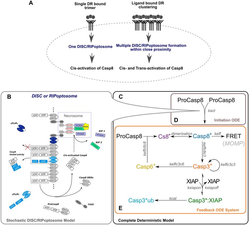
Random assembly of death receptor signalling platform explains cell death heterogeneity

Competing interests: The authors have declared
that no competing interests exist.                 binding within the platforms core we developed a stochastic mathematical model of the
                                                   death inducing signalling platform. Our model provided an explanation for delayed cell
                                                   death and fractional killing upon the death receptor stimulation. Additionally, we found
                                                   that the variability in the cell death response arises through the random assembly initiates
                                                   a slow noise-prone ramp activation of initiator Caspase-8 spontaneously triggering the
                                                   apoptotic cascade. Our computational simulations predicted high variation in the time
                                                   required for cell death induction at the single cell level and highlighted a significant role
                                                   of death receptor clustering in effective Caspase-8 activation. Our knowledge and data
                                                   driven model captures detailed processes governing the early events of cell death initiation
                                                   and can be used to guide the development of more rational combinational treatments
                                                   against cancer.

                                                 Introduction
                                                 Apoptotic signalling cascades are designed to irreversibly lead to cell death once specific death
                                                 thresholds are overcome [1,2]. Activation of caspases plays a central role in this process. In cer-
                                                 tain scenarios, apoptotic cell death signalling is interrupted. This may lead to the activation of
                                                 other forms of cell death or escape from cell death altogether.
                                                     Death ligands (DL) bind to death receptors (DR) at the plasma membrane and have been
                                                 developed as novel cancer therapeutics. However, many cells in our body are exposed from
                                                 time to time to endogenous DLs, such as TNF-α and TRAIL, without induction of cell death.
                                                 Several studies have shown that while binding of DLs to DRs can induce apoptosis, not all cells
                                                 will respond to DR stimulation with cell death, and only a fraction of the cell population will
                                                 undergo apoptosis even if DLs bind at death-inducing concentrations [2–6] (Fig 1A). Interest-
                                                 ingly, in vivo studies have shown that fractional death resistance has no direct association with
                                                 the amount of DRs expressed on the plasma membrane [7,8]. Therefore, cell signalling acti-
                                                 vated by extrinsic ‘death’ signals is rather encoded downstream of receptor binding.
                                                     Binding of DLs to dedicated DRs triggers either the formation of receptor-associated
                                                 Death-Inducing Signalling Complexes (DISC) (‘Complex I’) in proximity to the plasma mem-
                                                 brane, or RIPoptosome complexes (‘Complex II’) in the cytosol [5,9–17]. Both complexes
                                                 provide a platform for the activation of the initiator Caspase-8 (Casp8). For the activation of
                                                 Casp8, the inactive pro-form of Casp8 (ProCasp8) must undergo autocatalytic activation. This
                                                 is achieved through ProCasp8 dimerization and sequential inter- and intradimer cleavage, a
                                                 process which results in the release of active Casp8 [18–20]. The dimeric ProCasp8 associa-
                                                 tion-dissociation balance has been suggested to play a crucial role in the molecular control of
                                                 apoptotic responses after DR activation [21]. However, as demonstrated by mutagenesis stud-
                                                 ies, ProCasp8 dimerization alone is not sufficient to enhance apoptotic responses in vivo [22].
                                                 Instead, formation of the DISC or RIPoptosome platforms are necessary for effective ProCasp8
                                                 dimerization and Casp8 activation [10,23,24].
                                                     Apart from apoptosis initiation, DR-induced complexes also initiate necroptosis by accu-
                                                 mulating heterodimers of receptor-interacting proteins (RIPs), RIP1 and RIP3 (RIP1/3), and
                                                 the formation of filamentous scaffolds [25–28]. Formation of such ‘Necrosome’ platforms acti-
                                                 vates the mixed lineage kinase domain-like (MLKL) pseudokinase. MLKL activation triggers
                                                 necroptosis, a cell death distinct from apoptosis [29–31]. In theory, activation of DRs in indi-
                                                 vidual cells could lead to both apoptosis and necroptosis signalling through the formation
                                                 of different platforms. However, if RIP1/3 proteins are close to the site of Casp8 activation,
                                                 RIP1/3 is cleaved by Casp8 [32]. This cleavage eliminates the kinase activity of RIP1/3, and

PLOS Computational Biology | https://doi.org/10.1371/journal.pcbi.1007374 September 25, 2019                                                        2 / 22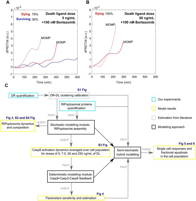
Random assembly of death receptor signalling platform explains cell death heterogeneity

                                Fig 1. New modelling approach developed in this study to explain vulnerable dynamics of Casp8 activation and fractional cell
                                death upon DR stimulation. (A, B) Casp8 activation dynamics tracked by the FRET reporter cleavage in Hela cells upon treatment
                                with low and high doses of the DR ligand (experimental data from Roux et al., 2015)[2] as evidence for stochastic fluctuation
                                dynamics and final switch like transition of the Casp8 activity which is the main cause of the fractional cell death. (A) Single cell
                                Casp8 activation upon 5 ng/mL rhTRAIL treatment with 100 nM Bortezomib. Fluctuating ramp in Casp8 activation which leads to
                                activity acceleration and MOMP within first 10 hours (in red) and fluctuating ramp which fails to trigger MOMP (in blue) causing
                                survival of 30% cell population (experimental data from Roux et al., 2015) [2]. (B) 100% cell death over population induced by 50 ng/
                                mL rhTRAIL with 100 nM Bortezomib treatment triggered by high acceleration rate of Casp8 activity (in red). (C) Schematic
                                diagram representing the methodological design of this study.
                                https://doi.org/10.1371/journal.pcbi.1007374.g001

                                              consequently necroptosis activation is suppressed [9,33–36] (Fig 2B). This suggests that if one
                                              type of cell death is triggered in a given cell, the other type of cell death is suppressed, i.e., that
                                              the two types of cell death are mutually exclusive.
                                                 Previous studies of the apoptotic signalling network activated by DRs have identified that
                                              variability in death signalling arises from the process preceding the mitochondrial outer mem-
                                              brane permeabilization (MOMP). This process triggers Casp8-mediated cleavage of the pro-

PLOS Computational Biology | https://doi.org/10.1371/journal.pcbi.1007374 September 25, 2019                                                                   3 / 22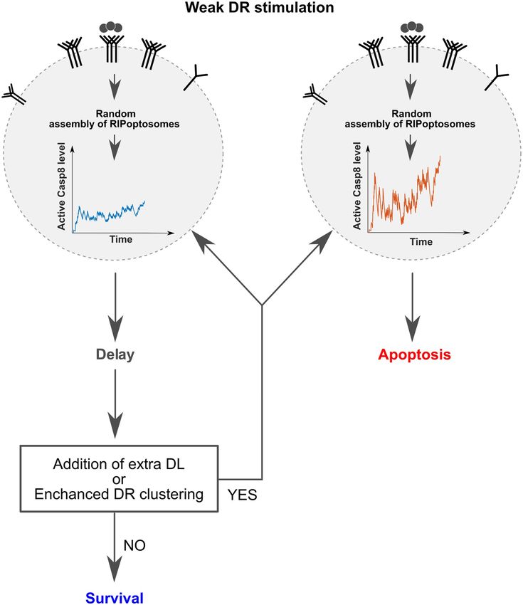
Random assembly of death receptor signalling platform explains cell death heterogeneity

                                Fig 2. New modelling approach developed in this study to explain vulnerable dynamics of Casp8 activation and fractional cell
                                death upon DR stimulation. (A) The DR clustering initiates the formation of a few RIPoptosomes in the closed proximity to each
                                other where Casp8, in addition to cis-activation, can undergo trans-activation. (B) The initial stochastic Casp8 activation on the
                                DISC/RIPoptosome platform is implemented by direct Gillespie stochastic simulation algorithm (SSA) which incorporates the
                                molecular assembly of FADD, RIP1, RIP3, ProCasp8, cFLIPs/l proteins. (C) Complete deterministic system comprised of initiation
                                ODE system (D) and feedback ODE system (E) used in the parameter scan and manual parameter adjustment. (D) Box represents
                                the deterministic approximation of the population kinetics of Casp8 activation on the DISC/RIPoptosome platform. (E)
                                Box represents the deterministic activation of the effector caspases, Casp3 and Casp6, which feedback to the rate of Casp8 activation
                                before MOMP.
                                https://doi.org/10.1371/journal.pcbi.1007374.g002

                                              apoptotic Bid protein [2,4,37], which mediates MOMP and leads to cytochrome-C release,
                                              apoptosome formation and executioner caspase activation [38].
                                                  To understand cell death decision making in more detail, we created a mathematical model
                                              which incorporates the central events prior to Bid cleavage. The model was constructed to
                                              estimate apoptotic and necroptotic pathway initiation through the random assembly of the
                                              DISC/RIPoptosome platform. As a multiprotein platform with diverse functionality, we
                                              hypothesised that the random and stochastic process of its assembly may lead to the heteroge-
                                              neous cellular responses (Fig 1A and 1B). Combining this model with experimentally derived
                                              sets of quantitative protein profiles and literature-based catalytic and binding rates, we simu-
                                              lated the heterogeneous responses of HeLa cells to DR activation. By modelling different

PLOS Computational Biology | https://doi.org/10.1371/journal.pcbi.1007374 September 25, 2019                                                                    4 / 22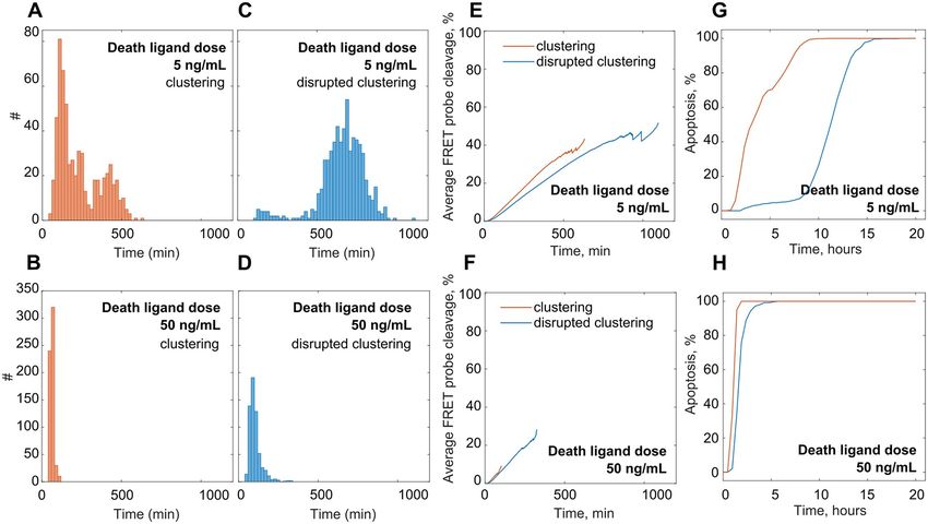
Random assembly of death receptor signalling platform explains cell death heterogeneity

                                            conditions of DR stimulation and clustering, we investigated in particular how heterogeneous
                                            apoptotic responses arise, which role the random assembly of DR-induced platforms play in
                                            determining death delay at the single cell level, and how DR clustering facilitates death signal-
                                            ling. Our analysis reveals that the noise in Casp8 activation exclusively caused by the stochastic
                                            molecular assembly of the DISC/RIPoptosome platform has a key function in the low level
                                            extrinsic apoptotic stimuli recognition.

                                            Results
                                            Quantitative estimation of death receptor abundance and clustering
                                            Apoptosis inducing DRs such as Tumour Necrosis Factor Receptor 1 (TNFR1) and Death
                                            Receptors 4 and 5 (DR4/5) are expressed at comparable protein levels in HeLa cells [39]. Addi-
                                            tionally, it is known that their protein expression level is correlated with the receptor abun-
                                            dance on the cell surface [8]. High variation in TNFR1 surface abundance were estimated in
                                            previous studies ranging from 300 to 3000 molecules per single HeLa cell [40,41]. To get more
                                            accurate estimates, we performed the single cell quantification of TNFR1 membrane expres-
                                            sion in HeLa cells employing the QuantiBRITE phycoerythrin beads based assay (see S1 File).
                                            We determined that the average number of TNFR1 does not exceed 905 receptors per cell.
                                            We further used this quantity as the reference in our comparative quantification of DR4/5
                                            receptors based on MS data set (S1 File). Thus, we calculated that DR4 and DR5 receptors
                                            are present on HeLa cell surface in an average amount of 769 and 926 monomeric receptors,
                                            respectively (Table A in S1 File).
                                               Next, we estimated the amount of the DR complexes associated with DL at the single cell
                                            level. Due to the fact that the DR-DL association is generally much quicker [42] than the
                                            downstream processes such as ProCasp8 dimerization and subsequent Casp8 activation [43],
                                            we applied the rapid equilibria approximation to calculate the amount of DL bound receptors.
                                            According to the law of mass action the time evolution of the amount of DR-DL complexes is
                                                                d½RL�
                                                                      ¼ kon ½R�½L�     koff ½RL�; where ½R� ¼ ½Rtotal �   ½RL�                     ð1Þ
                                                                  dt

                                               Where [Rtotal] is the total number of receptors per cell (Table A in S1 File), [RL] is the num-
                                            ber of DR-DL complexes and [L] is death ligand concentration (Table B in S1 File).
                                               Setting the RL to the rapid equilibrium
                                                                                         d½RL�
                                                                                               ¼0                                                  ð2Þ
                                                                                           dt

                                               From (1) we calculated the average number of DR-DL complexes per cell as a function of L,
                                            Rtotal and the DL dissociation constant Kd
                                                                                                ½Rtotal �
                                                                                     ½RL� ¼ �           �                                          ð3Þ
                                                                                                Kd
                                                                                                ½L�
                                                                                                      þ1

                                                The minimal unit of the active DR-DL complex is the trimer [44]. The trimeric DR-DL
                                            complex gives birth to a single DISC platform which internalizes within the subsequent 10–15
                                            minutes [45,46]. If the DISC has not bound to cellular Inhibitor of Apoptosis Proteins (cIAPs),
                                            cIAP1 or cIAP2, then it either releases active RIP1 protein into the cytosol [47] where it can
                                            form RIPoptosome or Necrosome platforms (Fig 2B) (as in case of TNFR1), or it makes active
                                            RIP1 protein accessible for further RIP1/3 and ProCasp8 proteins accumulation on the DISC
                                            itself (as in case of DR4/5 activation) [5,17]. Therefore, in the modelling routine each activated

PLOS Computational Biology | https://doi.org/10.1371/journal.pcbi.1007374 September 25, 2019                                                     5 / 22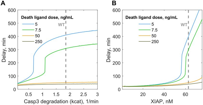
Random assembly of death receptor signalling platform explains cell death heterogeneity

                                            DISC was translated into a single RIP1 protein molecule which is available immediately after
                                            DL introduction to the cell culture.
                                                Trimeric DR-DL complexes tend to organise high order clusters in cellular membranes
                                            [44,48] and bring several associated DISC/RIPoptosomes into close proximity. Such clustering
                                            stimulates more efficient signalling [49] and enables ProCasp8 activation not only by dimer-
                                            ization on the single DISC/RIPoptosome but also by synchronised binding of two ProCasp8
                                            monomers with two independent DISC/RIPoptosomes within one cluster. To introduce
                                            DISC/RIPoptosomes clustering processes in the model, we estimated the number and the size
                                            of the DR-DL clusters based on the experimentally derived DR-DL probability distribution
                                            from a study published earlier by Fricke and co-workers [44]. We calibrated probability redis-
                                            tribution from the total pool of activated trimeric DR-DL complexes, calculated in the previous
                                            step, to the clusters of different size (see S1 File). Using these probabilities, we assigned for
                                            each random DISC/RIPoptosomes complex formed its associated cluster. The final algorithm
                                            assumes that DISC/RIPoptosomes complexes within one cluster are able to first encourage the
                                            activation of ProCasp8 by direct dimerization (cis-activation) and subsequently activate Pro-
                                            Casp8 via simultaneous binding within closed proximity (trans-activation) (Fig 2A). Thus, this
                                            information about the amount of the activated DR-DL complexes and their clustering confor-
                                            mation served as an important input for the model. The scenario of non-clustering DR signal-
                                            ling was studied as well by setting the probability of trans-activation of Casp8 within DISC/
                                            RIPoptosomes complexes cluster to zero. This scenario is hereafter referred to as disrupted
                                            clustering.

                                            Mathematical modelling of the initiation of the extrinsic apoptosis
                                            pathway
                                            We have developed a core model capturing the cascade of intracellular reactions that are essen-
                                            tial for the initiation of the apoptosis. The model reactions are partitioned into two modules: a
                                            stochastic and a deterministic module (Fig 2).
                                                The first stochastic module represents the process of stochastic assembly of DR-induced
                                            DISC/RIPoptosome multiprotein platform which facilitates initiation of ProCasp8 dimeriza-
                                            tion and self-activation by cleavage (Casp8� ; activated Casp8 dimer in Fig 2E). We imple-
                                            mented this module with the direct Gillespie stochastic simulation algorithm [50,51] which
                                            accounts for molecular fluctuations and slow association and dissociation rates following each
                                            component of the platform individually. It assigns the reaction propensities in probabilistic
                                            terms. The binding propensities of ProCasp8 together with its binding partner protein, Fas-
                                            associated death domain protein (FADD), and competitor protein RIP1/RIP3 that comprise
                                            the core scaffold of RIPoptosome are calculated from the concentrations that we quantified
                                            experimentally in HeLa cell culture (Table D in S1 File). FADD protein is crucial for apoptotic
                                            initiation [35,52]. This protein consists of both a Death Effector Domain (DED) and Death
                                            Domain (DD) which are specific motifs for ProCasp8 [53] and RIP1 [16,54] self-oligomeriza-
                                            tion respectively. Through these domains, ProCasp8 and RIP1 are bridged via FADD (as
                                            shown in grey in Fig 2B). RIP3 protein can form homo-oligomers, but can also associate with
                                            RIP1 scaffolds through the RIP homotypic interaction motif (RHIM), forming amyloid struc-
                                            tures [27,28] (Fig 2B). Intensive recruitment of RIP3 molecules to the amyloid triggers trans-
                                            phosphorylation of RIP3 by RIP1 with consequent transmission of phosphate groups to the
                                            MLKL pseudokinase. Phosphorylated MLKL executes necroptosis [25,30]. Therefore, in the
                                            absence of FADD and joint Casp8 activation platforms these structures spontaneously trigger
                                            necroptosis [35,55,56] (necrosome complex; purple in Fig 2B). Additionally, we quantified
                                            the concentrations of the cellular FLICE (FADD-like IL-1β-converting enzyme)-inhibitory

PLOS Computational Biology | https://doi.org/10.1371/journal.pcbi.1007374 September 25, 2019                                                     6 / 22
Random assembly of death receptor signalling platform explains cell death heterogeneity
protein (c-FLIP). As a DED-containing protein, cFLIP in its short (cFLIPs) and long (cFLIPl)
form, can be recruited to the ProCasp8 platform abrogating or restricting activation of Casp8
[53,57,58] (cFLIP molecules; light and dark blue in Fig 2B). In addition to this suppression,
Casp8 activation can be disrupted by binding its own processed DEDs which may remain in
the cytosol (DED1-DED2; white in Fig 2B).
The second deterministic module mimics the activation of two effector caspases, Caspase 3
(Casp3) and Caspase 6 (Casp6) which is triggered by stochastically activated Casp8. Pro-forms
of both caspases form stable dimers at physiological concentrations [59]. By cleavage, Casp8
activates Casp3 (Casp3� ; activated dimer of Casp3 in Fig 2E) [60]. Casp3� activates Casp6
(Casp6� ; activated dimer of Casp6 in Fig 2E) [61,62] and has autocatalytic function cleaving
ProCasp3 [63,64]. Finally, Casp6� can cleave free ProCasp8 (Casp8� ; cleaved monomer of
Casp8 in Fig 2E) [64–67] however Casp8 becomes active only after a very slow dimerization
(Casp8� ) [19,21]. Previous models suggest that this effector caspase feedback upon weak DR
stimulation probably can accelerate Casp8 activation which was initially started at the DISC or
RIPoptosome platform [68]. However, the feedback can be inhibited by X-linked IAP (XIAP)
which tightly binds Casp3 and, further, marks Casp3 with ubiquitin that leads to its proteaso-
mal degradation [69,70]. The overall dynamics of Casp8 activation can be tracked quantita-
tively with a Casp8-specific FRET cleavage probe (FRET, Fig 2E). The fixed threshold rate
of this FRET probe cleavage accurately determines the moment of MOMP in HeLa cells [2].
Based on the mass action and conservation laws, the time evolution of the variables that com-
prise this module were modelled by a deterministic system of ordinary differential equations
(ODE) (details in S1 File).
All protein concentrations and parameters used in the model are provided in Tables D and
E of Materials and Methods file (S1 File).
Stochastic initiation of Casp8 activation through DISC/RIPoptosome
assembly
The estimated weight of the RIPoptosome after short DR-targeted stimulation may exceed
2MDa [10,24,29]. To reproduce the RIPoptosome growth and composition we first employed
the stochastic modelling module simulating the assembly of the individual RIPoptosomes at
the single cell level. RIP1 on its own forms unlimited filaments in vitro [28], however, in the
cell the long-term RIPoptosomal filament growth is limited by the cell volume and the stiffness
of the cellular components. We followed unlimited filament growths without implementation
of these physical limits, focusing on the initial dynamics of RIPoptosome progression. Fig 3
and S2 Fig illustrates the simulated molecular composition of RIPoptosome in the single HeLa
cell treated with a dose of 5 ng/mL of the DL (rhTRAIL).
The composition and the time evolution of individual RIPoptosomes within single cell dif-
fered from one to another. Consequently, the size and, therefore, molecular weight of those
RIPoptosomes varies as well. As an example, we display the composition change in a few ran-
domly chosen RIPoptosomes over the first 20 min with 1 min step interval (Fig 3A, S2 Fig).
Next, we calculated the progression of the molecular weight of a complete cellular pool of
RIPoptosomes as simulated by the model. Interestingly, we found a high degree of variation
between the RIPoptosomes formed within the same cell (Fig 3B). Our simulations confirmed
that in HeLa cells, the most populated protein within each RIPoptosome is RIP1 through its
highly stable association mechanism. This is explained by the RHIM domain binding property
that shares homology with β-amyloids assembly domains. Simulation of the model revealed
that the RIP1 filaments formation is triggered immediately after DR stimulation (Fig 3A). The
PLOS Computational Biology | https://doi.org/10.1371/journal.pcbi.1007374 September 25, 2019 7 / 22Random assembly of death receptor signalling platform explains cell death heterogeneity
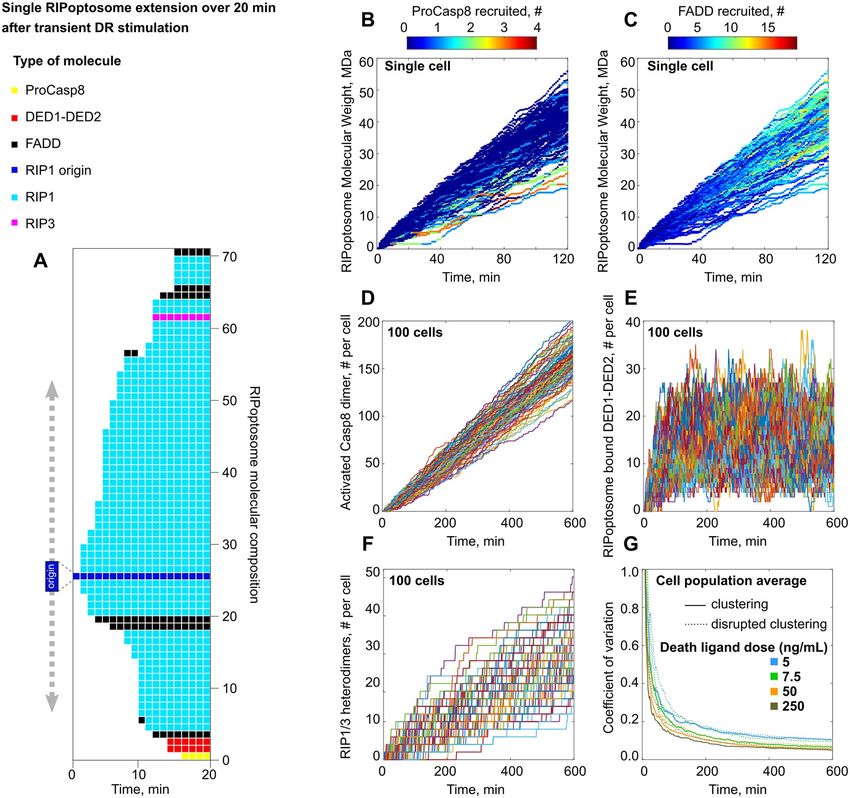
Fig 3. Simulated by model stochastic molecular assembly of RIPoptosome and Casp8, RIP1/RIP3 activation in a single cell. (A)
Model generated individual RIPoptosome growth over first 20 min after DR stimulation with 5ng/mL of DL assuming receptor
clustering scenario. (B,C) RIP1 filaments formation triggered immediately after DR stimulation which results in RIPoptosomes of
size 2 MDa and higher within first 5 min. (B) ProCasp8 recruitment is mostly abundant in the filaments of the lower molecular
weight. (C) FADD recruitment is proportional to the filament molecular weight and persistently increases with the filament
progression. (D) Single cell stochastic traces of RIPoptosome driven Casp8 activation, (E) accumulation of DEDs (DED1-DED2) and
(F) RIP1-RIP3 heterodimers induced by treatment with 5 ng/mL of the DL. 100 traces shown generated assuming DR clustering
scenario. (G) Time dependant variability in Casp8 activation over population of 100 cells stimulated with DL doses of 5, 7.5, 50 and
250 ng/mL for regular (solid line) and disrupted (dashed line) receptor clustering order.
https://doi.org/10.1371/journal.pcbi.1007374.g003
model also predicts that it would be possible to observe RIPoptosomes of size 2 MDa only 5
minutes after DR stimulation (Fig 3B).
FADD recruitment to the fraction of the high molecular weight complexes is persistently
increasing with post treatment time [29]. Our simulations show as well that the abundance of
FADD within a single RIPoptosome increases linearly with time progression (Fig 3C) in con-
junction with the filament growth. As a result, the abundance of FADD on average will not
exceed the amount of 10 molecules per origin within the first two hours. Moreover, this abun-
dance is independent of DL dose. Thus, a low dose of 5 ng/mL of the DL and a high dose of 50
ng/mL will result in similar FADD abundance (S4A and S4B Fig).
PLOS Computational Biology | https://doi.org/10.1371/journal.pcbi.1007374 September 25, 2019 8 / 22Random assembly of death receptor signalling platform explains cell death heterogeneity
On the contrary, ProCasp8 recruitment in the single cell is most abundant in the RIPopto-
some of the lower molecular weight (Fig 3B). The binding of the ProCasp8 or its DEDs domain
to the end of the filament blocks the RIP1 recruitment and therefore also blocks intensive fila-
ment growth by competition. The population average over 600 cells shows that ProCasp8
abundance per RIPoptosome (origin) saturates after 2 hours of stimulation (S4C and S4D Fig).
This relative abundance does not vary significantly for doses of 5 or 50 ng/mL of the DL and is
unaffected by the clustering or non-clustering assumption in the model. These rapid saturation
dynamics of ProCasp8 compared to linear FADD translocation has been observed earlier in
experiments where no co-binding of FADD and Casp8 has been observed after 1 hour of stim-
ulation but has become apparent at the second hour [29].
Single cell stochastic RIPoptosome assembly is the source of variation in
Casp8 and RIP1/3 protein activation
Molecular fluctuations in the RIPoptosome composition within single cells cause the fluctua-
tions in the active Casp8 abundance (Fig 3D). Stochastic single cell Casp8 activation traces for
5 ng/mL dose simulation with the corresponding per cell accumulation of Casp8 Pro domain
(DED1-DED2) are shown in Fig 3D and 3E. Interestingly, limited expression of RIP3 [28] pro-
tein in HeLa cell gives rise to very low and therefore heterogeneous distribution of RIP1-RIP3
heterodimers among the cells (Fig 3F) making the spontaneous event of the necroptosis less
probable to overtake the apoptotic course of the cell death.
Averaged over the population the Casp8 activation time course demonstrated high depen-
dence on the dose of the DL as well as the clustering capacity (S3 Fig). Thus, even low doses of
the DL with enhanced clustering property can activate Casp8. This result confirms the estab-
lished success in the application of combinational therapeutics where the DL has been com-
bined with the ligand specific cross-linking antibodies that enhance receptor clustering [49].
As expected, the overall variability in the Casp8 activation is a function of the treatment
dose (Fig 3G). Despite the coefficient of variation being within the limits of low-variance
(less than 1), the early Casp8 initiation dynamics can bring significant stochasticity into
triggering the downstream death pathway. Interestingly, the enhanced receptor clustering did
not reduce the variability in the individual HeLa cell Casp8 activation dynamics significantly.
We observed only a minor decrease in the coefficient of variation over all tested conditions
(Fig 3G).
Deterministic modelling of effector caspases feedback into Casp8
activation
Next we studied the downstream caspase cleavage cascade, the second deterministic modelling
module (Fig 2E), which feedbacks to the DISC/RIPoptosome based Casp8 production and is
potentially capable of boosting cell apoptotic capacity especially following treatment of low
doses of DL [68]. As an input we used the population average of the stochastic traces (Fig 2D,
S3 Fig) we simulated for the first module of the DISC/RIPoptosome based network initiation
assuming DR clustering (Fig 2B). Thus, we merged two modules into one complete determin-
istic system (Fig 2C) which enabled us to adjust undetermined parameters and estimate
parameter sensitivity, hence avoiding computationally expensive parameter scans of the full
stochastic formalism (see Materials and methods, S1 File).
The first undetermined parameter is the rate constant of Casp3 ubiquitin dependent degra-
dation (kcat). Ubiquitination of active Casp3, which is set by XIAP, will attract proteasomal
complex leading to Casp3 degradation. However, application of proteasome inhibitors does
not stabilise the pool of active Casp3 and consequently does not result in reduced Casp3
PLOS Computational Biology | https://doi.org/10.1371/journal.pcbi.1007374 September 25, 2019 9 / 22Random assembly of death receptor signalling platform explains cell death heterogeneity
Fig 4. Cell death delay sensitivity to the change in ProCasp6 concentration and Casp3 degradation kinetics in the clustering
scenario. (A) Dramatic increase in the cell death delay upon the increased Casp3 degradation rate constant (kcat) by XIAP mediated
ubiquitination which is a function of proteasomal protein degradation capacity as well as Casp3 self-cleavage for the cells stimulated
with lower DL doses (5, 7.5 ng/mL). (B) High sensitivity to the minor decrease in XIAP concentration from its mean estimated
concentration in HeLa cell 63 nM will lead to dramatic decrease in cell death delay for the cells stimulated with lower DL doses.
https://doi.org/10.1371/journal.pcbi.1007374.g004
proteasomal degradation. Instead, Casp3 catalytic activity is absolutely required for its own pro-
teasomal degradation [71,72]. Therefore, dynamics of Casp3 degradation triggered by XIAP
will not match the general degradation dynamics triggered by ubiquitin ligases for other types
of proteins and this specific rate constant needed to be identified. We estimated that kcat needs
to be significantly higher (1.75 min-1) from the general (basal) ubiquitin-dependent degradation
rate (0.04 min-1) [73] (Table E in S1 File). Again, low doses of the DL bring into play a switch-
like sensitive response to the change in kcat value (Fig 4A). In this case the cell death delay can
be initiated in a spontaneous fashion if the Casp3 degradation mechanism is perturbed.
Furthermore, the similar steep ultra-sensitive response can be also initiated by the mild
fluctuations in the XIAP concentration. We found that slight deviations from the mean XIAP
level, 63 nM, quantified earlier for HeLa [1] could speed up the cell death by more than 3-fold
in the case of low DL doses (Fig 4B). This decrease could be very sudden through this switch-
like type of response. Indeed, XIAP specific inhibitors such as Embelin, Mithramycin A are
able to overcome the DL resistance in different cancer types [74,75].
Semi-stochastic hybrid modelling of the complete initiation network
Finally, with the fully identified parameter set we formulated the new semi-stochastic hybrid
model of apoptotic pathway initiation in a single cell with the fixed partitioning of the whole
network into discrete (Fig 2B) and continuous reactions (Fig 2E). The slow discrete reactions
are the DISC/RIPoptosome assembly. The fast continuous reactions capture the caspase cleav-
age cascade.
The simulation results for a single cell response on the addition of low and high amounts of
the DL are demonstrated in Fig 5. We observed a prolonged ramp effect for all variables of the
network before the system switched to the rapid response. The ramp duration for the displayed
example exceeded 10 hours after treatment with the low dose of the DL (Fig 5A). Whereas the
PLOS Computational Biology | https://doi.org/10.1371/journal.pcbi.1007374 September 25, 2019 10 / 22Random assembly of death receptor signalling platform explains cell death heterogeneity
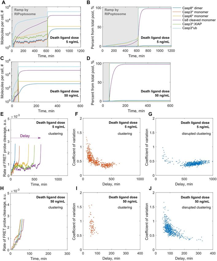
Fig 5. Simulated by semi-stochastic model single cell network activation. (A-D) Plots represent single cell responses to the low, 5
ng/mL, and high, 50 ng/mL, DL treatment doses. Responses begin with the slow ramp regime (in grey) characterised by high
stochastic noise explained by random assembly of RIPoptosome. Once a sufficient rate of Casp8 activation that can overcome XIAP
dependent Casp3 inactivation is reached, a rapid switch transition is triggered through the positive caspase feedback loop. The
molecular number of active Casp8 dimers; active Casp3, Casp6 and Casp8 monomers; XIAP and ubiquitin targeted Casp3 molecules
per HeLa cell are reflected (A, C) as well as theirs percentage from the total initial pool (B, D). (E-J) Plots represent cell death delay
driven by intrinsic noise. Single cell stochastic ramp traces of FRET probe cleavage are shown for the five randomly chosen cells
simulated with the low, 5 ng/mL, (E) and high, 50 ng/mL, (H) DL treatment doses upon receptor clustering. The trajectories were
terminated at the point of death threshold rate (see S1 File). Scatter plots represent the relation between individual cell ramp noise
and time of cell death delay within the population of 600 cells for the low, 5 ng/mL, (F-G) and high, 50 ng/mL, (I-J) DL doses with
the normal (F, I) and disrupted (G, J) receptor clustering order.
https://doi.org/10.1371/journal.pcbi.1007374.g005
PLOS Computational Biology | https://doi.org/10.1371/journal.pcbi.1007374 September 25, 2019 11 / 22Random assembly of death receptor signalling platform explains cell death heterogeneity
high dose treatment stimulates the ramp for shorter times, around one hour for a shown exam-
ple (Fig 5C). In a similar manner to the simulations with our entirely deterministic model, the
delay for the switch in the single cell response is a function of DL dose (Fig 4).
However, for both high and low doses we also observed very high dynamic noise in the
ramp (Fig 5A and 5C). This noise characterises the time course of dimeric Casp8 and active
Casp3 accumulation. In experiments both proteins are very unstable and hardly detectable in
the pre MOMP period of apoptotic initiation [19,71]. As we have shown earlier initial forma-
tion of new Casp8 dimer species can be limited by the vulnerability in molecular assembly of
the DISC/RIPoptosome platform (Fig 3). Moreover, active Casp8 dimer is unstable due to
high dissociation rate in the cytoplasm [19,20]. Indeed, Casp8 under physiological concentra-
tions is found mainly in monomeric form [18,20,59] (Fig 5B and 5D). Therefore, this process
prevents accumulation of the excess catalytically active pool of Casp8 for further downstream
apoptotic signalling in the pre MOMP period.
Casp3, as the main Casp8-dependent effector caspase [60], follows the noise in the dynamic
course of Casp8 dimer during the ramp. Besides, Casp3 is sacrificed in the pre MOMP period
due to the excess amount of XIAP which effectively [1,76] blocks Casp3 activity by binding
and subsequent ubiquitination which leads to Casp3 degradation.
To study how the ramp noise property in individual cells influences the cell death delay we
have performed 600 independent simulations of the semi-stochastic model mimicking the
overall cell culture response. These simulations were repeated for four different scenarios: low
and high dose treatment scenarios with or without receptor clustering order. The coefficient of
variation in Casp8 dependent FRET probe cleavage calculated over ramp period for each cell
was considered as the measure of the noise strength. As earlier, the moment of the individual
cell death was recorded once the rate of FRET probe cleavage exceeded the expected experi-
mental threshold rate [2] (Fig 5E and 5H). For the individual cells treated with low dose the
cell death delay varied from 1 to 10 hours if we integrated the receptor clustering order. Even
higher variability was observed when the clustering was absent. In this case the cell death time
could vary from 1 to 22 hours. Examples of FRET time traces for five individual cells are
shown in Fig 5E. By visualising the relationship between the single cell death delay and
dynamic ramp noise strength over a population, we found out that noise was an important
determinant of the delay. For both clustering and non-clustering scenarios this relationship
follows the same trend (Fig 5F and 5G). Moreover, this trend was independent of the treat-
ment dose (Fig 5I and 5J). Furthermore, for all tested scenarios coefficient of variation higher
than 0.5 strictly characterised early dying cells which commit apoptosis within the first 2
hours. Interestingly, receptor clustering enhanced ramp noise resulting in higher values of
coefficient of variation (Fig 5F and 5I).
Fractional cell killing was observed in DR-targeted treatments especially when applied in
low amounts [2]. As we have shown, the high dispersion of the death delays was the main rea-
son for fractional cell killing. What we found more interesting is that dispersion of the delays
could exhibit strong bimodality clearly distinguishing between the fraction of early and late
dying cells. Clear bimodality was predicted by our model particularly for the low ligand dose
upon receptor clustering order (Figs 6 and 5A). Taking this fact together with the ramp noise
analysis (Fig 5F) we can conclude that the high noise in the ramp sensitises cells for early death
which will take place within the first five hours at the latest. This fluctuation-enhanced sensi-
tivity has been called ‘stochastic focusing’ and allows quicker system relaxation to the station-
ary state when the noise is high. The bimodality breaks when receptor clustering is interrupted
(Fig 6C, see S1 File) and most of the cells would die only after 10 hours. On the population
average cell dynamics receptor clustering provides slightly quicker Casp8 activation for the
low dosage of the DL (Fig 6E). This may enable better coupling of this stochastic process with
PLOS Computational Biology | https://doi.org/10.1371/journal.pcbi.1007374 September 25, 2019 12 / 22Random assembly of death receptor signalling platform explains cell death heterogeneity
Fig 6. Low DL dose induced bimodal distribution of death delay within the cell culture. (A-D) Plots demonstrate the frequency
distribution of death delays over the population of 600 cells simulated under low (A,C) and high (B,D) DL treatment doses under
receptor clustering (A,B) and disrupted receptor clustering (C,D) assumption. (E-H) Casp8 activation characteristics calculated from
the population average are not indicative for the apoptotic rate. (E) and (F) show population average dynamics of the
Casp8-dependent FRET probe cleavage for the population of 600 cells simulated under low and high DL dose scenarios under
receptor clustering (red line) and disrupted receptor (blue line) clustering assumption. (G) and (H) show the cumulative apoptosis
event occurrence in the cell population.
https://doi.org/10.1371/journal.pcbi.1007374.g006
the continuous positive caspase feedback loop. Thus, stochastic focusing coupled with the pos-
itive feedback facilitates a more robust bimodal response without the need of multi-stability
encoded in the system itself. Finally, the overall cell survival can be dramatically reduced by
enhancing the receptor clustering mechanisms (Fig 6G).
Discussion
The roles of multiprotein signalling platforms assembled upon DR stimulation have been
broadly discussed in the context of the programmed cell death initiation [11,17,29,31] as well
as proliferation and proinflammatory signalling [5,77] over the last decades. The effect of DR
and apoptotic inhibitors targeting on the structure and function of these platforms were inves-
tigated in different experimental models. However, the mechanism through which these plat-
forms give rise to distinct functions is still poorly understood. Particularly, the mechanism
through which the heterogeneous apoptotic response to the DR targeted therapeutics is initi-
ated and how it can explain fractional cell death remains unclear. Our study shows that the
noise exclusively caused by the stochastic molecular assembly of the DISC/RIPoptosome plat-
form is able to explain fractional cell killing at low receptor level engagement. Furthermore,
this noise in conjunction with receptor clustering facilitates a more rapid apoptotic response.
Most of the variability in cell death delay raised upon DR stimulation originates from the
pre-MOMP phase. Individually, none of the proteins involved in the apoptosis activation prior
to MOMP can explain variation in cell death delays. Casp8 activation rate and consequently
the rate of Casp8-dependent BID cleavage are the only determinants of the process [2,4,78].
Casp8 activation is entirely dependent on the assembly of the multiprotein signalling platform
PLOS Computational Biology | https://doi.org/10.1371/journal.pcbi.1007374 September 25, 2019 13 / 22Random assembly of death receptor signalling platform explains cell death heterogeneity
such as RIPoptosome. Though there have been a few models developed none have explicitly
accounted for the stochastic nature of the signalling platform assembly [79]. Hence in this
study, we developed a novel mathematical model of the stochastic assembly of the RIPopto-
some in the single cell together with downstream effector-caspases cascade. Two of these
processes are paired together in the pre-MOMP phase of apoptotic pathway initiation. By
incorporating the absolute protein concentrations that we have measured in HeLa cells experi-
mentally, and using kinetic parameters derived from the literature we have simulated the
Casp8 activation dynamics in the single cell for various conditions: different DL doses, full and
disrupted DR clustering propensity. Our modelling simulations have shown that the random
and competitive multiprotein assembly of RIPoptosome allows prolonged and slow activation
of Casp8 in a ramp-like fashion which is prone to high stochastic fluctuations. Such fluctua-
tions in conjunction with downstream positive feedback loop of effector caspases after certain
delay can lead to the spontaneous acceleration of Casp8 accumulation. Because of these fluctu-
ations each cell behaves differently. We have found that the time the single cell will commit to
apoptosis depends on the amount of intrinsic noise level in the initial ramp Casp8 activation.
The higher ramp noise favours quicker cell death. By that we provide the evidence that the ran-
dom assembly of RIPoptosome on its own, without any contribution of extrinsic noise in pro-
tein expression may explain the heterogeneous cell death response.
Our modelling predictions confirm that the receptor clustering process is critical in the
extrinsic apoptotic response initiation [80]. Furthermore, a lower DL treatment dose will bene-
fit the most from the enhanced clustering capacity over all. However, the significant fraction
of the cell population will remain in the delayed apoptotic state. This new finding is clearly
reflected in the bimodality of the distribution of death delays initiated by low DL dose where
we demonstrated the clear split of the cell population into early and late responders (Fig 6A).
Despite the high affinity of XIAP to Casp3, their concentration balance in HeLa cell does
not ensure robust Casp3 inhibition prior to MOMP [76]. Additionally, XIAP stimulated Casp3
ubiquitination that leads to Casp3 degradation is critical to keeping the downstream execu-
tioner caspases cascade shut till the MOMP is set. We have shown that for the fixed XIAP level
in HeLa, Casp3 will play an important role in determination of cell death delay. Thus, suppres-
sion of the Casp3 ubiquitination/degradation rate at some point can trigger an ultra-sensitive
switch from late to early cell death (Fig 4A). This response is characteristic for the low doses
of DL and has been suggested in previous modelling studies [68]. However, experimentally
Casp3 proteasomal degradation is hard to inhibit unless the catalytic activity of Casp3 is
suppressed [71,72]. Instead, XIAP inhibition can initiate the same effect (Fig 4B) and as we
showed very minor suppression is needed to return rapid cell death response initiated by sub-
minimal DL doses. We believe that this ultra-sensitivity serves the best explanation for estab-
lished success in the application of XIAP specific inhibitors for DL dependant cell death
amplification [74,75,81,82]. Strikingly, we found that at the low DL doses an increase in XIAP
level exclusively would cause a tremendous linear increase in the time of cell death delay.
Indeed, exceptionally only XIAP overexpression, not cIAP1/2 or Smac up and down regulation
respectively, is the apoptosis resistance mechanism which can be developed in cancer cells in
response to the chemotherapeutics [83].
The content and dynamics of the RIPoptosome assembly predicted by model conform the
general knowledge that RIP1 is the most abundant protein among all that are comprising the
core RIPoptosome scaffold [10,23,24,54,84–86]. The engraftment of ProCasp8 molecules into
RIP1 oligomer can happen when the RIP1 filament growth is interrupted by binding of single
FADD molecule that occasionally can lead to the sequential binding of ProCasp8. Our simula-
tions have showed that this event is very rare for a given level of RIPoptosome proteins in
HeLa cell and we do not see strong oligomerization of ProCasp8 or its DEDs in HeLa cell.
PLOS Computational Biology | https://doi.org/10.1371/journal.pcbi.1007374 September 25, 2019 14 / 22Random assembly of death receptor signalling platform explains cell death heterogeneity
Fig 7. Representation of how stochastic assembly of the RIPoptosome yields kinetic proofreading of weak DR stimulation for
individual cell. Figure represents two cell decisions. One of which, apoptosis, is taken because of the RIPoptosomes higher capacity
to reconstitute Casp8 activation quicker and the signature of that is the high noise (red curve). The other decision is to wait or delay
apoptotic cell response till more sophisticated DR signalling because the current RIPoptosomes are not efficient enough to maintain
robust Casp8 activation rate which is confirmed by low noise (blue curve).
https://doi.org/10.1371/journal.pcbi.1007374.g007
Despite, overexpressed truncated form of ProCasp8 which includes only DED1-DED2 domain
is prone to form filamentous structure by oligomerisation [12,53,87,88], the full length protein
do not oligomerize [87–89]. Overall, the quantitative balance between the components may
dictate the structure of the RIPoptosome that vary between different cell types [12,84,85].
Therefore, we can conclude that the RIPoptosome formation in HeLa is a competitive process
PLOS Computational Biology | https://doi.org/10.1371/journal.pcbi.1007374 September 25, 2019 15 / 22Random assembly of death receptor signalling platform explains cell death heterogeneity
of RIP1, FADD and ProCasp8 assembly and the structure and function of this assembly varies
due to the noisy nature of the core protein binding and dissociation events.
In this context, the slow and probabilistic nature of Casp8 activation explained in our cur-
rent study by the random RIPoptosome assembly serves as the basis for caution mechanism of
kinetic proofreading. This mechanism needs to be in place to verify weak or temporal apopto-
tic stimuli. The cells which succeeded to assemble the pool of RIPoptosomes that can sustain
efficient Casp8 activation will proceed further down the apoptotic pathway triggering MOMP.
The high noise in the ramp of Casp8 activation, in this case, will signify the high RIPoptosome
efficiency showing that each moment the Casp8 activity is sacrificed the next moment it can be
reconstituted or even amplified (Fig 7).
The vulnerability of the apoptotic pathway and its susceptibility to adaptation are currently
the key limitation of therapeutics designed to kill cancer cells through the DR targeting thera-
peutics. In this paper, we have uncovered the original mechanism that explains inefficient cell
death stimulation through stochastic activation of apoptosis initiating caspase signalling, lead-
ing to heterogeneous responses. We believe that detailed understanding of basic principles of
early events of cell death initiation may also stimulate more rationalised approaches in the
development of combinational treatments against cancer.
Methods
DR amount, proteins concentrations and kinetic parameters determination
in HeLa
We quantified TNFR1 in HeLa cells by QuantiBRITE phycoerythrin beads based assay. The
amount of DR4/5 was calculated from TNFR1 level by comparative MS data analysis (Table A
in S1 File). Receptor clustering conformation was calculated from experimentally derived clus-
ter size probability distributions (S1 Fig). Initial protein concentrations were taken from the
literature (Table D in S1 File). Except FADD and RIP1, which we quantified with recombinant
protein comparative Western Blot and ProCasp6 concertation that we adjusted using the
complete deterministic model (S1 File). Most binding kinetics and catalytic enzymes activity
parameters were retrieved from the literature (Table E in S1 File). Hence FRET probe cleavage
rate and Casp3 degradation rate were adjusted in the simulations.
Mathematical model
Modelling formalism of Gillespie stochastic simulation algorithm (SSA) and ODE integration
as well as semi-stochastic hybrid model was implemented in the MATLAB 2017b environment
(see also S1 File).
Supporting information
S1 Fig. Trimeric DR clustering. Experimental frequency distribution of the TNFR1 cluster size
on the cellular membrane of unstimulated (black line) and TNFα stimulated HeLa cells derived
by Super-resolution PALM microscopy (Fricke et al., 2014). Distribution for ligand free (in
blue) and ligand bound receptors (in pink) in stimulated cells are followed separately. Distribu-
tion of bound receptors was approximated by splines (in red). Distribution for the trimeric
Vesicular Stomatitis Virus G protein (VSVG) (in yellow) was used for the peaks calibration.
Numbers above the peaks represent amount of monomeric receptor per corresponding cluster
and red dots below represent the trimeric receptor complexes. The table demonstrates the con-
version of the frequencies into the percentage of clusters from the total DR pool.
(TIF)
PLOS Computational Biology | https://doi.org/10.1371/journal.pcbi.1007374 September 25, 2019 16 / 22Random assembly of death receptor signalling platform explains cell death heterogeneity
S2 Fig. Simulated by model stochastic molecular assembly of RIPoptosome and Casp8,
RIP1/RIP3 activation in a single cell. Model generated individual RIPoptosome growth over
first 20 min after DR stimulation with 5ng/mL of DL assuming receptor clustering scenario.
Plots show eight individual RIPoptosomes randomly chosen from different randomly selected
cells.
(TIF)
S3 Fig. RIPoptosome based Casp8 activation on the cell population average level as com-
puted from ensemble simulations of stochastic model. Population average Casp8 activation
per cell simulated with model under receptor clustering and disrupted receptor clustering
assumption upon stimulation with 5, 7.5, 50 and 250 ng/mL of the DL. Single cell trajectories
have been averaged over 100 cells in each represented condition.
(TIF)
S4 Fig. FADD and ProCasp8 abundance within RIPoptosome. Average FADD abundance
per origin simulated for culture of 600 HeLa cells with clustering scenario (A) and without
clustering (B) for low (5 ng/mL) and high (50 ng/mL) concentrations of the DL. (C) and (D)
represent corresponding quantities for average abundance of ProCasp8 together with
DED1-DED2.s.
(TIF)
S5 Fig. Comparison of semi-stochastic hybrid model with full SSA. Result of semi-stochastic
model (A, B) and full stochastic model (C, D) simulation for 20 HeLa cells with clustering sce-
nario for 50 ng/mL concentration of the DL.
(TIF)
S1 File. Materials and methods.
(PDF)
S1 Appendix. A Matlab code for semi-stochastic model simulation.
(M)
Author Contributions
Conceptualization: Anna Matveeva, Jochen H. M. Prehn.
Data curation: Michael Fichtner, Katherine McAllister, Christopher McCann, Daniel B. Long-
ley, Jochen H. M. Prehn.
Formal analysis: Anna Matveeva, Michael Fichtner, Katherine McAllister, Christopher
McCann.
Funding acquisition: Daniel B. Longley, Jochen H. M. Prehn.
Investigation: Anna Matveeva, Michael Fichtner, Katherine McAllister, Christopher McCann.
Methodology: Anna Matveeva, Marc Sturrock.
Project administration: Jochen H. M. Prehn.
Software: Anna Matveeva.
Supervision: Jochen H. M. Prehn.
Visualization: Anna Matveeva.
Writing – original draft: Anna Matveeva, Jochen H. M. Prehn.
PLOS Computational Biology | https://doi.org/10.1371/journal.pcbi.1007374 September 25, 2019 17 / 22Random assembly of death receptor signalling platform explains cell death heterogeneity
Writing – review & editing: Anna Matveeva, Marc Sturrock, Daniel B. Longley, Jochen H. M.
Prehn.
References
1. Rehm M, Huber HJ, Dussmann H, Prehn JHM. Systems analysis of effector caspase activation and its
control by X-linked inhibitor of apoptosis protein. EMBO J. 2006; 25: 4338–4349. https://doi.org/10.
1038/sj.emboj.7601295 PMID: 16932741
2. Roux J, Hafner M, Bandara S, Sims JJ, Hudson H, Chai D, et al. Fractional killing arises from cell-to-cell
variability in overcoming a caspase activity threshold. Mol Syst Biol. 2015; 11: 803. https://doi.org/10.
15252/msb.20145584 PMID: 25953765
3. Flusberg DA, Roux J, Spencer SL, Sorger PK. Cells surviving fractional killing by TRAIL exhibit transient
but sustainable resistance and inflammatory phenotypes. Mol Biol Cell. 2013; 24: 2186–2200. https://
doi.org/10.1091/mbc.E12-10-0737 PMID: 23699397
4. Spencer SL, Gaudet S, Albeck JG, Burke JM, Sorger PK. Non-genetic origins of cell-to-cell variability in
TRAIL-induced apoptosis. Nature. 2009; 459: 428–432. https://doi.org/10.1038/nature08012 PMID:
19363473
5. Henry CM, Martin SJ. Caspase-8 acts in a non-enzymatic role as a scaffold for assembly of a pro-
inflammatory “FADDosome” complex upon TRAIL stimulation. Mol Cell. Elsevier; 2017; 65: 715–729.
6. Osanai M, Petkovich M. Expression of the retinoic acid-metabolizing enzyme CYP26A1 limits pro-
grammed cell death. Mol Pharmacol. 2005; 67: 1808–1817. https://doi.org/10.1124/mol.104.005769
PMID: 15703382
7. Saturno G, Valenti M, De Haven Brandon A, Thomas G V, Eccles S, Clarke PA, et al. Combining trail
with PI3 kinase or HSP90 inhibitors enhances apoptosis in colorectal cancer cells via suppression of
survival signaling. Oncotarget. 2013; 4: 1185–1198. https://doi.org/10.18632/oncotarget.1162 PMID:
23852390
8. Mohr A, Yu R, Zwacka RM. TRAIL-receptor preferences in pancreatic cancer cells revisited: Both
TRAIL-R1 and TRAIL-R2 have a licence to kill. BMC Cancer. 2015; 15: 494. https://doi.org/10.1186/
s12885-015-1508-2 PMID: 26138346
9. Feltham R, Vince JE, Lawlor KE. Caspase-8: not so silently deadly. Clin Transl Immunol. 2017; 6: e124.
https://doi.org/10.1038/cti.2016.83 PMID: 28197335
10. Tenev T, Bianchi K, Darding M, Broemer M, Langlais C, Wallberg F, et al. The Ripoptosome, a signaling
platform that assembles in response to genotoxic stress and loss of IAPs. Mol Cell. 2011; 43: 432–448.
https://doi.org/10.1016/j.molcel.2011.06.006 PMID: 21737329
11. Micheau O, Tschopp J. Induction of TNF receptor I-mediated apoptosis via two sequential signaling
complexes. Cell. 2003; 114: 181–190. https://doi.org/10.1016/s0092-8674(03)00521-x PMID:
12887920
12. Dickens LS, Boyd RS, Jukes-Jones R, Hughes MA, Robinson GL, Fairall L, et al. A death effector
domain chain DISC model reveals a crucial role for caspase-8 chain assembly in mediating apoptotic
cell death. Mol Cell. 2012; 47: 291–305. https://doi.org/10.1016/j.molcel.2012.05.004 PMID: 22683266
13. Park Y-H, Jeong MS, Jang SB. Death domain complex of the TNFR-1, TRADD, and RIP1 proteins for
death-inducing signaling. Biochem Biophys Res Commun. 2014; 443: 1155–1161. https://doi.org/10.
1016/j.bbrc.2013.12.068 PMID: 24361886
14. Wajant H. TRAIL- and TNF-induced signaling complexes-so similar yet so different. EMBO J. 2017;
https://doi.org/10.15252/embj.201796997 PMID: 28400401
15. Antoniou N, Vlachakis D, Memou A, Leandrou E, Valkimadi P-E, Melachroinou K, et al. A motif within
the armadillo repeat of Parkinson’s-linked LRRK2 interacts with FADD to hijack the extrinsic death path-
way. Sci Rep. 2018; 8: 3455. https://doi.org/10.1038/s41598-018-21931-8 PMID: 29472595
16. Park Y-H, Jeong MS, Park HH, Jang SB. Formation of the death domain complex between FADD and
RIP1 proteins in vitro. Biochim Biophys Acta. 2013; 1834: 292–300. https://doi.org/10.1016/j.bbapap.
2012.08.013 PMID: 22922561
17. Lafont E, Kantari-Mimoun C, Draber P, De Miguel D, Hartwig T, Reichert M, et al. The linear ubiquitin
chain assembly complex regulates TRAIL-induced gene activation and cell death. EMBO J. 2017;
https://doi.org/10.15252/embj.201695699 PMID: 28258062
18. Boatright KM, Renatus M, Scott FL, Sperandio S, Shin H, Pedersen IM, et al. A unified model for apical
caspase activation. Mol Cell. 2003; 11: 529–541. https://doi.org/10.1016/S1097-2765(03)00051-0
PMID: 12620239
19. Pop C, Fitzgerald P, Green DR, Salvesen GS. Role of proteolysis in caspase-8 activation and stabiliza-
tion. Biochemistry. ACS Publications; 2007; 46: 4398–4407.
PLOS Computational Biology | https://doi.org/10.1371/journal.pcbi.1007374 September 25, 2019 18 / 22You can also read