Keshan Disease: A Potentially Fatal Endemic Cardiomyopathy in Remote Mountains of China - Frontiers
←
→
Page content transcription
If your browser does not render page correctly, please read the page content below
REVIEW
                                                                                                                                               published: 09 March 2021
                                                                                                                                         doi: 10.3389/fped.2021.576916

                                                Keshan Disease: A Potentially Fatal
                                                Endemic Cardiomyopathy in Remote
                                                Mountains of China
                                                Ying Shi 1 , Wei Yang 2 , Xianwen Tang 3 , Quanhao Yan 3 , Xiaojing Cai 3 and Fenfang Wu 1*
                                                1
                                                  Department of Central Laboratory, Shenzhen Hospital, Beijing University of Chinese Medicine, Shenzhen, China,
                                                2
                                                  Department of Physical Examination, Shenzhen Hospital, Beijing University of Chinese Medicine, Shenzhen, China,
                                                3
                                                  Department of Cardiovascular Medicine, Shenzhen Hospital, Beijing University of Chinese Medicine, Shenzhen, China

                                                Keshan disease (KD) as an endemic, highly lethal cardiomyopathy, first reported in
                                                northeast China’s Keshan County in 1935. The clinical manifestations of patients with KD
                                                include primarily congestive heart failure, acute heart failure, and cardiac arrhythmia. Even
                                                though some possible etiologies, such as viral infection, fungal infection, microelement
                                                deficiency, and malnutrition, have been reported, the exact causes of KD remain poorly
                                                known. The endemic areas where KD is found are remote and rural, and many are poor
                                                and mountainous places where people are the most socioeconomically disadvantaged
                                                in terms of housing, income, education, transportation, and utilization of health services.
                                                To date, KD is a huge burden to and severely restricts the economic development of
                                                the local residents and health systems of the endemic areas. Although efforts have
                           Edited by:           been made by the government to control, treat, and interrupt disease transmission, the
                 Andrew Landstrom,
                                                cure for or complete eradication of KD still requires global attention. For this reason, in
        Duke University, United States
                                                this review, we systematically describe the etiological hypothesis, clinical manifestations,
                        Reviewed by:
                      Consolato Sergi,          incidence characteristics, and treatment of KD, to facilitate the better understanding of
University of Alberta Hospital, Canada          and draw more attention to this non-representative cardiovascular disease, with the aim
                       Kamilu Karaye,
      Bayero University Kano, Nigeria           of accelerating its elimination.
                  *Correspondence:              Keywords: Keshan disease, cardiomyopathy, endemic, etiological, environment
                         Fenfang Wu
               wufenfang19@126.com
                                                INTRODUCTION
                    Specialty section:
          This article was submitted to         Keshan disease (KD) is an endemic cardiomyopathy with high fatality rates, first reported in Keshan
                   Pediatric Cardiology,        County in China in 1935 (1). Nationwide, KD has been reported in 2,953 towns in 327 counties in
                a section of the journal
                                                16 provinces (municipalities and autonomous regions) from northeast to southwest, a band area.
                  Frontiers in Pediatrics
                                                These KD-endemic areas contain approximately 60.487 million people (2). The average annual
             Received: 07 July 2020             incidence was 10/100,000 population (3). In 1960, the worst incidence of KD in the Chuxiong
          Accepted: 25 January 2021
                                                region of Yunnan Province exceeded 100/100,000, and the mortality rate exceeded 98%.
           Published: 09 March 2021
                                                    The clinical manifestations of KD are acute or chronic episodes of heart disease characterized
                               Citation:
                                                by cardiogenic shock, congestive heart failure, and arrhythmia, along with cardiomegaly (4). Based
  Shi Y, Yang W, Tang X, Yan Q, Cai X
 and Wu F (2021) Keshan Disease: A
                                                on the onset, cardiac function, clinical manifestations, or pathological results, the etiology of KD
              Potentially Fatal Endemic         is defined as follows, divided into four types: acute KD, subacute KD, chronic KD, and latent KD
Cardiomyopathy in Remote Mountains              (2). For acute KD, the onset is sudden, manifesting as acute heart function, cardiac insufficiency
   of China. Front. Pediatr. 9:576916.          such as pulmonary edema, severe arrhythmia, and cardiogenic shock (5). Electrocardiogram (ECG)
      doi: 10.3389/fped.2021.576916             commonly reveals ST–T changes. In subacute cases, the onset is slower than in acute patients, and

Frontiers in Pediatrics | www.frontiersin.org                                         1                                           March 2021 | Volume 9 | Article 576916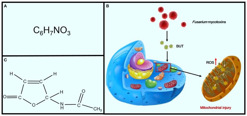
Shi et al.                                                                                             Keshan Disease, Cardiomyopathy, Environment

most cases show a “galloping” heart rhythm and facial edema (6).          Until 1964, white muscle disease caused by Se deficiency was
In recent years, chronic and latent KD have been the most two             found in animals in areas where KD was reported, with the
prevalent types reported. In chronic KD, the onset is slow. The           main pathological changes being similar to those of KD (16).
patient presents with chronic heart failure, ventricular dilation,        Therefore, it was hypothesized that the myocardial lesions of KD
myocardial fibrosis, and a thinning heart wall. In latent KD, the         are associated with Se deficiency.
episode is disguised, and the patient’s cardiac function is fairly           It is known that KD has regional distribution, with population
good [New York Heart Association (NYHA) class I]. Ventricular             and seasonal fluctuations of incidence.
contractions and changes in the right bundle-branch block or
ST–T are common. However, the etiology of KD is unknown (7).
   In the last few decades, numerous investigators have explored          REGIONAL DISTRIBUTION OF KD
the causes of KD, and the main etiology is believed to be selenium
                                                                          KD has been reported in 2,953 townships in 327 counties in 15
(Se) deficiency. This is primarily because KD usually occurs in a
                                                                          provinces from the northeast to the southwest, only in certain
specific region of China and has affected individuals reporting
                                                                          areas in China, a belt zone (Figure 2). The KD-endemic areas
similar Se deficiency conditions (8). Se is a trace mineral that
                                                                          are all remote rural mountain areas where people are the most
plays a crucial role in protecting the body against oxidants,
                                                                          socioeconomically disadvantaged (2). Interestingly, in the KD-
serving as an essential component of several antioxidant enzymes
                                                                          endemic provinces, only a few counties and towns are KD-
such as glutathione peroxidase (GPx) and glutathione reductase.
                                                                          endemic (4, 16). According to the KD Endemic Area Definition
An Se deficiency has also been known to contribute to
                                                                          and Classification, KD-endemic areas are often adjacent to Se-
coxsackievirus B3 (CVB3)–induced myocarditis in acute and
                                                                          rich regions, and the affected areas within the endemic zones
subacute phases of infection. Thus, KD may have a dual etiology,
                                                                          present as small foci (16). KD-endemic and non-endemic areas
with both CVB3 infection and Se deficiency being responsible
                                                                          are usually separated by a hill or a river. Amid KD-endemic
for KD of the heart (9, 10). Recent studies have also indicated
                                                                          areas there are likely to be small non-endemic areas, called
that CYP1A1 and CYP2C19 are highly expressed (ratios ≥2.0)
                                                                          “safety islands.”
in patients with KD (11). These genes belong to the cytochrome
                                                                             In the non-endemic areas, staple foods were mixed, suggesting
P450 isoforms, and their metabolites are biologically active and
                                                                          that people who had been eating a “single type of food” all year
critical for the maintenance of essential bodily functions (12,
                                                                          round, harvested in an endemic area with its particular water-
13). KD is widely considered a multifactorial environment–
                                                                          soil conditions, were liable to suffer from KD (17, 18). The soil
gene interaction complex disease. Since an outbreak of KD in
                                                                          erosion in endemic areas is serious, leading to lower levels of trace
1935, resulting in speculation about and efforts to determine the
                                                                          elements, minerals, vitamins, and amino acids essential for good
etiology of the disease, KD still haunts the health of poor farmers
                                                                          cardiac metabolism (19). Endemic foci can always be explained
in historically serious endemic areas.
                                                                          by Se distribution. The survey found that the Se content of
   KD is regarded as the cardiovascular disease most responsible
                                                                          local residents’ food samples was lower in endemic areas than in
for the high morbidity and mortality rates in China (14).
                                                                          nearby non-endemic areas (20, 21).
All KD-endemic areas are rural, where people are the most
socioeconomically disadvantaged. Patients with KD are usually
among the poorest. Although KD has appeared in China for                  SEASONAL PREVALENCE OF KD
nearly a century, it has received less attention, especially by
Western medical scientists, and only a few articles on the                The incidence of KD is seasonal, with acute KD showing peaks
pathology of KD have been published in English. Herein,                   in winter or spring in northeast rural areas and in summer
we describe the etiological hypothesis, clinical manifestations,          in southwest rural areas in China (22). Chronic and subacute
incidence characteristics, and treatment of KD, to facilitate the         KD shows year-round prevalence. Therefore, some experts have
understanding of and draw more attention to the eradication of            speculated that temperature may accelerate the onset of this
this non-representative and challenging cardiovascular disease.           disease (23). The 2008 cold spell of South China was widely
                                                                          considered to be the most extreme of the past five decades. In an
                                                                          investigation reported in Guangdong, the southern province of
INCIDENCE CHARACTERISTICS                                                 China, three cities were selected for a study of the impact of the
                                                                          cold spell on cardiovascular incidents from January 2006 through
KD was first identified in Keshan County, Heilongjiang Province,          December 2009. This study showed 66.2, 66.5, and 39.7% more
China, during the winter of 1935 (Figure 1). KD was prevalent             deaths than the average for the corresponding days of the three
from 1950 to 1970, with three major outbreaks, in 1959, 1964,             preceding years in the three cities, indicating that temperature
and 1970. In 1956, Guo et al. tested the bacterial content                challenges directly affect cardiovascular health. Similarly, heat
of Fusarium beads in the grains in both endemic and non-                  waves also play a key role in the occurrence of cardiovascular
endemic areas. The content of bacteriocin in the endemic areas            diseases (24). A survey in Sydney, Australia, showed that there
was found to be much higher, and the fungal infection theory              was a statistically significant increase in the ratios of hospital
was proposed (15). Four years later, the coxsackievirus was               admissions for cardiovascular reasons on hot days (25). However,
found through enterovirus isolation and serum antibody tests of           doctors at Jilin Medical College reported that deaths occurring
patients with KD, and the virus infection hypothesis prevailed.           from acute KD and newly formed pathologic changes in the heart

Frontiers in Pediatrics | www.frontiersin.org                         2                                       March 2021 | Volume 9 | Article 576916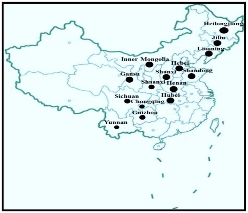
Shi et al.                                                                                                  Keshan Disease, Cardiomyopathy, Environment

  FIGURE 1 | A brief history of KD.

  FIGURE 2 | The main endemic areas of KD in China (highlighted in black).

are found all year round. This indicates that the onset of KD is not             after lactation. In the southwestern area, child-bearing females
dependent only on seasonal changes (26).                                         are the most susceptible. According to recent reports, the KD
                                                                                 incidence rate was also higher in females (2.20%) than in males
                                                                                 (1.98%) (6, 28). The higher KD incidence rates in women might
PREVALENT POPULATION                                                             be caused by their lower immunity compared with that of men
                                                                                 (29). However, others living in the same endemic areas, such
Most patients with KD are among the poorest of peasants (27).                    as foresters, coal miners, and railway workers, who consume
Ninety-nine percent live on grains from their own fields, and                    commercial agricultural products rather than food produced by
80% of these are young females of child-bearing age and infants                  local peasants, did not have KD (30). Thus, KD leads to a vicious

Frontiers in Pediatrics | www.frontiersin.org                                3                                     March 2021 | Volume 9 | Article 576916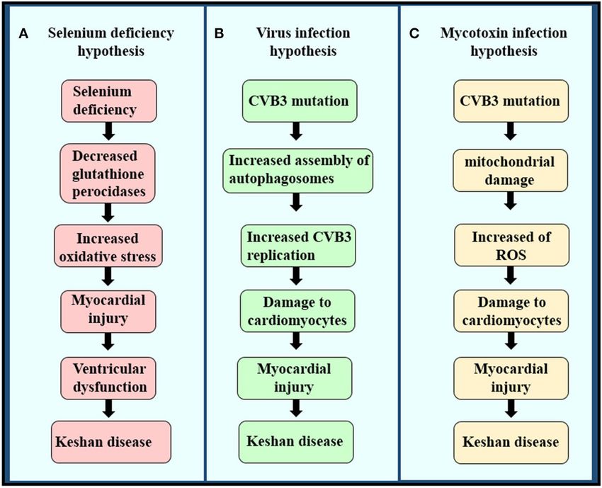
Shi et al. Keshan Disease, Cardiomyopathy, Environment
cycle of poverty and illness in remote mountain regions. Today, sequence, SECI-binding protein 2 (SBP2), SecP43, nuclease-
KD still afflicts poor farmers in remote mountainous endemic sensitive element-binding protein 1 (NSEP1), and DNA-binding
areas, causing great damage to their health and economy. protein B. Se deficiency seriously affects the synthesis of
selenoprotein, and Se is a critical component of a central
antioxidant enzyme (41, 45).
ETIOLOGY OF KD GPx and thioredoxin reductase, two selenoproteins, are
important members of the body’s antioxidant system (46). GPx-1
Although KD has been reported in China for nearly a century,
activity decreases dramatically in Se deficiency and increases
its cause has not been clear until now. Multiple etiological
during Se supplementation. GPx-1 activity is also associated with
hypotheses have been proposed, including intoxication with
GPx-1 (Pro198Leu) polymorphism (47, 48). GPx-1 is the most
mycotoxins or environmental toxins (31, 32), viral infection, and
prevalent of the GPx family and is found in the cytosol of all cells.
trace element deficiency caused by a monotonous diet lacking
The deficiency of GPx-1 always exacerbates ischemia-reperfusion
minerals or vitamins (32), such as magnesium, iron, or thiamin
injury in cardiomyocytes (49, 50). GPx-3 is a key antioxidant
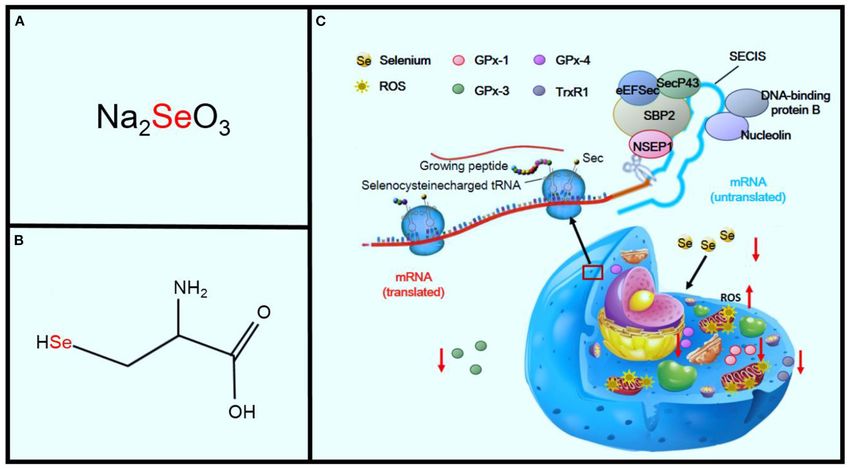
(Figures 3A–C) (18). Among the many underlying causes of
enzyme in the extracellular environment. A deficiency in GPx-3
KD, the hypothesis of Se deficiency is considered the most
leads to increased thrombosis as a result of the increased
convincing (33).
oxidative inactivation of nitric oxide (NO). NO also plays a
key role in the regulation of cardiovascular homeostasis and is
Se DEFICIENCY related to many cardiovascular diseases, including hypertension,
atherosclerosis, stroke, and heart failure (51). There is evidence
In 1964, Chen et al. obtained samples from patients with KD that reactive oxygen species (ROS) are responsible for the
and showed a myocardial pallor due to patchy necrosis and reduced NO bioavailability in cardiac and vascular pathologies
fibrosis, and sarcolemmal outlines indicative of myocytolysis, (52). GPx-4 is an essential regulator of membrane oxidation. A
a morphology similar to that of white muscle disease (34), a deficiency in GPx-4 decreases proatherothrombotic actions of
non-inflammatory degenerative muscle disease observed mostly these peroxidated species. Thioredoxin reductase 1 (TrxR1) is an
in cattle fed cereals and forage from Se-deficient areas (35). isozyme of thioredoxin reductase and is an antioxidant enzyme
Therefore, some Chinese scientists have considered that high KD expressed in the cytoplasm (53). It has catalytic reduction
incidence may also be associated with Se deficiency. Se as an activity and thus plays an important role in the oxidative
indispensable trace element that plays an important role in many stress reaction. Research has shown that TrxR1 knockout mice
aspects of human health, such as antioxidant defenses, thyroid showed increased oxidative stress leading to the emergence
hormone metabolism, and the immune system (36, 37). The of heart diseases. Meanwhile, the expression of TrxR1 was
detection of topsoil Se has suggested that Se concentrations are significantly lower in cases of KD (54, 55). Se deficiency
typically below 0.125 mg/kg, with concentrations of >3 mg/kg significantly promotes oxidant stress and injury, which may
in non-endemic areas. Nutritionists have found that the mean also potentiate the oxidant injury of other contributing
Se contents in hair were 0.200 mg/kg in non-endemic KD zones (38). In 2020, a study (Figure 4C) (56).
investigated serum Se levels in 571 individuals and found that SE deficiency can also lead to other cardiomyopathies, such
levels in those living in KD-endemic areas were only 0.97 µmol/L, as dilated cardiomyopathy (DCM) (57). In 2016, a Galveston
which was significantly lower than the levels in those living (TX) investigator reported a rare case of DCM caused by severe
in non-endemic areas (1.01 µmol/L) (39, 40). Further, results malnutrition combined with Se deficiency in a 14-year-old boy
of urinary Se loading tests showed that the population in the (58). The echocardiogram (echo) showed a globular dilated left
affected areas was Se-poor (38). The Se status of heart, liver, ventricle with a severely depressed systolic function [ejection
kidney, and muscle compared with that of individuals with KD fraction (EF)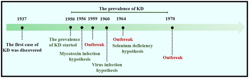
Shi et al.                                                                                                                Keshan Disease, Cardiomyopathy, Environment

  FIGURE 3 | The pathogenesis of the three etiologies of KD. (A) Selenium deficiency hypothesis, (B) virus infection hypothesis, and (C) mycotoxin infection hypothesis.

  FIGURE 4 | Effects of Se deficiency on KD. Panel (A) shows sodium selenite, the primary inorganic form of SE. Panel (B) shows the amino acid, L-selenocysteine.
  Panel (C) shows the sequence of mechanisms leading from Se deficiency to oxidative stress in the cell.

Frontiers in Pediatrics | www.frontiersin.org                                       5                                            March 2021 | Volume 9 | Article 576916
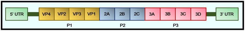
Shi et al.                                                                                              Keshan Disease, Cardiomyopathy, Environment

infection, but the mechanism is still unclear (61). More recent              and are frequently detected in foodstuffs and cereals (74,
studies have suggested that CVB3 may be a contributing factor                75). Butenolide (4-acetamido-4-hydroxy-2-butenoic acid γ-
in KD and that Se deficiency has additional, wide-ranging effects            lactone; BUT) is one of the most common of the Fusarium
(62). Li et al. detected the CVB3 RNA by in situ hybridization               mycotoxins, which are found in cereals from KD-endemic areas
(3). They found that the positive rates in patients with acute,              (Figures 6A,B) (75). It was first isolated in 1971 by Yates from
subacute, and chronic types of KD were 83, 67, and 80%,                      Fusarium tricinctum NRRL 3249 (76). Increasing evidence has
respectively (63). Their results confirmed that CVB3 might                   indicated that treatment of rats with BUT (10 and 20 mg/kg per
play a key role in the pathogenesis of KD (64). In addition,                 day) for 2 months induces serious myocardial injuries, which are
the distribution of the positive signal of CVB3 was related to               characterized by fragmentation of myofibers and necrosis of the
the occurrence of KD. CVB3 in acute and subacute KD has                      myocardium (74). BUT can also induce a variety of cytotoxicities,
been located in the surviving myocardium in or around the                    such as swelling of the mitochondria, fragmentation of cristae,
necrotic focus, but is dispersed in all cardiomyocytes in chronic            and phospholipid bilayer rupture, all of which are similar to
KD (65, 66).                                                                 the characteristics of mitochondrial injuries in patients with KD,
   The genome of CVB3 is a single-stranded, polyadenylated                   leading to speculation that BUT may be one of the etiological
RNA molecule consisting of 7,400 nucleotides (Figure 5). The                 factors for KD and that oxidative damage may be essential for
single open reading frame (ORF) is located on the side of 5′                 myocardial mitochondria (Figure 6C) (77). The mitochondrial
UTR and 3′ UTR and is divided into three areas, P1 to P3 (67).               respiratory chain is the main source of ROS (78). Furthermore,
The P1 region encodes viral capsid proteins (VP1, VP2, VP3,                  excessive generation of ROS will attack unsaturated fatty acids
and VP4) (68). The P2 and P3 regions encode non-structural                   in mitochondrial membrane lipids and exacerbate mitochondrial
viral proteins that are important for processing, replication, and           damage (79). Therefore, the mitochondrial lipid peroxidation
translation of multiple proteins. Some studies have shown that               observed in this study may be the consequence of ROS attack
the 5′ UTR enteroviruses may play a key role in generating or                produced by mitochondria, eventually leading to damage to
maintaining myocardial virulence, since the U→ C mutation of                 cardiomyocytes (80, 81).
nt 234 in CVB3 5′ UTR will lead to the attenuated cardiac toxicity
phenotype in mice (69). Subsequent analysis of various clinical              CLINICAL MANIFESTATIONS
CVB3 isolates and other enteroviruses indicated that nt 234 is
always U regardless of the cardiovirulence phenotype of the virus,           Patients with KD are categorized into four types: acute, subacute,
consistent with 234C being an artificial mutation (70). This result          chronic, and latent (Table 1).
indicates that the mutation of the virus genome might play an
important role in the pathogenesis of KD.                                    Acute KD
   Although the enterovirus infection rate is remarkably high                The onset is rapid. Patients present acute heart failure, severe
in the myocardial tissue of patients with KD, the hypothesis                 arrhythmia, cardiogenic shock, cardiogenic fainting, and acute
of viral infection is still questioned (63, 65), because the                 pulmonary symptoms and may have cardiomegaly (82). Fibrosis
enterovirus infection phenomenon can also be found in patients               is rare. The most common clinical manifestations include
without myocardial damage. Recently, research has reported                   dizziness, loss of appetite, malaise, nausea, chilly sensations,
that, in some cases, such as trace element deficiency, a non-                substernal discomfort, and dyspnea. The main physiological
cardiovirulent strain can change into a cardiovirulent strain                symptoms of this type of KD are those caused by cardiogenic
or, under these conditions, can increase cardiac toxicity. The               shock, such as pallor, venous stenosis of the extremities, and low
accelerated mutations seen in CVB in Se-deficient mice are the               arterial pressure (
Shi et al.                                                                                                           Keshan Disease, Cardiomyopathy, Environment

  FIGURE 5 | Gene structure of coxsackievirus B3.

  FIGURE 6 | Effects of Fusarium mycotoxins on KD. Panel (A) shows the chemical formula of BUT. Panel (B) shows the chemical structural formula of BUT. Panel (C)
  shows the sequence of mechanisms leading from Fusarium mycotoxins to mitochondrial injury in cells.

May in the north and from May to August in the south (87).                           be aware of the disease until death, when it may be discovered
Only a few cases of acute or subacute KD onset have been seen                        only as an incidental finding after a regular physical examination
in recent years, mainly in chronic and latent-type patients found                    or routine autopsy. The most common complaints are dizziness,
in KD-endemic areas (1).                                                             fatigue, and heart palpitations after physical activity or work.
                                                                                     These symptoms are associated with a minor enlarged heart and
Chronic KD                                                                           abnormal ECG changes, including repolarization abnormalities,
This is usually characterized by insidious onset and slow                            such as wide QRS/T angle, QT prolongation, and high QRS non-
progression. Chronic cases may appear spontaneously or as                            dipolar voltage. Ventricular extrasystole and right bundle-branch
the consequence of acute and subacute types (88). The                                block or ST–T changes are common (90). Cardiomegaly is rarely
symptoms of chronic KD usually vary according to the degree                          observed in latent KD, but some minor cardiac abnormalities,
of cardiac insufficiency. The patients present chronic heart                         with compensating heart function, are always involved.
failure, congestive heart failure, dilated chambers of the heart,
and severe myocardial fibrosis. The main manifestations of                           TREATMENT OF KD
the patients include unconsciousness, cough with hemoptysis,
shortness of breath, oliguria, and edema. The changes in ECG                         Acute KD
are even more pronounced in chronic cases, such as atrial                            For acute cases, mortality can be significantly reduced by early
fibrillation and right bundle-branch block with left anterior                        treatment, although there is no specific therapy. Xian Medical
hemiblock (89).                                                                      College first introduced a regimen whereby large doses of
                                                                                     ascorbic acid administered intravenously were found to be an
Latent KD                                                                            effective treatment for cardiogenic shock. The first dose of 5–
The onset is disguised, and these types of patients usually have                     10 g, with not
Shi et al. Keshan Disease, Cardiomyopathy, Environment
TABLE 1 | Clinical manifestations of four types KD. TABLE 2 | Common diuretics and their dosage.
Type Symptoms ECG Name Initial dose Common dose Maximum dose
Acute KD Dizziness, malaise, nausea, Proximal tachycardia Furosemide 20–40 mg/d 20–40 mg 120–160 mg
loss of appetite, chilly diminished voltage of QRS Bumetanide 0.5–1.0 mg/d 1–4 mg 6–8 mg
sensation, projectile waves, prolongation in the
Torasemide 10 mg/d 10–40 mg 100 mg
vomiting, precardia, or atrioventricular conduction
substernal discomfort and time and Q–T intervals, A–V Hydrochlorothiazide 12.5–25 mg, qd 25–50 mg 100 mg
dyspnea Block, right bundle-branch Amiloride 2.5 mg, qd–bid 5–10 mg 20 mg
Cardiogenic shock, pallor, block, changes in S–T Triamterene 25 mg, qd 100 mg 200 mg
constricted veins in the segment and inversion of T
Spironolactone 20 mg, bid 50–100 mg 100 mg
extremities, and low arterial wave
pressure (Shi et al. Keshan Disease, Cardiomyopathy, Environment
Latent KD Despite the fact that Se deficiency was closely related to the
Patients with latent KD should monitor their lifestyle, prevent prevalence of both KD and KBD, oversupplementation can
infection, and balance nutrition. At the same time, it is important have toxic effects on the human body. Characteristic features
for such patients to consult their physicians regularly, so that of Se toxicity include brittle hair, hair loss, and stratified
underlying symptoms can be detected and therapy can be nails, along with an odor of garlic on the breath and skin
initiated as early as possible (21). Additionally, low doses of drugs (19). More acute Se poisoning cases can include pulmonary
and Se could also be used to improve myocardial compensation. challenges and vomiting (105). For the effective prevention
of KD and Se poisoning, supplementary Se intake should be
monitored appropriately.
PREVENTION OF KD Is Se deficiency really the etiology of KD? The hypothesis
that Se deficiency causes KD is still disputed by many
As the causes of KD remain unclear, its elimination or prevention
scholars (7). The epidemiological and clinical features of
depends on economic developments, attention to KD, and the
KD are not fully addressed by Se deficiency alone (21).
improvement of living standards in endemic areas, all of which
First, not all Se-deficient individuals in the endemic area
are difficult to achieve in a short period of time (6, 96). One
have the disease. Moreover, cases of KD have been found
successful program for KD prevention is to supply Se salts to
in some areas with normal Se content, such as Wenshang
the population in KD-endemic areas. Vitamin E has strong
County in Shandong Province. Second, the incidence of
antioxidant properties and is involved in the protection of the
KD cannot be fully controlled by Se supplementation only
membrane structure by preventing the free radical attack of
(20). Further studies investigating the relationship between
unsaturated fatty acids. Nutritional balance should be actively
Se and KD are needed to determine not only Se status
emphasized for those living in KD-endemic areas.
but also genotype in relation to selenoproteins and related
As described previously, KD is most common in rural,
pathways (106).
remote, mountainous areas in China, where the majority of
Although KD has been known for nearly a century, its
the population are low-income, poorly educated, and in poor
therapeutic strategy has been severely hampered by critical
physical health. Most patients with KD are low-income peasants
shortcomings of donor patient samples and pathological models
and underserved in the utilization of medical facilities. Therefore,
(107). Therefore, generating human-cell-based functional and
inexpensive and effective treatment is very important for them.
personalized disease models on a sufficiently large scale to
The endemic areas’ health politics should focus on contributing
meet the demand for drug efficacy and toxicity tests is
to disease control and the interruption of disease transmission by
a major challenge that must be overcome. The generation
(i) relief of treatment costs for KD patients, (ii) improving clinical
of induced pluripotent stem cells (iPSCs) may provide an
diagnosis and case management and sharing information about
efficient platform for pathogenesis research and drug screening.
KD, and (iii) training health personnel to facilitate diagnosis and
With the advancement of hiPSC technologies, attention has
medical care.
been devoted to the study of an organoid, i.e., a three-
dimensional tissue in a dish. Multiple organoids have been
FUTURE PERSPECTIVES successfully developed, such as the myocardium, liver, stomach,
and pancreas (108, 109). Recently, a scaffold-free cardiac
Although the etiology of KD is not yet fully clear, Se organoid differentiation from hiPSCs has been reported,
deficiency is the most convincing hypothesis (41), based which functionally and structurally resembles the lumenized
primarily on the following: first, low soil Se concentrations vascular network in the developing myocardium (110). In
in KD-endemic areas lead to deficient nutritional status of the future, we intend to reprogram myocardial cells from
Se in local residents through the food chain; second, Se patients with KD to iPSCs to construct a KD organoid model,
concentrations in urine, blood, and hair of patients with KD which will contribute to the etiology and drug screening
and living in endemic areas were significantly lower than of KD.
those of healthy people living in non-endemic areas (100); Since the first case of KD was discovered, early diagnosis
third, the Se contents of patients with KD were positively has been the focus of KD research. Several medical methods,
related to the prevalence of KD; and last, the incidence of such as ECG, X-ray, and ultrasonography, are also commonly
KD could be decreased by Se supplementation in KD-endemic used for this purpose. However, their specificities are limited,
areas (101). although ultrasonography is useful for distinguishing KD
Similar to KD, Kashin-Beck disease (KBD) always occurs in from hypertrophic cardiomyopathy, rheumatic heart disease,
areas with a low Se eco-environment (102). This disease further and pericarditis. It is difficult to distinguish early stages of
confirms that Se deficiency seriously affects human health (103). KD from idiopathic DCM, as both show an enlarged heart,
KBD is an endemic chronic osteochondral disease, with main systolic dysfunction, and arrhythmias (40, 111). Currently,
symptoms including symmetrical enlargement of the phalanges, studies have increasingly focused on the use of saliva and
joint deformity, and even dwarfism (104). SE supplementation serum for screening potential lectins as differential diagnostic
measures have been widely implemented in KD- and KBD- biomarkers of patients with KD. Wang et al. demonstrated
endemic areas throughout the country, which has led to a that Solanum tuberosum (potato) lectin (STL) may be used as
significant decrease in the incidence of KD and KBD (42). a biomarker for the diagnosis of male chronic KD and latent
Frontiers in Pediatrics | www.frontiersin.org 9 March 2021 | Volume 9 | Article 576916Shi et al. Keshan Disease, Cardiomyopathy, Environment
KD and female latent KD. Triticum vulgaris (WGA) may be AUTHOR CONTRIBUTIONS
useful in distinguishing between the two different stages (28).
Subsequently, the content of LDH isoenzyme and HRAb in the FW and YS: study concept and design and drafting of the
serum of patients with KD may be used in the diagnosis of KD. manuscript. FW, QY, and XC: critical revision and final approval
KD is mainly caused by repeated injury to the myocardium. of the manuscript. FW, WY, and XT: administrative and
When cardiomyocytes are damaged, LDH isozymes and HRAb technical support. FW: obtained funding and study supervision.
may be released into the blood. Thus far, there is no gold All authors contributed to the article and approved the
standard for the diagnosis of KD, and it is hoped that there submitted version.
will be a major breakthrough in the diagnosis of KD in the
near future. FUNDING
In conclusion, KD remains of great concern in
endemic areas in China, and complete eradication of this This study was supported by the National Natural Science
endemic myocardial disease requires worldwide attention. Foundation of China (31871244), and Natural Science
Thus, an effective approach is essential to address this Foundation of Guangdong Province (2020A1515011314),
challenging disease, for better control strategies, the Natural Science Foundation of Shenzhen City (Grant No.
development of new diagnostic tools and medications, JCYJ20190807101401682), and Medical Health Science
and investigation and treatment of different types and Technology Project of Longgang District Health
of KD. Commission (LGKCYLWS2019000361).
REFERENCES 13. Elbekai RH, El-Kadi AOS. Cytochrome P450 enzymes: central players
in cardiovascular health and disease. Pharmacol Ther. (2006) 112:564–
1. Wang S, Yan R, Wang B, Meng P, Tan W, Guo X. The functional 87. doi: 10.1016/j.pharmthera.2005.05.011
analysis of selenium-related genes and magnesium-related genes 14. Yang F. Keshan disease and mitochondrial cardiomyopathy. Sci China C Life
in the gene expression profile microarray in the peripheral blood Sci. (2006) 49:513–8. doi: 10.1007/s11427-006-2041-y
mononuclear cells of Keshan Disease. Biol Trace Elem Res. (2019) 15. Zhou B, Wang X, Li F, Wang Y, Yang L, Zhen X, et al. Mitochondrial activity
192:3–9. doi: 10.1007/s12011-019-01750-2 and oxidative stress functions are influenced by the activation of AhR-
2. Li Q, Liu M, Hou J, Jiang C, Li S, Wang T. The prevalence of Keshan disease induced CYP1A1 overexpression in cardiomyocytes. Mol Med Rep. (2017)
in China. Int J Cardiol. (2013) 168:1121–6. doi: 10.1016/j.ijcard.2012.11.046 16:174–80. doi: 10.3892/mmr.2017.6580
3. Li Y, Peng T, Yang Y, Niu C, Archard LC, Zhang H. High prevalence 16. Liu Q, Cai J, Gao Y, Yang J, Gong Y, Zhang Z. miR-2954 inhibits PI3K
of enteroviral genomic sequences in myocardium from cases of endemic signaling and induces autophagy and apoptosis in myocardium selenium
cardiomyopathy (Keshan disease) in China. Heart. (2000) 83:696– deficiency. Cell Physiol Biochem. (2018) 51:778–92. doi: 10.1159/000495332
701. doi: 10.1136/heart.83.6.696 17. Gauntt CJ, Trousdale MD, LaBadie DR, Paque RE, Nealon T. Properties of
4. Wang S, Fan Z, Zhou B, Wang Y, Du P, Tan W, et al. Roles of glycoproteins in Coxsackievirus 83 variants which are amyocarditic or myocarditic for mice.
the diagnosis and differential diagnosis of chronic and latent Keshan disease. J Med Virol. (1979) 3:207–20. doi: 10.1158/0008-5472.CAN-17-0307
Molecules. (2017) 22:746. doi: 10.3390/molecules22050746 18. Malumbres M. Cyclin-dependent kinases. Genome Boil. (2014)
5. Zhu Y, Lai B, Niu X, Wei J, Tan W, Wang X. Long-term prognostic 15:122. doi: 10.1186/gb4184
value of major and minor ECG abnormalities in latent Keshan disease 19. MacFarquhar, JK, Broussard DL, Melstrom P, Hutchinson R, Wolkin A,
with suspect chronic Keshan disease. J Epidemiol. (2014) 24:385– Martin C et al. Acute selenium toxicity associated with a dietary supplement.
91. doi: 10.2188/jea.JE20130180 Arch Int Med. (2010) 170:256–61. doi: 10.1001/archinternmed.2009.495
6. Ge K, Xue A, Bai J, Wang S. Keshan disease-an endemic cardiomyopathy 20. Wang S, Lv Y, Wang Y, Du P, Tan W, Lammi MJ, et al. Network analysis of
in China. Virchows Arch A Pathol Anat Histopathol. (1983) 401:1– Se-and Zn-related proteins in the serum proteomics expression profile of the
15. doi: 10.1007/BF00644785 endemic dilated cardiomyopathy Keshan disease. Biol Trace Elem Res. (2017)
7. Vinceti M, Filippini T, Wise, LA. Environmental selenium and 183:40–8. doi: 10.1007/s12011-017-1063-6
human health: an update. Curr Environ Health Rep. (2018) 21. Sun Y, Gao C, Wang X, Yuan Y, Liu Y, Jia J. Serum quantitative
5:464–85. doi: 10.1007/s40572-018-0213-0 proteomic analysis of patients with Keshan disease based on iTRAQ labeling
8. Steinbrenner H, Al-Quraishy S, Dkhil MA, Wunderlich F, Sies H. Dietary technique: a first term study. J Trace Elem Med Biol. (2017) 44:331–
selenium in adjuvant therapy of viral and bacterial infections. Adv Nutr. 8. doi: 10.1016/j.jtemb.2017.09.012
(2015) 6:73–82. doi: 10.3945/an.114.007575 22. Yang GQ, Chen JS, Wen ZM, Ge KY, Zhu LZ, Chen XC, et al.
9. Guillin O, Vindry C, Ohlmann T, Chavatte L. Selenium, selenoproteins The role of selenium in Keshan disease. Adv Nutr Res. (1984) 6:203–
and viral infection. Nutrients. (2019) 11:2101. doi: 10.3390/nu110 31. doi: 10.1007/978-1-4613-2801-8_8
92101 23. Chen C, Chen S, Pang L, Yan H, Luo M, Zhao Q, et al. Analysis of
10. Post M, Lubiński W, Lubiński J, Krzystolik K, Baszuk P, Muszyńska M, et al. the expression of cell division cycle-associated genes and its prognostic
Serum selenium levels are associated with age-related cataract. Ann Agric significance in human lung carcinoma: a review of the literature databases.
Environ Med. (2018) 25:443–8. doi: 10.26444/aaem/90886 Biomed Res Int. (2020) 2020:6412593. doi: 10.1155/2020/6412593
11. Zhou B, He S, Wang XI, Zhen X, Su X, Tan W. Metabolism of 24. Kang SH, Oh IY, Heo J, Lee H, Kim J, Lim WH, et al. Heat, heat
arachidonic acid by the cytochrome P450 enzyme in patients with chronic waves, and out-of-hospital cardiac arrest. Int J Cardiol. (2016) 221:232–
Keshan disease and dilated cardiomyopathy. Biomed Rep. (2016) 4:251– 7. doi: 10.1016/j.ijcard.2016.07.071
5. doi: 10.3892/br.2015.563 25. Parry M, Green D, Zhang Y, Hayen A. Does particulate matter
12. He SL, Tan WH, Zhang ZT, Zhang F, Qu CJ, Lei YX, et al. Mitochondrial- modify the short-term association between heat waves and hospital
related gene expression profiles suggest an important role of PGC-1alpha in admissions for cardiovascular diseases in greater Sydney, Australia? Int
the compensatory mechanism of endemic dilated cardiomyopathy. Exp Cell J Environ Res Public Health. (2019) 16:3270. doi: 10.3390/ijerph1618
Res. 319:2604–16. doi: 10.1016/j.yexcr.2013.07.018 3270
Frontiers in Pediatrics | www.frontiersin.org 10 March 2021 | Volume 9 | Article 576916Shi et al. Keshan Disease, Cardiomyopathy, Environment
26. Chen X, Yang G., Chen J, Chen X, Wen Z, Ge K. Studies on the 46. Wall SB, Wood R, Dunigan K, Li Q, Li R, Rogers LK, et al.
relations of selenium and Keshan disease. Biol Trace Elem Res. (1980) Thioredoxin reductase-1 inhibition augments endogenous glutathione-
2:91–7. doi: 10.1007/BF02798589 dependent antioxidant responses in experimental bronchopulmonary
27. Pandey RM, Gupta R, Misra A, Misra P, Singh V, Agrawal A, et al. dysplasia. Oxid Med Cell Long. (2019) 2019:1–10. doi: 10.1155/2019/
Determinants of urban–rural differences in cardiovascular risk factors in 7945983
middle-aged women in India: a cross-sectional study. Int J Cardiol. (2013) 47. Lei C, Niu X, Wei J, Zhu J, Zhu Y. Interaction of glutathione peroxidase-1
163:157–62. doi: 10.1016/j.ijcard.2011.06.008 and selenium in endemic dilated cardiomyopathy. Clin Chim Acta. (2009)
28. He S, Tan W, Wang S, Wu C, Wang P, Wang B, et al. Genome-wide study 399:102–8. doi: 10.1016/j.cca.2008.09.025
reveals an important role of spontaneous autoimmunity, cardiomyocyte 48. Wang S, Nong X, Yang G. Selenium-rich diet induces myocardial structural
differentiation defect and anti-angiogenic activities in gender-specific and functional abnormalities by activating caspase-9 and caspase-3 in Gpx-
gene expression in Keshan disease. Chin Med J (Engl). (2014) 127:72– 1P198L-overexpression transgenic mice. Med Sci Monit. (2019) 25:61–
8. doi: 10.3760/cma.j.issn.0366-6999.20131167 70. doi: 10.12659/msm.911120
29. Anand SS, Islam S, Rosengren A, Franzosi MS, Steyn K, Yusufali 49. Loscalzo J. Keshan disease, selenium deficiency, and the selenoproteome. N
AH, et al. Risk factors for myocardial infarction in women and men: Engl J Med. (2014) 370:1756–60. doi: 10.1056/NEJMcibr1402199
insights from the INTERHEART study. Eur Heart J. (2008) 29:932– 50. Lubos E, Loscalzo J, Handy DE. Glutathione peroxidase-1 in health and
40. doi: 10.1093/eurheartj/ehn0181 disease: from molecular mechanisms to therapeutic opportunities. Antioxid
30. Yusuf S, Hawken S, Ounpuu S, Dans T, Avezum A, Lanas F, et al. Effect of Redox Signal. (2011) 15:1957–97. doi: 10.1089/ars.2010.3586
potentially modifiable risk factors associated with myocardial infarction in 51. Garoffolo G, Pesce M. Mechanotransduction in the cardiovascular system:
52 countries (the INTERHEART study): case-control study. Lancet. (2004) from developmental origins to homeostasis and pathology. Cells. (2019)
364:937–52. doi: 10.1016/s0140-6736(04)17018-9 8:1607. doi: 10.3390/cells8121607
31. Li C, Niu X, Lei C. Circulating adhesion molecules in patients with Keshan 52. Ritchie RH, Drummond GR, Sobey CG, De Silva TM, Kemp-Harper BK.
disease and their relationship with Coxsackie B virus infection. J Huazhong The opposing roles of NO and oxidative stress in cardiovascular disease.
Univ Sci Technol Med Sci. (2009) 29:173–6. doi: 10.1007/s11596-009-0207-0 Pharmacol Res. (2017) 116:57–69. doi: 10.1016/j.phrs.2016.12.017
32. Lin NF, Tang J, Bian JM. Geochemical environment and health 53. Prigge JR, Coppo L, Martin SS, Ogata F, Miller CG, Bruschwein MD,
problems in China. Environ Geochem Health 2004. (2004) et al. Hepatocyte hyperproliferation upon liver-specific co-disruption of
26:81–8. doi: 10.1023/b:egah.0000020987.74065.1d thioredoxin-1, thioredoxin reductase-1, and glutathione reductase. Cell Rep.
33. Zhu YH, Wang, XF, Yang G, Wei J, Tan WH, Wang LX, et al. Efficacy (2017) 19:2771–81. doi: 10.1016/j.celrep.2017.06.019
of long-term selenium supplementation in the treatment of chronic 54. Yamamoto M, Yang G., Hong C, Liu J, Holle E, Yu X, et al.
Keshan disease with congestive heart failure. Curr Med Sci. (2019) 39:237– Inhibition of endogenous thioredoxin in the heart increases
42. doi: 10.1007/s11596-019-2025-3 oxidative stress and cardiac hypertrophy. J Clin Invest. (2003)
34. Chen X, Xu J, Liu D, Sun Y, Qian G, Xu S, et al. The aggravating 112:1395–406. doi: 10.1172/JCI1770010.1172/JCI200317700
effect of selenium deficiency on T-2 toxin-induced damage on primary 55. Arnér ESJ. Targeting the selenoprotein thioredoxin reductase
cardiomyocyte results from a reduction of protective autophagy. Chem Biol 1 for anticancer therapy. Adv Cancer Res. (2017) 136:139–
Interact. (2019) 300:27–34. doi: 10.1016/j.cbi.2019.01.009 51. doi: 10.1016/bs.acr.2017.07.005
35. Löfstedt J. White muscle disease of foals. Vet Clin North Amer Equine Pract. 56. Pei J, Fu W, Yang L, Zhang Z, Liu Y. Oxidative stress is involved in the
(1997) 13:169–85. doi: 10.1016/s0749-0739(17)30262-6 pathogenesis of Keshan disease (an endemic dilated cardiomyopathy) in
36. Wang, SQ, Niu XL, Liu ZW, Zhu, YH, Gao DF. Selenium China. Oxid Med Cell Long. (2013) 2013:1–5. doi: 10.1155/2013/474203
deficiency is associated with endoplasmic reticulum stress in a 57. Bomer N, Grote Beverborg N, Hoes MF, Streng KW, Vermeer M, Dokter
rat model of cardiac malfunction. Biol Trace Elem Res. (2013) MM, et al. Selenium and outcome in heart failure. Eur J Heart Fail. (2019)
156:196–201. doi: 10.1007/s12011-013-9834-1 22:1415–23. doi: 10.1002/ejhf.1644
37. Cheng WH, Ho YS, Valentine BA, Ross DA, Combs Jr GF, Lei XG. 58. Dasgupta S, Aly AM. Dilated cardiomyopathy induced by chronic
Cellular glutathione peroxidase is the mediator of body selenium to protect starvation and selenium deficiency. Case Rep Pediatr. (2016) 2016:1–
against paraquat lethality in transgenic mice1. J Nutr. (1998) 128:1070– 4. doi: 10.1155/2016/8305895
6. doi: 10.1093/jn/128.7.1070 59. Li Z, Wei J, Zhang Y, Li G, Zhu H, Lei N, et al. Risk factors for Keshan disease:
38. Fairweather-Tait SJ, Bao Y, Broadley MR, Collings R, Ford D, Hesketh JE, a prospective cohort study protocol of gut flora. BMC Cardiovasc Disord.
et al. Selenium in human health and disease. Antioxid Redox Signal. (2011) (2020) 20. doi: 10.1186/s12872-020-01765-x
14:1337–83. doi: 10.1089/ars.2010.3275 60. Kuffner M, Pawlak A, Przybylski M. Viral infection of the
39. Liu X, Wang Y, Han S, Zhang Y, Zou Y, Su S, et al. A spatial ecological study heart: pathogenesis and diagnosis. Pol J Microbiol. (2017)
on serum selenium and Keshan disease in Heilongjiang province, China. Biol 65:391–8. doi: 10.5604/17331331.1227664
Trace Elem Res. (2020). doi: 10.1007/s12011-020-02478-0. [Epub ahead of 61. Song JH, Ahn JH, Kim SR, Cho S, Hong EH, Kwon BE, et al.
print]. Manassantin B shows antiviral activity against coxsackievirus B3 infection
40. Xiang Y, Xu Q, Tan W, He S, Shi X, Zhang W, et al. Serum biomarkers of by activation of the STING/TBK-1/IRF3 signalling pathway. Sci Rep. (2019)
Keshan disease assessed using a protein profiling approach based on ClinProt 9:9413. doi: 10.1038/s41598-019-45868-8
technique. Prot J. (2014) 33:344–53. doi: 10.1007/s10930-014-9567-9 62. Levander OA, Beck MA. Interacting nutritional and infectious etiologies
41. Zhou H, Wang T, Li Q, Li D. Prevention of Keshan disease by selenium of Keshan disease. Insights from coxsackie virus B-induced myocarditis
supplementation: a systematic review and meta-analysis. Biol Trace Elem Res. in mice deficient in selenium or vitamin E. Biol Trace Elem Res. (1997)
(2018) 186:98–105. doi: 10.1007/s12011-018-1302-5 56:5–21. doi: 10.1007/BF02778980
42. Whanger PD. Selenium interactions with carcinogens. Fundam Appl Toxicol. 63. Beck MA, Kolbeck PC, Rohr LH, Shi Q, Morris VC, Levander OA. Benign
(1983) 3:424–30. doi: 10.1016/s0272-0590(83)80016-5 human enterovirus becomes virulent in selenium-deficient mice. J Med Virol.
43. Kuganesan M, Samra K, Evans E, Singer M, Dyson A. Selenium and (1994) 43:166–70. doi: 10.1002/jmv.1890430213
hydrogen selenide: essential micronutrient and the fourth gasotransmitter? 64. Beck MA, Shi Q, Morris VC, Levander OA. Rapid genomic evolution
Intensive Care Med Exp. (2019) 7:71. doi: 10.1186/s40635-019-0281-y of a non-virulent Coxsackievirus B3 in selenium-deficient mice results
44. Rose AH, Hoffmann PR. Part–selenoproteins and cardiovascular stress. in selection of identical virulent isolates. Nat Med. (1995) 1:433–
Thromb haemost. (2015) 113:494–504. doi: 10.1160/TH14-07-0603 6. doi: 10.1038/nm0595-433
45. Chen CS, Stadtman TC. Selenium-containing tRNAs from Clostridium 65. Ren LQ, Li X-J, Li GS, Zhao ZT, Sun B, Sun F. Coxsackievirus B3 infection
sticklandii: cochromatography of one species with L-prolyl-tRNA. Proc Natl and its mutation in Keshan disease. World J Gastroenterol. (2004) 10:3299–
Acad Sci USA. (1980) 77:1403–7. doi: 10.1073/pnas.77.3.1403 302. doi: 10.3748/wjg.v10.i22.3299
Frontiers in Pediatrics | www.frontiersin.org 11 March 2021 | Volume 9 | Article 576916Shi et al. Keshan Disease, Cardiomyopathy, Environment
66. Dan M, Chantler, JK. A genetically engineered attenuated Coxsackievirus 85. Utiger RD. Kashin-Beck disease–expanding the spectrum of
B3 strain protects mice against lethal infection. J Virol. (2005) 79:9285– iodine-deficiency disorders. N Engl J Med. (1998) 339:1156–
95. doi: 10.1128/jvi.79.14.9285-9295.2005 8. doi: 10.1056/NEJM199810153391611
67. Okonko IO, Adebiyi AA, Ogah OS, Adu FD. Enteroviruses as a possible 86. Sun Y, Gao C, Wang X, Liu Y. Preliminary quantitative proteomics
cause of hypertension, dilated cardiomyopathy (DCM) and hypertensive analysis in chronic and latent Keshan disease by iTRAQ labeling approach.
heart failure (HHF) in South western Nigeria. Afr Health Sci. (2014) Oncotarget. (2017) 8:105761–74. doi: 10.18632/oncotarget.22397
13:1098. doi: 10.4314/ahs.v13i4.34 87. Li M, Wu M, Qin Y, Liu H, Tu C, Shen B, et al. Differentially expressed
68. Garmaroudi FS, Marchant D, Hendry R, Luo H, Yang D, Ye X, et al. serum proteins in children with or without asthma as determined using
Coxsackievirus B3 replication and pathogenesis. Future Microbiol. (2015) isobaric tags for relative and absolute quantitation proteomics. PeerJ. (2020)
10:629–53. doi: 10.2217/fmb.15.5 8:e9971. doi: 10.7717/peerj.9971
69. Huber SA, Gauntt CJ, Sakkinen P. Enteroviruses and 88. Mirlean N, Seus-Arrache ER, Vlasova O. Selenium deficiency in subtropical
myocarditis: viral pathogenesis through replication, cytokine littoral pampas: environmental and dietary aspects. Environ Geochem Health.
induction, and immunopathogenicity. Adv Virus Res. (1998) (2017) 40:543–56. doi: 10.1007/s10653-017-9951-4
51:35-80. doi: 10.1016/s0065-3527(08)60783-6 89. Li Q, Li XZ, Wang T, Zhou LW, Feng HQ, Gao L, et al. Selenoprotein P and
70. Tao Z, Song Y., Li Y, Liu Y, Jiang P, Lin X, et al. Coxsackievirus B3, Yunnan endemic sudden cardiac death—an ecological study. Biol Trace Elem
Shandong Province, China, 1990–2010. Emerg Infect Dis. (2012) 18:1865– Res. (2012) 151:14–17. doi: 10.1007/s12011-012-9530-6
7. doi: 10.3201/eid1811.120090 90. Yang J, Wang T, Wu C, Liu C. Selenium level surveillance for the year
71. Beck MA, Kolbeck PC, Rohr LH, Shi Q, Morris VC, Levander OA. 2007 of Keshan disease in endemic areas and analysis on surveillance
Vitamin E deficiency intensifies the myocardial injury of coxsackievirus results between 2003 and 2007. Biol Trace Elem Res. (2010) 138:53–
B3 infection of mice. J Nutr. (1994) 124:345–58. doi: 10.1093/jn/ 9. doi: 10.1007/s12011-010-8609-1
124.3.345 91. Zozina VI, Covantev S, Goroshko OA, Krasnykh LM, Kukes
72. Sun S. Chronic exposure to cereal mycotoxin likely citreoviridin VG. Coenzyme Q10 in cardiovascular and metabolic diseases:
may be a trigger for Keshan disease mainly through oxidative stress current state of the problem. Curr Cardiol Rev. (2018) 14:164–
mechanism. Med Hypothes. (2010) 74:841–2. doi: 10.1016/j.mehy.2009. 74. doi: 10.2174/1573403x14666180416115428
11.043 92. Andrews R, Charlesworth A, Evans A, Cowley AJ. A double-blind, cross-over
73. Ge K, Yang G. The epidemiology of selenium deficiency in the comparison of the effects of a loop diuretic and a dopamine receptor agonist
etiological study of endemic diseases in China. Am J Clin Nutr. (1993) as first line therapy in patients with mild congestive heart failure. Eur Heart
57:259S−63S. doi: 10.1093/ajcn/57.2.259S J. (1997) 18:852–7. doi: 10.1093/oxfordjournals.eurheartj.a015351
74. Creppy EE. Update of survey, regulation and toxic effects of mycotoxins 93. Vazir A, Cowie MR. Decongestion: diuretics and other
in Europe. Toxicol Lett. (2002) 127:19–18. doi: 10.1016/s0378-4274(01) therapies for hospitalized heart failure. Ind Heart J. (2016)
00479-9 68:S61–8. doi: 10.1016/j.ihj.2015.10.386
75. Yoshizawa T, Yamashita A, Luo Y. Fumonisin occurrence in corn 94. Liu M, Ai J, Feng J, Zheng J, Tang K, Shuai Z, et al. Effect of paeoniflorin on
from high- and low-risk areas for human esophageal cancer in China. cardiac remodeling in chronic heart failure rats through the transforming
Appl Environ Microbiol. (1994) 60:1626–9. doi: 10.1128/AEM.60.5.1626-16 growth factor β1/Smad signaling pathway. Cardiovasc Diagn Ther. (2019)
29.1994 9:272–80. doi: 10.21037/cdt.2019.06.01
76. Wang YM, Liu JB, Peng SQ. Effects of Fusarium mycotoxin butenolide on 95. Cheng CJ, Rodan AR, Huang CL. Emerging targets of diuretic therapy. Clin
myocardial mitochondria in vitro. Toxicol Mech Methods. (2009) 19:79– Pharmacol Therap. (2017) 102:420–35. doi: 10.1002/cpt.754
85. doi: 10.1080/15376510802322802 96. Yancy CW, Jessup M, Bozkurt B, Butler J, Casey Jr DE, Drazner MH,
77. Liu JB, Wang YM, Peng SQ, Han G, Dong YS, Yang HY, et al. et al. 2013 ACCF/AHA guideline for the management of heart failure:
Toxic effects of Fusarium mycotoxin butenolide on rat myocardium executive summary: a report of the American College of Cardiology
and primary culture of cardiac myocytes. Toxicon. (2007) 50:357– Foundation/American Heart Association Task Force on practice guidelines.
64. doi: 10.1016/j.toxicon.2007.04.014 Circulation. (2013) 128:1810–52. doi: 10.1161/CIR.0b013e31829e8807
78. Turrens JF. Mitochondrial formation of reactive oxygen species. J. Physiol. 97. DeFilippis, EM Desai, AS. Treatment of hyperkalemia in heart failure. Curr
(2003) 552:335–44. doi: 10.1113/jphysiol.2003.049478 Heart Fail Rep. (2017) 14:266–74. doi: 10.1007/s11897-017-0341-0
79. Nezelof C, Bouvier R, Dijoud F. Multifocal myocardial necrosis: a 98. Granger CB, McMurray JVJ, Yusuf S, Held P, Michelson EL, Olofsson
distinctive cardiac lesion in cystic fibrosis, lipomatous pancreatic B, et al. Effects of candesartan in patients with chronic heart failure
atrophy, and Keshan disease. Pediatr Pathol Mol Med. (2002) and reduced left-ventricular systolic function intolerant to angiotensin-
21:343–52. doi: 10.1080/0277093029005655 converting-enzyme inhibitors: the CHARM-Alternative trial. Lancet. (2003)
80. Liu J, Shen HM, Ong CN. Role of intracellular thiol depletion, mitochondrial 362:772–6. doi: 10.1016/s1062-1458(03)00562-2
dysfunction and reactive oxygen species in Salvia miltiorrhiza-induced 99. Sarwar CMS, Papadimitriou L, Pitt B, Piña I, Zannad F, Anker SD, et al.
apoptosis in human hepatoma HepG2 cells. Life Sci. (2001) 69:1833– Hyperkalemia in heart failure. J Amer College Cardiol. (2016) 68:1575–
50. doi: 10.1016/s0024-3205(01)01267-x 89. doi: 10.1016/j.jacc.2016.06.060
81. Cassarino DS, Parks LK, Parker Jr WD, Bennett Jr JP. The 100. Ye Y, Qu J, Pu Y, Rao S, Xu F, Wu C, et al. Selenium biofortification
Parkinsonian neurotoxin MPP+ opens the mitochondrial permeability of crop food by beneficial microorganisms. J Fungi. (2020)
transition pore and releases cytochrome c in isolated mitochondria 6:59. doi: 10.3390/jof6020059
via an oxidative mechanism. Biochim Biophys Acta. (1999) 101. Beck MA, Levander LO, Handy J. Selenium deficiency and viral infection. J
1453:49–62. doi: 10.1016/s0925-4439(98)00083-0 Nutr. (2003) 133:1463S−7S. doi: 10.1093/jn/133.5.1463S
82. Crompton M. On the involvement of mitochondrial intermembrane 102. Xie D, Liao Y, Yue J, Zhang C, Wang Y, Deng C, et al. Effects of five
junctional complexes in apoptosis. Curr Med Chem. (2003) 10:1473– types of selenium supplementation for treatment of Kashin-Beck disease in
84. doi: 10.2174/0929867033457197 children: a systematic review and network meta-analysis. BMJ Open. (2018)
83. Aggarwal R, Harling L, Efthimiou, E, Darzi A, Athanasiou T, Ashrafian 8:e017883. doi: 10.1136/bmjopen-2017-017883
H. The effects of bariatric surgery on cardiac structure and function: a 103. Liu H, Yu F, Shao W, Ding D, Yu Z, Chen F, et al. Associations between
systematic review of cardiac imaging outcomes. Obes Surg. (2015) 26:1030– selenium content in hair and Kashin-Beck disease/Keshan disease in children
40. doi: 10.1007/s11695-015-1866-5 in Northwestern China: a prospective cohort study. Biol Trace Elem Res.
84. Alfthan G, Xu GL, Tan WH, Aro A, Wu J, Yang YX, et al. Selenium (2017) 184:16–23. doi: 10.1007/s12011-017-1169-x
supplementation of children in a selenium-deficient area in China: blood 104. Zhang Y, He Y, Zhang D, Zhang M, Wang M, Zhang Y, et al. Death of
selenium levels and glutathione peroxidase activities. Clin Trial. (2000) chondrocytes in Kashin-Beck disease: apoptosis, necrosis or necroptosis? Int
73:113–25. doi: 10.1385/BTER:73:2:113 J Exp Pathol. (2018) 99:312–22. doi: 10.1111/iep.12297
Frontiers in Pediatrics | www.frontiersin.org 12 March 2021 | Volume 9 | Article 576916Shi et al. Keshan Disease, Cardiomyopathy, Environment
105. Zhang X, Wang T, Li S, Ye C, Hou J, Li Q, et al. A spatial ecology study of functional human cardiac organoids. Biomaterials. (2017)
of Keshan disease and hair selenium. Biol Trace Elem Res. (2018) 189:370– 142:112–23. doi: 10.1016/j.biomaterials.2017.07.021
8. doi: 10.1007/s12011-018-1495-7 111. Yu WH. A study of nutritional and bio-geochemical factors in the occurrence
106. Lech T. Suicide by sodium tetraoxoselenate(VI) poisoning. Forensic Sci Int. and development of Keshan disease. Jpn Circ J. (1982) 46:1201–7.
(2002) 130:44–8. doi: 10.1016/s0379-0738(02)00303-1
107. Steinnes E. Soils and geomedicine. Environ Geochem Health. (2009) 31:523– Conflict of Interest: The authors declare that the research was conducted in the
35. doi: 10.1007/s10653-009-9257-2 absence of any commercial or financial relationships that could be construed as a
108. Lancaster MA, Knoblich JA. Organogenesis in a dish: modeling potential conflict of interest.
development and disease using organoid technologies. Science. (2014)
345:1247125. doi: 10.1126/science.1247125 Copyright © 2021 Shi, Yang, Tang, Yan, Cai and Wu. This is an open-access article
109. Tanaka A, Yuasa S, Node K, Fukuda K. Cardiovascular disease distributed under the terms of the Creative Commons Attribution License (CC BY).
modeling using patient-specific induced pluripotent stem cells. The use, distribution or reproduction in other forums is permitted, provided the
Int J Mol Sci. (2015) 16:18894–922. doi: 10.3390/ijms1608 original author(s) and the copyright owner(s) are credited and that the original
18894 publication in this journal is cited, in accordance with accepted academic practice.
110. Richards DJ, Coyle RC, Tan Y, Jia J, Wong K, Toomer K, et al. No use, distribution or reproduction is permitted which does not comply with these
Inspiration from heart development: biomimetic development terms.
Frontiers in Pediatrics | www.frontiersin.org 13 March 2021 | Volume 9 | Article 576916You can also read