Structure of the chromatin remodelling enzyme Chd1 bound to a ubiquitinylated nucleosome - bioRxiv
←
→
Page content transcription
If your browser does not render page correctly, please read the page content below
bioRxiv preprint first posted online Mar. 30, 2018; doi: http://dx.doi.org/10.1101/290874. The copyright holder for this preprint
 (which was not peer-reviewed) is the author/funder, who has granted bioRxiv a license to display the preprint in perpetuity.
                                It is made available under a CC-BY-NC 4.0 International license.

             Structure of the chromatin remodelling enzyme Chd1 bound
                                       to a ubiquitinylated nucleosome.

                    Ramasubramanian Sundaramoorthy1, Amanda L. Hughes1, Hassane El-
                                Mkami2, David Norman3 and Tom Owen-Hughes1.

            1
                Centre for Gene Regulation and Expression, School of Life Sciences, University of
                                                Dundee, Dundee, DD1 5EH, UK.

                2
                    School of Physics and Astronomy, University of St Andrews, St Andrews KY16
                                                                9SS, UK

                      3
                       Nucleic Acids Structure Research Group, University of Dundee, Dundee
                                                             DD1 5EH, UK.

                                                                      1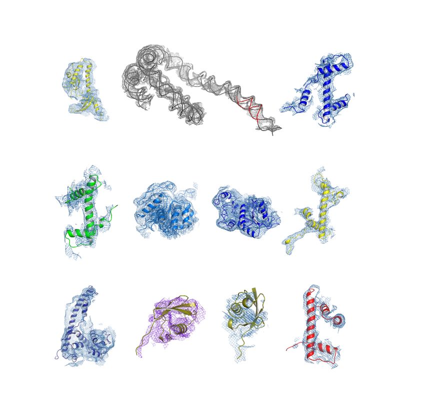
bioRxiv preprint first posted online Mar. 30, 2018; doi: http://dx.doi.org/10.1101/290874. The copyright holder for this preprint
 (which was not peer-reviewed) is the author/funder, who has granted bioRxiv a license to display the preprint in perpetuity.
                                It is made available under a CC-BY-NC 4.0 International license.

          Abstract

          ATP-dependent chromatin remodelling proteins represent a diverse family of
          proteins that share ATPase domains that are adapted to regulate protein-DNA
          interactions. Here we present structures of the yeast Chd1 protein engaged
          with nucleosomes in the presence of the transition state mimic ADP-beryllium
          fluoride. The path of DNA strands through the ATPase domains indicates the
          presence of contacts conserved with single strand translocases and additional
          contacts with both strands that are unique to Snf2 related proteins. The
          structure provides connectivity between rearrangement of ATPase lobes to a
          closed, nucleotide bound state and the sensing of linker DNA. Two turns of
          linker DNA are prised off the surface of the histone octamer as a result of
          Chd1 binding, and both the histone H3 tail and ubiquitin conjugated to lysine
          120 are re-orientated towards the unravelled DNA. This indicates how
          changes to nucleosome structure can alter the way in which histone epitopes
          are presented.

          Introduction

          The extended family of ATPases related to the yeast Snf2 protein act to alter
          DNA protein interactions (Flaus et al., 2006; Narlikar et al., 2013). They act on
          a diverse range of substrates. For example, while the Mot1 protein acts on
          complexes between the TATA box binding protein BP and DNA (Wollmann et
          al., 2011), the Snf2 protein carries out ATP dependent nucleosome disruption
          (Cote et al., 1994). At the heart of all these proteins are paired domains
          capable of rearranging during the ATP hydrolysis cycle to create a ratchet like
          motion along DNA in single base increments (Clapier et al., 2017; Gu & Rice,
          2010; Velankar et al., 1999).

                                                                      2
bioRxiv preprint first posted online Mar. 30, 2018; doi: http://dx.doi.org/10.1101/290874. The copyright holder for this preprint
 (which was not peer-reviewed) is the author/funder, who has granted bioRxiv a license to display the preprint in perpetuity.
                                It is made available under a CC-BY-NC 4.0 International license.

          The yeast Chd1 protein is a member of this protein family and acts to
          organise nucleosomes over coding regions (Gkikopoulos et al., 2011;
          Ocampo et al., 2016; Pointner et al., 2012; Tran et al., 2000). Consistent with
          this Chd1 is known to interact with elongation factors including the Spt4-Spt5
          proteins, Paf1 and FACT (Kelley et al., 1999; Krogan et al., 2002; Simic et al.,
          2003).The partially redundant functions of Chd1 and Isw1 in organising
          nucleosomes over coding regions are in turn required to prevent histone
          exchange and non-coding transcription (Hennig et al., 2012; Radman-Livaja
          et al., 2012; Smolle et al., 2012).

          In addition to the positioning of nucleosomes, the distribution of many histone
          modifications is ordered with respect to promoters (Liu et al., 2005; Mayer et
          al., 2010). For example histone H3 K4 methylation is frequently observed at
          promoters, while histone H3 K79 and K36 trimethylation are detected in
          coding regions (Kizer et al., 2005; Li et al., 2003; Pokholok et al., 2005).
          Histone H2B is also observed to be ubiquitinylated within coding regions
          (Fleming et al., 2008; Xiao et al., 2005). Ubiquitinylation of histone H2B at
          lysine 123 in budding yeast, H2B K120 (H2BK120ub) in mammals, is
          dependent on the E2 ligase Rad6 (Robzyk et al., 2000) and the E3 ligase
          Bre1 (Hwang et al., 2003; Wood et al., 2003) and removed by the
          deubiquitinases Ubp8 and Ubp10 (Bonnet et al., 2014; Schulze et al., 2011;
          Wyce et al., 2007). A specific reader of H2BK120ub has not been identified.
          However, H2BK120ub does assist the histone chaperone FACT in enabling
          transcription through chromatin (Pavri et al., 2006), and has been found to be
          required for methylation of histone H3 K4 and K79 (Sun & Allis, 2002). An
          intriguing aspect of H2BK120ub is that while mutation of the writer enzymes
          or K120 itself disrupts nucleosome organisation, deletion of the
          deubiquitinylases increases chromatin organisation (Batta et al., 2011). One
          way in which H2BK120 may influence nucleosome organisation is via effects
          on enzymes responsible for chromatin organisation. Consistent with this
          H2BK120 increases nucleosome repositioning mediated by Chd1
          (Levendosky et al., 2016).

                                                                      3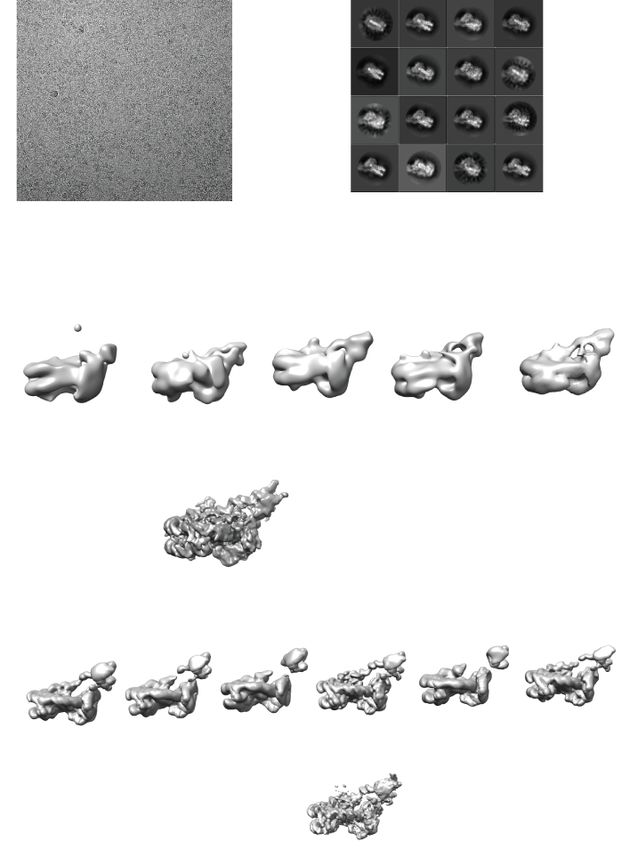
bioRxiv preprint first posted online Mar. 30, 2018; doi: http://dx.doi.org/10.1101/290874. The copyright holder for this preprint
 (which was not peer-reviewed) is the author/funder, who has granted bioRxiv a license to display the preprint in perpetuity.
                                It is made available under a CC-BY-NC 4.0 International license.

          Yeast Chd1 serves as a useful paradigm in that it functions predominantly as
          a single polypetide. In addition, the catalytic core of the enzyme has been
          crystallised in association with the adjacent tandem chromodomains (Hauk et
          al., 2010). Similarly the C-terminal region of the protein has been crystalized
          revealing that this region includes SANT and SLIDE domains that comprise
          the DNA binding domain (DNABD) (Ryan et al., 2011b; Sharma et al., 2011)
          and are also present in ISWI proteins (Grune et al., 2003). Chd1 enzyme
          engages nucleosomes in a conformation in which the SANT and SLIDE
          domains bind linker DNA, while the ATPase domains engage DNA at super
          helical location (SHL) 2 (Nodelman et al., 2017; Sundaramoorthy et al., 2017).
          Higher resolution structures of both Chd1 (Farnung et al., 2017) and Snf2
          (Liu et al., 2017) show that the ATPase domains make contacts with DNA via
          residues that are conserved in ancestral single stranded ATPases and some
          unique to Snf2 related ATPases. The binding of the Chd1 DNABD unravels
          two turns of DNA from the surface of nucleosomes in a nucleotide stimulated
          reaction (Farnung et al., 2017; Sundaramoorthy et al., 2017). Here we report
          a structure for the yeast Chd1 protein in association with a nucleosome,
          bearing modifications that are found to occur within coding regions, where
          Chd1 is known to act. Interestingly, nucleosomal epitopes are observed to be
          reconfigured specifically on the side of the nucleosome on which DNA is
          unwrapped. This indicates the potential for changes to nucleosome structure
          to reconfigure the way in which histone epitopes are presented.

          Results

          The structure of Chd1 nucleosome complexes

          As Chd1 functions on transcribed genes, it is of interest to understand the
          interplay between Chd1 and histone modifications observed in coding region
          chromatin. As a result nucleosomes were prepared in which histone H3 K36
          was alkylated to mimic trimethylation (Figure 1 – Figure supplement 1) and
          H2B cross-linked to ubiquitin (Figure 1 – Figure supplement 2). Conditions
          were established to favour binding of a single Chd1 to modified nucleosomes
          that included an asymmetric linker DNA extension of 14bp (Figure 1 – figure
                                                                      4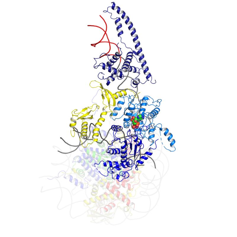
bioRxiv preprint first posted online Mar. 30, 2018; doi: http://dx.doi.org/10.1101/290874. The copyright holder for this preprint
(which was not peer-reviewed) is the author/funder, who has granted bioRxiv a license to display the preprint in perpetuity.
It is made available under a CC-BY-NC 4.0 International license.
supplement 2B) in the presence of ADP-BeF. Purified complexes were frozen
onto EM grids.
2D classification of some 893000 particles revealed 16 classes in which
nucleosomes with the Chd1 molecule attached could be identified (Figure 1 –
Figure supplement 3B). Initial 3D classification resulted in 5 related classes
(Figure 1 - Figure supplement 3C). Three of these were combined and
reclassified as six classes, one of which was selected for refinement. This
resulted in the generation of a map with a resolution of 4.5Å (FSC 0.143)
(Figure 1 – Figure supplement 4A). The resolution varies within the map, with
resolution close to 4 Å in the region occupied by the nucleosome and ATPase
lobes and lower resolution in the vicinity of the DNABD and ubiquitin peptides
(Figure 1 – figure supplement 4B). The nucleosome particles exhibited a
preferred orientation which may limit the resolution (Figure 1 Figure
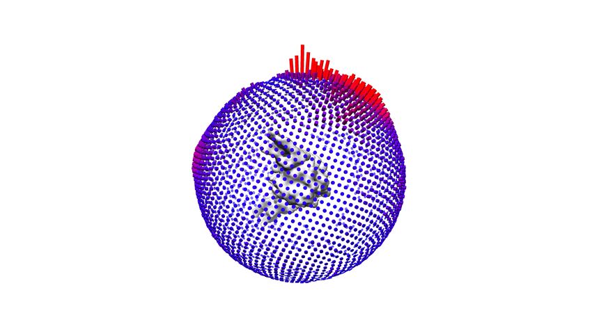
supplement 4C). A structural model was generated to fit the density map
making use of the structures of a nucleosome assembled on the 601 DNA
sequence, Chd1 chromoATPase, and DNABD (Figure 1). The fit for individual
components of the structure to the electron density is shown in Figure 1 -
Figure supplement 5.
The overall organisation of Chd1 is similar to that observed previously by cryo
EM (Farnung et al., 2017; Sundaramoorthy et al., 2017) and directed cross-
linking (Nodelman et al., 2017). The ATPase domains are bound at the SHL-2
location. Of the two SHL2 locations within nucleosomes, the bound site is in
closest proximity to SANT-SLIDE domain bound linker DNA in physical space,
but distal on the unwrapped linear DNA sequence (Figure 1). Chd1
predominantly contacts the nucleosome via contacts with DNA, via the
DNABD in the linker and ATPase lobes at SHL2, contacts with histones are
limited to the histone H3 and H4 N-terminal regions discussed below.
We previously showed that Chd1 binding results in nucleotide-dependent
unwrapping of nucleosomal DNA resulting from the interaction of the DNABD
with linker DNA (Sundaramoorthy et al., 2017). The higher resolution of the
current structure shows that precisely two turns of nucleosomal DNA are
5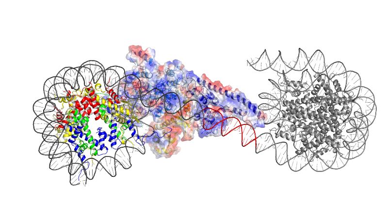
bioRxiv preprint first posted online Mar. 30, 2018; doi: http://dx.doi.org/10.1101/290874. The copyright holder for this preprint
 (which was not peer-reviewed) is the author/funder, who has granted bioRxiv a license to display the preprint in perpetuity.
                                It is made available under a CC-BY-NC 4.0 International license.

          unravelled (Figure 1). The extent of DNA unwrapping observed here when
          Chd1 is bound to nucleosomes flanked by a 14 base pair linker DNA is
          identical to that observed when Chd1 is bound to the opposite surface of the
          601 nucleosome positioning sequence with a 63 base pair linker (Farnung et
          al., 2017). As the interaction of histones with the two sides of the 601
          positioning sequence differ quite dramatically (Chua et al., 2012; Hall et al.,
          2009; Levendosky et al., 2016; Ngo et al., 2015), this suggests that the extent
          of unwrapping is dominated by the properties of Chd1 rather than the affinity
          of DNA for the octamer. The path of this unwrapped DNA is oriented away
          from the plane of the wrapped DNA gyre and is kinked at the location where
          contacts are made with the SANT-SLIDE domains (Figure 1). Other than DNA
          unwrapping we do not detect additional changes in the organisation of DNA
          on Chd1 bound nucleosomes at this resolution.

          The orientation of the DNABD is critical in determining the extent of DNA
          unwrapping. The only contacts detected between the DNABD and the
          remainder of Chd1 are contacts with the chromodomains (Figure 2). The first
          of these is the interaction between K329 of chromodomain II and D1201
          P1202 in the SLIDE component of the DNABD and has been observed
          previously (Farnung et al., 2017; Nodelman et al., 2017)(Figure 2 – Figure
          supplement 1A). The second contact is between S344 and K345 in the linker
          helix between chromodomain II and ATPase lobe I with the SANT component
          of the DNA binding domain at D1033-D1038 (Figure 2 – Figure supplement
          1A). Given that chromodomains are present in Chd1 enzymes but not ISWI
          and Snf2 remodellers it makes sense that the residues contacted in the SANT
          and SLIDE domains are most highly conserved in Chd1 proteins (Figure 2 –
          Figure supplement 1B). The interaction of the DNABD with linker DNA prises
          off two turns of nucleosomal DNA, a process that likely requires substantial
          force (Hall et al., 2009; Meng et al., 2015). As a result it is somewhat
          surprising that the area of contact between the chromodomains and DNABD
          is so small. It is possible that additional regions of Chd1 including the N-
          terminus that is not resolved in the density map also contribute to this
          interaction as suggested by previous studies of Chd1 proteins (Liu et al.,
          2015; Zhou et al., 2018)(Sundaramoorthy et al., 2017).
                                                                      6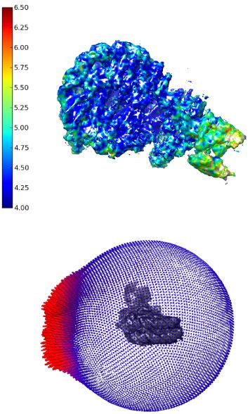
bioRxiv preprint first posted online Mar. 30, 2018; doi: http://dx.doi.org/10.1101/290874. The copyright holder for this preprint
 (which was not peer-reviewed) is the author/funder, who has granted bioRxiv a license to display the preprint in perpetuity.
                                It is made available under a CC-BY-NC 4.0 International license.

          Repositioning of Chd1 ATPase lobes to a closed ATP-bound state drives
          repositioning of chromodomains.

          The position of chromodomains is determined by each of the four contacts
          made with other components of the complex (Figure 2). When not bound to
          nucleosomes, the tandem chromodomains of Chd1 are observed to impede
          DNA binding to the ATPase domains (Hauk et al., 2010). This gave rise to the
          prediction that these domains would be rearranged in the nucleosome bound
          state (Hauk et al., 2010). This is indeed the case as the chromodomains
          undergo an 18 degree rotation when compared to the orientation observed in
          the crystal structure of Chd1 in the open state (Figure 3). Following
          repositioning chromodomain I interacts with nucleosomal DNA at SHL1
          (Figure 2 – Figure supplement 2) as observed previously (Farnung et al.,
          2017; Nodelman et al., 2017).

          Coincident with repositioning of the chromodomains, ATPase lobe II is
          repositioned closer to lobe I. This results in residues including those
          contributing to the conserved Walker box motifs (K407 and R804, R807)
          being brought into an arrangement compatible with ATP catalysis. Density for
          ADP-BeF within the pocket formed by conserved residues from ATPase
          domains I and II is well defined (Figure 2 – Figure supplement 3).

          The repositioning of ATPase lobe II enables contacts to be made with
          nucleosomal DNA (see below), the histone H4 tail and the histone H3 alpha 1
          helix (Figure 2 – figure supplement 4).These are the only direct contacts with
          histone components of the nucleosome. The contact with the H4 tails is
          conserved in mtISWI and Snf2 (Liu et al., 2017; Yan et al., 2016). D729 and
          E669 are conserved across all classes of remodelling enzyme but D725 is not
          as well conserved in Snf2 related enzymes (Figure 2 – figure supplement 4B).
          The conservation of this contact in Chd1 enzymes is consistent with the H4
          tail playing an important role in regulating Chd1 activity; deletion or mutation
          of the H4 tail has been shown to reduce nucleosome sliding and ATPase
          activity (Ferreira et al., 2007). The additional helices that make up the
                                                                      7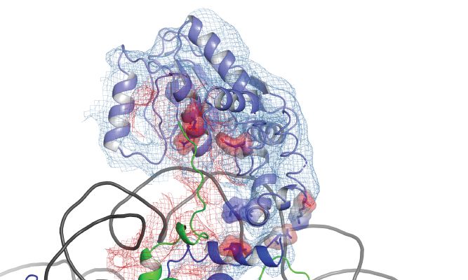
bioRxiv preprint first posted online Mar. 30, 2018; doi: http://dx.doi.org/10.1101/290874. The copyright holder for this preprint
 (which was not peer-reviewed) is the author/funder, who has granted bioRxiv a license to display the preprint in perpetuity.
                                It is made available under a CC-BY-NC 4.0 International license.

          protrusion 2 region of ATPase lobe 2 in Chd1 are conserved in chromatin
          remodeling ATPases, but not within all SF2 DNA translocases. Within this
          region residues 638-642 interact with the alpha 1 helix of histone H3 (Figure 2
          – figure supplement 4A). The residues participating in the interaction are
          progressively less well conserved in ISWI and SNF2 related proteins (Figure 2
          – figure supplement 4C). A loop from Phe1033 to Leu 1045 in an equivalent
          region of the yeast Snf2 protein is not assigned in the Snf2-nucleosome
          structure, but this region is positioned such that a related contact with histone
          H3 could be made.

          The structure, also provides clues as to how these conformational changes
          are driven. A central event is likely to be the closure of the cleft between
          ATPase domains driven by ATP binding (Figure 2 – Figure supplement 3).
          The 40o rotation of ATPase lobe II required to form the ATP binding pocket
          results in a negatively charged surface, observed to interact with an acidic
          surface on the long helix of chromodomain I (Figure 3A)(Hauk et al., 2010),
          being replaced by an acidic surface likely to repel chromodomain I (Figure
          3B). As a result closure of the ATPase domains is anticipated to drive
          nucleotide dependent repositioning of the chromodomains. Pulsed EPR was
          used to directly measure repositioning of the chromodomains in the absence
          of nucleosomes (Figure 4). The distance between engineered label at V256C
          in chromodomain I and S524C in ATPase lobe1 is 4.4nm in the open state,
          consistent with that observed in the crystal structure of the Chd1
          chromoATPase domains (Hauk et al., 2010). In the presence of ADP-BeF the
          4.4nm distance predominates, but a shoulder is observed consistent with a
          proportion of molecules adopting a new conformation with a distance of 5.6nm
          (Figure 4) which is similar to that observed in the ADP-BeF bound
          nucleosome by cryo-EM. This indicates that ATP binding is a driving event for
          repositioning of the chromodomains.

          The partial repositioning of the chromodomains observed in free Chd1 is likely
          to be stabilised by additional favourable interactions formed when this
          repositioning occurs within the context of nucleosome bound Chd1. These
          include the formation of contacts between chromodomain I and DNA at SHL1,
                                                                      8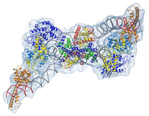
bioRxiv preprint first posted online Mar. 30, 2018; doi: http://dx.doi.org/10.1101/290874. The copyright holder for this preprint
 (which was not peer-reviewed) is the author/funder, who has granted bioRxiv a license to display the preprint in perpetuity.
                                It is made available under a CC-BY-NC 4.0 International license.

          between ATPase lobe II and the H3 alpha 1 helix, between ATPase lobe II
          and the histone H4 tail and most significantly the formation of a substantial
          interaction interface between ATPase lobe II and nucleosomal DNA at SHL2.
          The repositioning of the chromodomains in turn acts as a lever to reposition
          the DNA binding domain. In the context of nucleosomes this results in
          nucleotide dependent unwrapping of two turns of nucleosomal DNA
          (Sundaramoorthy et al., 2017). Conversely, the interaction of the DNABD
          requires linker DNA to be accessible.

          In order to investigate how the ability of the DNABD to interact with linker DNA
          is affected by the presence of an adjacent nucleosome, interactions between
          dinucleosomes with different separations were modelled. With a linker length
          of 19 bp Chd1 can be modelled binding the linker between adjacent
          nucleosomes (Figure 5). However, as the linker between nucleosomes is
          reduced, steric clashes become increasingly prohibitive. The requirement for
          a 19bp linker is likely to provide a limit below which engagement of the
          DNABD will be less stable. As this lower limit is set by clashes between the
          DNABD and the adjacent nucleosome, it is different from the length of linker
          required to occupy the DNA binding surface of the SANT and SLIDE domains
          on a mononucleosome with a free DNA linker. In this latter case 7 base pairs
          of DNA make contact with the DNABD (Figure 1). The c19 bp separation
          below which access of the DNABD to linker becomes progressively more
          difficult resonates with the average inter-nucleosome spacing of 19 bp
          observed in Saccharomyces cerevisiae (Tsankov et al., 2010). As the
          conformation of the DNABD is connected via the chromodomains to the
          ATPase domains, the structure of Chd1 provides molecular connectivity
          between the availability of nucleosomal linker DNA in excess of 19bp and the
          generation of closed nucleotide bound motor domains. This potentially
          provides a mechanism via which linker DNA length regulates the rate of
          nucleosome movement (Yang et al., 2006).

          Organisation of the Chd1 ATPase domains

                                                                      9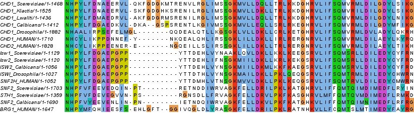
bioRxiv preprint first posted online Mar. 30, 2018; doi: http://dx.doi.org/10.1101/290874. The copyright holder for this preprint
 (which was not peer-reviewed) is the author/funder, who has granted bioRxiv a license to display the preprint in perpetuity.
                                It is made available under a CC-BY-NC 4.0 International license.

          Nucleosome repositioning is likely to be driven by the ability of the ATPase
          domains to drive ATP dependent DNA translocation. This has been observed
          directly for several Snf2 family proteins (Deindl et al., 2013; Lia et al., 2006;
          Sirinakis et al., 2011; Zhang et al., 2006) and is conserved within a wider
          family of superfamily II ATPases (Singleton et al., 2007). Structures of
          superfamily II single stranded translocases, such as herpes virus NS3, in
          different NTP bound states illustrate how the ratcheting motion of the ATPase
          domains drives translocation (Gu & Rice, 2010). To date such a series of
          structures does not exist for a double strand specific translocase. This raises
          the question as to whether structures of NS3 can be used to inform key
          aspects of the mechanism of Chd1 such as identifying the tracking strand. To
          do this we first align the ATPase lobes of Chd1 individually with NS3. The
          ATPase lobes of Chd1 like other Snf2 related proteins contain additional
          helices not conserved with NS3 (Durr et al., 2005; Liu et al., 2017; Thoma et
          al., 2005). As a result the alignment is restricted to conserved helices. In the
          case of lobe I and II the RMSD of the fit is 4.9 Å and 6.5 Å respectively
          (Figure 6 – figure supplement 1A). In the closed state alignment of both
          domains with the structure of NS3 in the ADP.BeF bound state results in an
          RMSD of 9.8 Å. Using this alignment the ssDNA bound by NS3 can be
          docked into the Chd1-Nucleosome structure (Figure 6A). This ssDNA aligns
          with the top strand of nucleosomal DNA (Figure 6). Conserved motif Ia in
          ATPase lobe 1 and motifs IV and V from ATPase lobe 2 contact this strand.
          These residues undergo a ratcheting motion during the course of ATP
          hydrolysis that drives the ssDNA through NS3 (Gu & Rice, 2010). Similar
          motion between these residues would be anticipated to drive nucleosomal
          DNA across the nucleosome dyad in the direction of the longer linker (Figure
          6B).

          It is notable that within Chd1 additional DNA contacts are made that differ
          from those observed in NS3. Firstly, motifs II and III within lobe 1 contact the
          opposite DNA strand (Figure 6A). As these motifs are intimately associated
          with motif Ia they would be anticipated to undergo a similar ratcheting motion
          with respect to the contacts made by lobe 2. Secondly, the ATPase lobes of
          Chd1 like those of Snf2 contain additional helices including the protrusions to
                                                                      10
bioRxiv preprint first posted online Mar. 30, 2018; doi: http://dx.doi.org/10.1101/290874. The copyright holder for this preprint
(which was not peer-reviewed) is the author/funder, who has granted bioRxiv a license to display the preprint in perpetuity.
It is made available under a CC-BY-NC 4.0 International license.
the helical lobes and the brace helix that are unique to chromatin remodelling
enzymes (Figure 6A)(Farnung et al., 2017; Flaus et al., 2006; Liu et al., 2017).
These extend the binding cleft between the ATPase lobes and make
additional contacts with both DNA strands.
The structure of a fragment of the yeast Snf2 protein bound to a nucleosome
revealed contacts between ATPase lobe 1 with DNA at SHL2 and the
adjacent DNA gyre at SHL 6 (Liu et al., 2017)(Figure 6 – Figure supplement
2A). The basic surface of lobe 1 responsible for this interaction is not
conserved in Chd1, and the acidic residues D464 and E468 make a similar
interaction with DNA unlikely. In addition, DNA is not present in this location
as it is lifted off the surface of the octamer (Figure 6 – Figure supplement 2B).
In the case of the Snf2 protein the interaction with the adjacent DNA gyre is
proposed to anchor the translocase preventing it from transiting around the
octamer surface (Liu et al., 2017). Chd1 has a relatively small interaction
interface with histones, so there is a similar requirement for DNA interactions
to constrain motion of the whole protein. In the case of Chd1 this could
instead be provided through the interaction of the chromodomains with DNA
at SHL1 and through the interaction of the DNA binding domain with linker
DNA. Amino acids 476 to 480 of lobe 1 also interact with DNA in the
unravelled state (Figure 6 – figure supplement 2B). These residues are not
conserved even in Chd1 proteins so the significance of this contact is not
clear.
Two molecules of Chd1 can bind a single nucleosome using the same
mode of binding.
Chromatin organising motor proteins are capable of catalysing bidirectional
nucleosome repositioning that can occur as a result of the binding of two or
one enzyme (Blosser et al., 2009; Qiu et al., 2017; Racki et al., 2009; Willhoft
et al., 2017). As Chd1 binds to one side of the nucleosome, no steric clashes
are anticipated should a second Chd1 bind linker DNA on the opposite side of
the nucleosome. To investigate this further, complexes consisting of two Chd1
molecules bound to one nucleosome were prepared using nucleosomal DNA
11bioRxiv preprint first posted online Mar. 30, 2018; doi: http://dx.doi.org/10.1101/290874. The copyright holder for this preprint
(which was not peer-reviewed) is the author/funder, who has granted bioRxiv a license to display the preprint in perpetuity.
It is made available under a CC-BY-NC 4.0 International license.
with symmetrical linkers of 14 base pairs and the images processed as
indicated (Figure 7 – Figure supplement 1). Most particles were assigned to
2D classes in which two bound Chd1 molecules are discernible, though one is
often more dominant likely as a result of the projections of the dominant
orientations observed. All 3D classes have two bound Chd1 molecules and
the most abundant classes refine to provide an envelope with 15 Å resolution
(Figure 7). Two Chd1 molecules bound in the same mode observed in the 1:1
complex can be docked into this volume. There are no direct contacts
between the two Chd1 proteins suggesting that the two bound enzymes are
likely to function independently. Previously, negative stain EM of two Chd1
molecules bound to a single nucleosome indicated that the DNA binding
domain interacted with linker DNA on only one side of the nucleosome
(Nodelman et al., 2017). Our envelope shows that both DNA binding domains
can bind to linker DNA simultaneously and that the extent of DNA unwrapping
is similar on both sides of the nucleosome. This provides further evidence that
the differences in DNA binding to the two sides of the 601 nucleosome
positioning sequence do not influence the extent of DNA unwrapping. Any
differences between the two bound Chd1 molecules must be localised and not
detectable at this resolution.
The trajectory of the histone H3 tail is altered by DNA unwrapping.
On the fully wrapped side of the nucleosome the H3 tail can be traced to
proline 38, emerging between the DNA gyres at SHL1. In contrast, on the
unwrapped side of the nucleosome the H3 tail can be traced to alanine 26
indicating that on this side of the nucleosome the H3 tail is better ordered. In
addition, the trajectory of the tail is different to that observed in previous
structures (Figure 8A). This altered trajectory was also not observed in a
previous structure of a Chd1 bound nucleosome, however, this structure was
made in the presence of PAF1 and FACT which may result in some noise in
this region that is not apparent in our structure (Farnung et al., 2017). A
potential explanation for the defined and altered trajectory of the histone H3
tail on the unwrapped side of the nucleosome is that amino acids within
extreme N-terminal region, that are not resolved in our structure, interact with
12bioRxiv preprint first posted online Mar. 30, 2018; doi: http://dx.doi.org/10.1101/290874. The copyright holder for this preprint
(which was not peer-reviewed) is the author/funder, who has granted bioRxiv a license to display the preprint in perpetuity.
It is made available under a CC-BY-NC 4.0 International license.
the unravelled DNA. The 25 N-terminal residues include eight lysine and
arginine residues that could interact with DNA at different locations along the
unravelled linker.
Ubiquitin interacts with unravelled nucleosomal DNA
The electron density for ubiquitin molecules is not as well defined as other
components of the complex, and limiting for the overall resolution (Figure 1).
This is likely to reflect mobility of the ubiquitin peptides. Consistent with this
the electron density determined from X-ray diffraction on crystallised
nucleosomes with ubiquitin conjugated at this location resulted in no density
attributable to ubiquitin (Machida et al., 2016). On the wrapped side of the
nucleosome, ubiquitin is located adjacent to the acidic patch that is widely
used as an interface for nucleosome binding proteins (McGinty & Tan, 2016)
(Figure 8B). This is also the location that ubiquitin conjugated to H2A K15 has
been observed to occupy on unbound nucleosomes (Wilson et al., 2016) and
likely represents a favourable conformation for ubiquitin when coupled at
different sites within this locality (Vlaming et al., 2014).
On the unwrapped side of the nucleosome, the ubiquitin peptide is displaced
across the lateral surface towards the DNA. The unwrapped DNA is oriented
away from the lateral surface and this positions the DNA backbone at SHL6 in
contact with the repositioned ubiquitin (Figure 8B). In 2D classes, ubiquitin is
more prominent on the unwrapped side suggesting it is more tightly
constrained. The residues interacting with DNA include Lys 48 Arg 54 and
Asp 60. It is likely this interaction stabilises DNA in the unwrapped state,
perhaps explaining why H2B K123 ubiquitinylation directly stimulates
remodelling by Chd1(Levendosky et al., 2016).
Discussion
13bioRxiv preprint first posted online Mar. 30, 2018; doi: http://dx.doi.org/10.1101/290874. The copyright holder for this preprint
(which was not peer-reviewed) is the author/funder, who has granted bioRxiv a license to display the preprint in perpetuity.
It is made available under a CC-BY-NC 4.0 International license.
A striking feature of the Chd1 nucleosome complex is the limited number of
contacts with histones. The two direct contacts with histones are with the H3
alpha one helix and with the histone H4 tail (Figure 2 – Figure supplement 4).
The contact is with the H4 tail is required for efficient remodelling by Chd1
(Ferreira et al., 2007) as it is for ISWI subfamily enzymes (Clapier et al., 2001;
Hamiche et al., 2001). Aside from these two contacts, the interaction of Chd1
with nucleosomes is dominated by interactions with DNA. This leaves the
majority of the nucleosome accessible for binding by additional factors. We
show that a second Chd1 molecule can bind the opposite side of the
nucleosome using a similar mode of interaction (Figure 7). Even in the case of
a nucleosome bound by two Chd1 molecules the lateral surfaces of the
nucleosome are accessible for binding by other factors.
Despite a lack of direct contacts with histones H2A and H2B, Chd1 activity is
dependent on histone dimers (Levendosky et al., 2016). The requirement for
histone dimers may arise as a result of dimer loss affecting DNA wrapping.
Loss of a histone dimer will result in a loss of histone DNA contacts at
SHL3.5, 4.5 and 5.5. More extensive unwrapping of DNA to SHL3.5 would
require major repositioning of DNABD in order to retain the interaction with
linker DNA while the chromoATPase is engaged at SHL-2. This provides a
potential explanation for the dependency of Chd1activity on histone dimers.
Conversely, association of a histone dimer with a histone tetramer or hexamer
around which DNA is initially significantly unwrapped, could be stabilised by
the binding of Chd1 rewrapping DNA to SHL5. The stabilisation of the SHL5
wrapped state may facilitated correct docking of histone dimers into
chromatin. This provides a mechanistic basis for the observed activities of
Chd1 in H2A/H2B transfer (Lusser et al., 2005) and chromatin assembly (Fei
et al., 2015; Lee et al., 2012; Torigoe et al., 2013). Repositioning of the Chd1
DNA binding domain towards the major orientation observed in free solution,
the Apo state reported by (Sundaramoorthy et al., 2017), would guide linker
DNA towards the fully wrapped state. As a result Chd1 has the potential to
function in multiple stages of chromatin assembly and the generation of
organised chromatin (Lusser et al., 2005; Robinson & Schultz, 2003; Torigoe
et al., 2013).
14bioRxiv preprint first posted online Mar. 30, 2018; doi: http://dx.doi.org/10.1101/290874. The copyright holder for this preprint
(which was not peer-reviewed) is the author/funder, who has granted bioRxiv a license to display the preprint in perpetuity.
It is made available under a CC-BY-NC 4.0 International license.
The Chd1 enzyme has the ability to organise spaced arrays of nucleosomes
both in vitro and in vivo (Gkikopoulos et al., 2011; Lusser et al., 2005;
Robinson & Schultz, 2003). Enzymes that exhibit this organising activity
typically reposition nucleosomes away from the ends of short DNA fragments.
This is also true for Chd1 (McKnight et al., 2011; Stockdale et al., 2006). As a
result we would anticipate that Chd1 would be most likely to reposition
nucleosomes away from the short (exit) linker, encroaching into the long
(entry) linker. Repositioning of nucleosomes with this directionality conflicts
with the directionality of translocation inferred from docking the tracking strand
of NS3 into Chd1(Farnung et al., 2017)(Figure 6). Tracking along this strand
with 3’-5’ directionality would instead be anticipated to draw DNA into the
nucleosome from the exit side.
Inferring the mechanism of Chd1 from NS3 is complicated by the fact these
enzymes are not so closely related. Conserved motifs are difficult to align
based on sequence alone. In addition some aspects of nucleic acid binding by
both Snf2 and Chd1 profoundly differ from NS3. Notably, motifs II and III
within lobe 1 contact the opposite, 3’-5’ stand, which is not present in NS3. In
addition, Snf2 related chromatin remodelling enzymes contain features that
extend the nucleic acid binding cleft between the two ATPase lobes and make
contacts with both strands (Figure 6). As a result of the extensive contacts
with both strands, it is possible that the assignment of guide and tracking
strands within remodelling ATPases is not absolute as tracking may be
coupled to both strands. Consistent with this experiments that have probed
the action of remodelling enzymes using short gaps in either strand of
nucleosomal DNA have found them to be sensitive to lesions in either strand
(Saha et al., 2005; Zofall et al., 2006).
The introduction of gaps in nucleosomal DNA has also been used to infer the
directionality with which ATPases’s move along DNA. Introduction of gaps
distal to the SHL 2 location closest to the entry linker DNA has been observed
to impede the action of Snf2, Iswi and Chd1 enzymes (McKnight et al., 2011;
Saha et al., 2005; Zofall et al., 2006). This has been used as evidence that
15bioRxiv preprint first posted online Mar. 30, 2018; doi: http://dx.doi.org/10.1101/290874. The copyright holder for this preprint
(which was not peer-reviewed) is the author/funder, who has granted bioRxiv a license to display the preprint in perpetuity.
It is made available under a CC-BY-NC 4.0 International license.
the enzyme translocation that drives repositioning initiates from the SHL 2
located distal to the entry DNA. As a consequence it has been proposed that
Chd1 bound in the cross gyres conformation, that we and others observe,
represents an inactive state in which the interaction of the DNABD with linker
DNA is inhibitory (Farnung et al., 2017; Nodelman et al., 2017). If this were
the case, then Chd1 directed repositioning might be expected to be limited
once a nucleosome has moved far enough from a DNA end to enable the
DNABD to contact linker DNA. On Chd1 bound nucleosomes the DNABD
contacts extend to 7bp from the nucleosomes edge. A stall to repositioning
after 7bp is not observed, instead Chd1 repositions nucleosomes 23-39 base
pairs into a 54 base pair linker in a very similar way to Isw1a and Isw2
(Stockdale et al., 2006).
There is further evidence to support the conformation of Chd1 with the
DNABD bound to linker DNA as an active state. Firstly, in the ADP.BeF bound
state, the ATPase domains are repositioned to a closed conformation and
conserved residues are positioned for catalysis (Figure 2 – figure supplement
3). The closure of the ATPase domains in the Chd1-nucleosome complex is
connected to the nucleotide-dependent unwrapping of DNA via repositioning
of the chromodomains, which in turn levers the DNABD position. Secondly,
efficient nucleosome repositioning by Chd1 is dependent on the DNABD
(Ryan et al., 2011b). This is anticipated if the DNA binding domain acts to
generate an active conformation, but not if sensing of exit linker DNA is
repressive. Thirdly, the activity of chimeric Chd1 proteins in which DNA
binding is provided via a heterologous domain is greatest when the cognate
binding site is placed in the entry linker mimicking the arrangement observed
in the Chd1-nuceosome structure (McKnight et al., 2011; Patel et al., 2012).
An additional confounding factor in assigning the directionality with which
Chd1 translocates is the recent observation that a single Chd1 molecule can
direct bidirectional nucleosome movement (Qiu et al.). As a result, further
studies are required to resolve how Chd1 acts to drive DNA across the
octamer surface.
16bioRxiv preprint first posted online Mar. 30, 2018; doi: http://dx.doi.org/10.1101/290874. The copyright holder for this preprint
(which was not peer-reviewed) is the author/funder, who has granted bioRxiv a license to display the preprint in perpetuity.
It is made available under a CC-BY-NC 4.0 International license.
Both budding yeast Chd1 and human Chd2 are found to be enriched within
coding regions (de Dieuleveult et al., 2016; Gkikopoulos et al., 2011; Lee et
al., 2017). Histone H3 K36me3 is a hallmark of coding region nucleosomes,
so we prepared nucleosomes alkylated to mimic trimethylation at this position.
Alkylation modestly stimulates Chd1 activity (Figure 1 – Figure supplement 1),
raising the possibility that this modification is recognised by the enzyme,
possibly via the chromodomains. However, we observe electron density for
the histone H3 tail to residue 26, indicating that H3K36 does not stably
interact with the chromodomains or any other component of Chd1 in the
structure reported here. Furthermore, for this interaction to occur, either the
chromodomains would need to be repositioned, or the structure of the N-
terminus of H3 reconfigured for example by unfolding of the alpha–N helix
(Elsasser et al., 2012; Liu et al., 2012).
The improved density for the H3 tail on the unwrapped side of the
nucleosome is most likely to result from the interaction of the basic N-terminal
region of the H3 tail, which is not resolved, with DNA. It is notable that in the
fully wrapped state the H3 tail would need to follow a very different path in
order to interact with DNA. Consistent with this the trajectory of the H3 tail on
the unwrapped side of the nucleosome is different to that observed in
structures of intact nucleosomes (Wilson et al., 2016). This raises the
possibility that changes to DNA wrapping could affect the way in which
histone tail epitopes are displayed. In principle, such effects could be positive
or negative. For example the tudor domain of PHF1 preferentially interacts
with trimethylated H3K36 on partially unwrapped nucleosomes (Gibson et al.,
2017). The interaction of the PHD domains of Chd4 with DNA is also inhibited
by nucleosomal DNA (Gatchalian et al., 2017). As a result if Chd4 generates
unwrapped structures similar to those observed with Chd1 the interaction of
these domains would be enhanced. The reconfiguration of the H3 tail by Chd1
has the potential to affect the interaction of histone reader, writer and eraser
enzymes with the tail and as a result the distribution of these modifications in
chromatin. Such effects have been observed, as Chd1 contributes to the
establishment of boundaries between H3K4me3 and H3K36me3 at most
transcribed genes (Lee et al., 2017).
17bioRxiv preprint first posted online Mar. 30, 2018; doi: http://dx.doi.org/10.1101/290874. The copyright holder for this preprint
(which was not peer-reviewed) is the author/funder, who has granted bioRxiv a license to display the preprint in perpetuity.
It is made available under a CC-BY-NC 4.0 International license.
H2BK120 ubiquitination is also enriched in coding region chromatin, and has
previously been observed to stimulate Chd1 activity (Levendosky et al., 2016).
Although the level of Chd1 stimulation is only 2-fold, we have previously
observed that mutations exerting a 2-fold effect on Chd1 activity in vitro affect
nucleosome organisation in vivo (Sundaramoorthy et al., 2017). It has also
been observed that H2BK120Ub negatively affects the activity of some ISWI
containing enzymes (Fierz et al., 2011). As organisation of coding region
nucleosomes involves these and other enzymes (Krietenstein et al., 2016;
Ocampo et al., 2016; Parnell et al., 2015), H2BK120Ub has the potential to
regulate interplay between different enzymes.
Ubiquitin on the unwrapped side of the nucleosome is repositioned such that it
interacts directly with DNA. As in the case of the H3 tail, the repositioning of
the ubiquitin resulting from Chd1-directed DNA unwrapping could potentially
affect interactions with the factors involved in the placing, removal or
recognition of H2BK120ub. The most striking functional evidence for this
interplay is that H2BK120ub is greatly reduced in Chd1 mutants (Lee et al.,
2012). One possible explanation for this effect is that Chd1 sequesters
ubiquitin in a conformation less accessible for removal. Consistent with this
the position of ubiquitin on the unwrapped side of Chd1 bound nucleosomes
is incompatible with interaction with the SAGA DUB module (Morgan et al.,
2016). Interestingly, the paradigm for trans regulation between histone
modifications stems from the interplay between H2BK120ub and H3 K4
methylation (Sun & Allis, 2002), both of which are influenced by Chd1 binding.
While Chd1 is not required for H3K4me3 (Lee et al., 2012) it does influence
the distribution of this histone modification (Lee et al., 2017).
H2BK120Ub has previously been observed to directly affect chromosome
structure at the level of chromatin fibre formation (Debelouchina et al., 2017;
Fierz et al., 2011). Our observations show a new role for H2BK120Ub at the
level of nucleosomal DNA wrapping. The specific relocation of ubiquitin on the
unravelled side of the nucleosome, the local distortion of H2B at the site of
attachment and the presence of lysine and arginine residues at the site of
18bioRxiv preprint first posted online Mar. 30, 2018; doi: http://dx.doi.org/10.1101/290874. The copyright holder for this preprint
(which was not peer-reviewed) is the author/funder, who has granted bioRxiv a license to display the preprint in perpetuity.
It is made available under a CC-BY-NC 4.0 International license.
interaction with DNA all indicate this is a favourable interaction that stabilises
DNA in the unwrapped state. The outer turns of nucleosomal DNA rapidly
associate and dissociate on millisecond time scales, with occupancy of the
unwrapped state estimated at 10% (Li et al., 2005). The ubiquitin interaction
we have observed would be anticipated to stabilise the transiently unwrapped
state increasing its abundance. It is however, unlikely that the unwrapped
state predominates in the absence of Chd1 or other factors that promote
unwrapping as the structure of isolated ubiquitinylated nucleosomes is
unchanged (Machida et al., 2016). Nonetheless, increased occupancy of the
transiently unwrapped state would be anticipated to facilitate access to
nucleosomal DNA. Chromatin folding to form higher order structures is likely
to be favoured by fully wrapped, nucleosomes and so an increase in the
proportion of unwrapped nucleosomes could potentially contribute to the
effects of H2BK120Ub on chromatin fibre formation (Fierz et al., 2011). Many
other processes involving chromatin dynamics are linked to H2BK120Ub
including transcription (Bonnet et al., 2014), DNA repair (Moyal et al., 2011;
Nakamura et al., 2011) and DNA replication (Lin et al., 2014). A more stable
unwrapped state could also provide an explanation for the association of
factors that lack recognised ubiquitin interaction domains, with ubiquitinylated
chromatin (Shema-Yaacoby et al., 2013). Interestingly, H2BK120Ub
associating proteins include human Chd1, SWI/SNF complex, pol II and the
elongation factors NELF and DISF (Shema-Yaacoby et al., 2013).
The change in the position of ubiquitin also has the potential to indirectly
affect the way in which other factors interact with ubiquitinylated
nucleosomes. On the wrapped side of the nucleosome ubiquitin is positioned
such that it occludes access to the acidic patch formed by the cleft between
histones H2A and H2B. This provides surface via which many proteins
including LANA peptides (Barbera et al., 2006), RCC1 (Makde et al., 2010),
Sir3 (Armache et al., 2011), PRC1 (McGinty et al., 2014) and the SAGA DUB
module (Morgan et al., 2016) interact with nucleosomes. The repositioning of
ubiquitin away from the acidic patch on the unwrapped side of the
nucleosome improves access to the acidic patch. In this way H2BK120ub may
19bioRxiv preprint first posted online Mar. 30, 2018; doi: http://dx.doi.org/10.1101/290874. The copyright holder for this preprint
(which was not peer-reviewed) is the author/funder, who has granted bioRxiv a license to display the preprint in perpetuity.
It is made available under a CC-BY-NC 4.0 International license.
provide a means of regulating access to the acidic patch that is sensitive to
changes in nucleosome structure.
Although the repositioning of the H3 tail and ubiquitin were observed on Chd1
bound nucleosomes, the potential for reconfiguration of histone epitopes may
be more general. All processes that generate local DNA unwrapping would be
anticipated to result in similar repositioning of histone tail epitopes. In
particular, where combinations of modifications are recognised bivalently, the
spatial alignment of epitopes will be important for recognition by coupled
reader domains. This potentially provides a means of tuning signalling via
histone modifications to regions of transient histone dynamics.
Acknowledgements
We thank Daniel Claire electron Bio-Imaging Centre (eBIC), Diamond light
source Ltd, UK for data collection with respect to the 4.5 Å structure. We
thank Rebecca Thompson and Neil Ranson for assistance with collection of
2Chd1 bound to one nucleosome structure at the Astbury Biostructre
Laboratory, University of Leeds. The ubiquitin expression plasmid was kindly
provided by Ron Hay, University of Dundee. This work was funded by
Wellcome Senior Fellowship 095062, Wellcome Trust grants 094090, 099149
and 097945. ALH was funded by and EMBO long term fellowship ALTF 380-
2015 co-funded by the European Commission (LTFCOFUND2013, GA-2013-
609409). We Thank Dale Wigley and Chris Aylett for useful discussions.
Methods
Cloning, protein expression and purification
ScChd1 C-terminal and N-terminal truncations were made from the full length
clone described in Ryan et al, using an inverse PCR strategy (Ryan et al.,
2011a). Site directed mutagenesis was used to introduce cysteine residues at
strategic locations on ScChd1 1-1305ΔC using standard cloning procedure. All
proteins were expressed in Rosetta2 (DE3) pLysS Escherichia Coli cells at 20°
20bioRxiv preprint first posted online Mar. 30, 2018; doi: http://dx.doi.org/10.1101/290874. The copyright holder for this preprint
(which was not peer-reviewed) is the author/funder, who has granted bioRxiv a license to display the preprint in perpetuity.
It is made available under a CC-BY-NC 4.0 International license.
C in Auto-induction media and the purification of the protein was carried out
typically as described in Ryan et al. After the purification of the protein the GST
tag was cleaved with precision protease and the tag cleaved proteins were
subjected to size exclusion chromatography using Superdex S200 10/300 GL
columns (GE Healthcare). Expression and purification of Xenopus laevis
histones were carried out as described previously (Luger et al., 1999).
Installation of Methyl-lysine analogues in H3 K36
Alkylation of cysteine-mutant histones to generate histones modified with
methyl-lysine analogues was performed as in (Simon M.D. et al, Cell 2007).
Approximately 10mg of lyophilised cysteine mutant histone was resuspended
in 800uL (me3) or 900uL (me0) degassed alkylation buffer (1M HEPES, 10mM
D,L-methionine, 4M Guanidine HCl, pH7.8). Histones were reduced with fresh
30mM DTT for 30 minutes at room temperature.
For trimethyl-lysine analogues, the reduced histone was added to
approximately 125mg of (2-Bromoethyl) trimethylammonium bromide (Sigma
117196-25G) in 200uL of DMF and incubated in the dark at 50⁰C for 3 hours.
An additional 10uL of DTT was added, and the reaction was allowed to proceed
overnight at room temperature.
For generation of the unmethylated lysine analogue, 75uL of 1M 2-
Bromoethylamine hydrobromide (Fluka 06670-100G) was added to the
reduced histone and was incubated at room temperature in the dark for 3 hours.
An additional 10uL of DTT was added for 30 minutes prior to the addition of an
extra 75uL of alkylating agent, and the reaction was allowed to proceed
overnight at room temperature in the dark.
The reaction was terminated with the addition of 50uL 2-mercaptoethanol for
30 minutes and the alkylated histone was desalted either by dialysis into water
with 2mM 2-mercaptoethanol or on a PD-10 desalting column (GE 52130800).
The shift in molecular weight associated was confirmed via MALDI-TOF mass
spectrometry.
In vitro ubiquitination
Recombinant expression of xH2B K120C and His-TEV-Ubiquitin G76C mutant
proteins was induced with IPTG for 4 hours in Rosetta 2 DE3 pLysS cells grown
21bioRxiv preprint first posted online Mar. 30, 2018; doi: http://dx.doi.org/10.1101/290874. The copyright holder for this preprint
(which was not peer-reviewed) is the author/funder, who has granted bioRxiv a license to display the preprint in perpetuity.
It is made available under a CC-BY-NC 4.0 International license.
at 37⁰C. Inclusion body purification followed by cation exchange
chromatography was performed to isolate the histone protein. Ubiquitin was
purified using HisPur cobalt resin with 150mM sodium chloride/20mM Tris pH8
buffer and eluted with 350mM imidazole. Histones and ubiquitin were desalted
by dialysis into water with 2mM 2-mercaptoethanol and lyophilised for storage.
Proteins were re-suspended in 50mM ammonium bicarbonate pH 8 and treated
with 2mM TCEP for 1 hour. Ellman’s reagent was used to ascertain the
concentration of free sulfhydryls, and xH2b and ubiquitin were combined at
equimolar ratios, as defined by the Ellman’s assay, and diluted with 50mM
ammonium bicarbonate to 200-250uM each protein. The proteins were
crosslinked at room temperature with four hourly additions of ¾ molar ratio of
1,3 dichloroacetone (freshly prepared in DMF). An equal volume of denaturing
buffer (7M Guanidine HCl, 350mM sodium chloride, 25mM Tris pH7.5) was
added to the reactions, which were purified using HisPur cobalt resin, pre-
equilibrated in denaturing buffer. The His-TEV-Ub-xH2B crosslinked product
was eluted with 350mM imidazole and dialysed into SAUDE200 buffer (7M
Urea, 20mM sodium acetate, 200mM sodium chloride, 1mM EDTA, 5mM 2-
mercaptoethanol) overnight. The ubiquitinated histone was further purified over
a cation exchange column, as before, and fractions were dialysed into water
with 2mM 2-mercaptoethanol and lyophilised for storage.
Preparation of recombinant nucleosomes
Xenopus H2B-K120 ubiquitinylated histones were refolded in equimolar ratios
with H2A and similarly H3 K36 methyl analogue histones were refolded in
equimolar ratios with histone H4 to obtain dimers and tetramers as described
previously for wild type histones Dyer et al., and purified on a size exclusion
chromatography using S200 gel filtation column. The peak fractions were
analysed with SDS-PAGE gel and pooled. 601 DNA fragments of respective
lengths for recombinant nucleosome assembly were generated by PCR method
as described previously (Sundaramoorthy et al., 2017). Nucleosomes were
generated by salt dialysis as described previously by combining H2A/H2B-
K120 ubiquitin dimer, H3K36 methyl lysine analogue tetramer (2:1 ratio) with
DNA containing PCR amplified Widom 601 DNA sequence.
22bioRxiv preprint first posted online Mar. 30, 2018; doi: http://dx.doi.org/10.1101/290874. The copyright holder for this preprint
(which was not peer-reviewed) is the author/funder, who has granted bioRxiv a license to display the preprint in perpetuity.
It is made available under a CC-BY-NC 4.0 International license.
Nucleosome Repositioning Assay
Nucleosomes were reconstituted on Cy3 (me0) and Cy5 (me3) labelled DNA,
based on the 601 sequence, with a 47bp extension. Repositioning by Chd1 was
performed in 40mM Tris pH7.4, 50mM KCl, 3mM MgCl2, 1mM ATP, 100nM
each nucleosome, and 10nM Chd1; 10uL was removed at each time point (T=0,
4, 8, 16, 32, and 64 minutes), placed on ice, and stopped with the addition of
100ng/uL competitor DNA, 200mM NaCl, and 1.6% sucrose. Repositioned
nucleosomes were run on 6% PAGE/0.2X TBE gels in recirculating 0.2X TBE
buffer for 3-4 hours at 300V. The percent of repositioned nucleosomes was
analysed using Aida image analysis software. Data were fit to a hyperbola in
Sigma Plot, to determine the initial rate of repositioning.
Nucleosome binding.
Xenopus laevis nucleosomes (20nM), reconstituted on Cy3 labelled 0W11
DNA, were bound to titrations of Chd1 enzymes (concentration specified in
figure legend) in 50mM Tris pH 7.5, 50mM sodium chloride, and 3mM
magnesium chloride supplemented with 100ug/mL BSA. Unbound and bound
nucleosomes were separated on a pre-run 6% polyacrylamide gel (49:1
acrylamide: bis-acrylamide) in 0.5X TBE buffer for 1 hour at 150V. The gel shift
was scanned on Fujifilm FLA-5100 imaging system at 532nm.
Spin labelling of ScChd1, PELDOR measurements and modelling
MTSL was conjugated to introduce cysteines immediately following size
exclusion purification as described in Hammond et al (Hammond et al., 2016).
Excess unreacted labels were removed from the sample by dialysis. PELDOR
experiments were conducted at Q-band (34 GHz) operating on a Bruker
ELEXSYS E580 spectrometer with a probe head supporting a cylindrical
resonator ER 5106QT-2w and a Bruker 400 U second microwave source unit
as described previously (Hammond et al., 2016). All measurements reported
here were made at 50K. Data analysis was carried out using the DeerAnalysis
2013 package (Jeschke and Polyhach, 2007). The dipolar coupling evolution
data were first corrected to remove background decay. Tikhonov regularisation
was then used to determine distance distributions from each dataset.
23bioRxiv preprint first posted online Mar. 30, 2018; doi: http://dx.doi.org/10.1101/290874. The copyright holder for this preprint
(which was not peer-reviewed) is the author/funder, who has granted bioRxiv a license to display the preprint in perpetuity.
It is made available under a CC-BY-NC 4.0 International license.
To model the distance distribution for the open conformation of Chd1 helicase
lobes crystal structure of chromo helicase (PDB Code: 3MWY) (Hauk et al.,
2010) was used. For the closed conformation refined cryoEM structure of Chd1
bound to nucleosome in the presence of ADP.BeFx described in this study was
used as a model. For each structure, R1 spin labels were added and the
distribution simulated for each position using MTSSL wizard in Pymol. Also the
average distance from the distribution from a pair of spin labels were calculated
using MTSSL wizard in Pymol.
Sample preparation, Cryo Electron Microscopy data collection and
analysis
The appropriate ratio of ScChd1(1-1305∆57-88) to nucleosome for 1:1 and the
2:1 complex formation in the presence of 5-fold molar excess of ADP-BeFx was
determined by titration and native PAGE analysis. The formed complex was
then purified by size exclusion gel filtration using a PC 3.2/30 superdex 200
analytical column in 20mM Tris, 50mM NaCl and 250 µM ADP.BeFx . In a
typical run 50uLs of 20 µM of sample was injected using Dionex autoloader.
50uLs fractions were collected and analysed in 6% Native PAGE gel and
appropriate fractions containing ScChd1-nucleosome complexes were pooled
together. A 4 µl drop of sample was then applied to C-flat Holey carbon foil
(400 mesh R1.2/1.3 uM) pre-cleaned with glow discharge (Quorum
technologies). After 15 second incubation, grids were double side blotted for 4
s in a FEI cryo-plunger (FEI Mark III) at 90% humidity and plunge frozen into
−172 °C liquefied ethane. Standard vitrobot filter paper Ø 55/20 mm, Grade
595 was used for blotting.
The prepared grids are initially checked for its ice quality and the particle
distribution using a JEOL 2010 microscope with side-entry cryo-holder
operated at 200 keV and equipped with a gatan 4k × 4k CCD camera. Cryo-
grids were then stored in liquid nitrogen and dry-shipped to respective centre
for data collection. For the 1:1 complex the data was acquired on a FEI Titan
Krios transmission electron microscope (TEM) operated at 300 keV,
equipped with a K2 summit direct detector (Gatan). Automated data
24You can also read