Coordinated crosstalk between microtubules and actin by a spectraplakin regulates lumen formation and branching - eLife
←
→
Page content transcription
If your browser does not render page correctly, please read the page content below
RESEARCH ARTICLE

                                     Coordinated crosstalk between
                                     microtubules and actin by a spectraplakin
                                     regulates lumen formation and branching
                                     Delia Ricolo1,2, Sofia J Araujo1,2*
                                     1
                                      Department of Genetics, Microbiology and Statistics, School of Biology, University
                                     of Barcelona, Barcelona, Spain; 2Institute of Biomedicine University of Barcelona
                                     (IBUB), Barcelona, Spain

                                     Abstract Subcellular lumen formation by single-cells involves complex cytoskeletal remodelling.
                                     We have previously shown that centrosomes are key players in the initiation of subcellular lumen
                                     formation in Drosophila melanogaster, but not much is known on the what leads to the growth of
                                     these subcellular luminal branches or makes them progress through a particular trajectory within
                                     the cytoplasm. Here, we have identified that the spectraplakin Short-stop (Shot) promotes the
                                     crosstalk between MTs and actin, which leads to the extension and guidance of the subcellular
                                     lumen within the tracheal terminal cell (TC) cytoplasm. Shot is enriched in cells undergoing the
                                     initial steps of subcellular branching as a direct response to FGF signalling. An excess of Shot
                                     induces ectopic acentrosomal luminal branching points in the embryonic and larval tracheal TC
                                     leading to cells with extra-subcellular lumina. These data provide the first evidence for a role for
                                     spectraplakins in single-cell lumen formation and branching.

                                     Introduction
*For correspondence:                 Cell shape is intrinsically connected with cell function and varies tremendously throughout nature.
sofiajaraujo@ub.edu                  Tissue and organ morphogenesis rely on cellular branching mechanisms that can be multicellular or
                                     arise within a single-cell. Through extensive cellular remodelling, this so-called single-cell or subcellu-
Competing interests: The
                                     lar branching, transforms an initially relatively symmetrical unbranched cell into an elaborate
authors declare that no
                                     branched structure. These cellular remodelling events are triggered by widespread cytoskeletal
competing interests exist.
                                     changes and cell membrane growth, which allow these branched cells to span very large areas and
Funding: See page 26                 accomplish their final function. Despite this clear link between morphology and function, not much is
Received: 15 July 2020               known about the signalling events that trigger the formation of these subcellular branches or what
Accepted: 27 October 2020            makes them choose a particular trajectory within the cytoplasm of the cell.
Published: 28 October 2020               In Drosophila melanogaster, tracheal system terminal cells (TCs) and nervous system dendrites
                                     are models for these subcellular branching processes. During tracheal embryonic through larval
Reviewing editor: Derek
Applewhite, Reed College,
                                     development, the generation of single-cell branched structures by TCs is characterised by extensive
United States                        remodelling of the MT network and actin cytoskeleton, followed by vesicular transport and mem-
                                     brane dynamics (Best, 2019; Gervais and Casanova, 2010; Sigurbjörnsdóttir et al., 2014). During
   Copyright Ricolo and Araujo.
                                     embryonic development, TCs, as tip-cells, lead multicellular branch migration and extension in
This article is distributed under
                                     response to Bnl-Btl signalling, which induces the expression of Drosophila serum response factor
the terms of the Creative
Commons Attribution License,         (DSRF/blistered (bs)) and its downstream effectors (Affolter et al., 1994; Posern and Treisman,
which permits unrestricted use       2006). Although epithelial in origin, TCs do not have a canonical apical-basal polarity, and, as they
and redistribution provided that     migrate, extend numerous filopodia on their basolateral membrane, generating transient protrusive
the original author and source are   branches at the leading edge (Lebreton and Casanova, 2014). As a consequence, they display a
credited.                            polarity similar to that of a migrating mesenchymal cell (Fischer et al., 2019).

Ricolo and Araujo. eLife 2020;9:e61111. DOI: https://doi.org/10.7554/eLife.61111                                                         1 of 29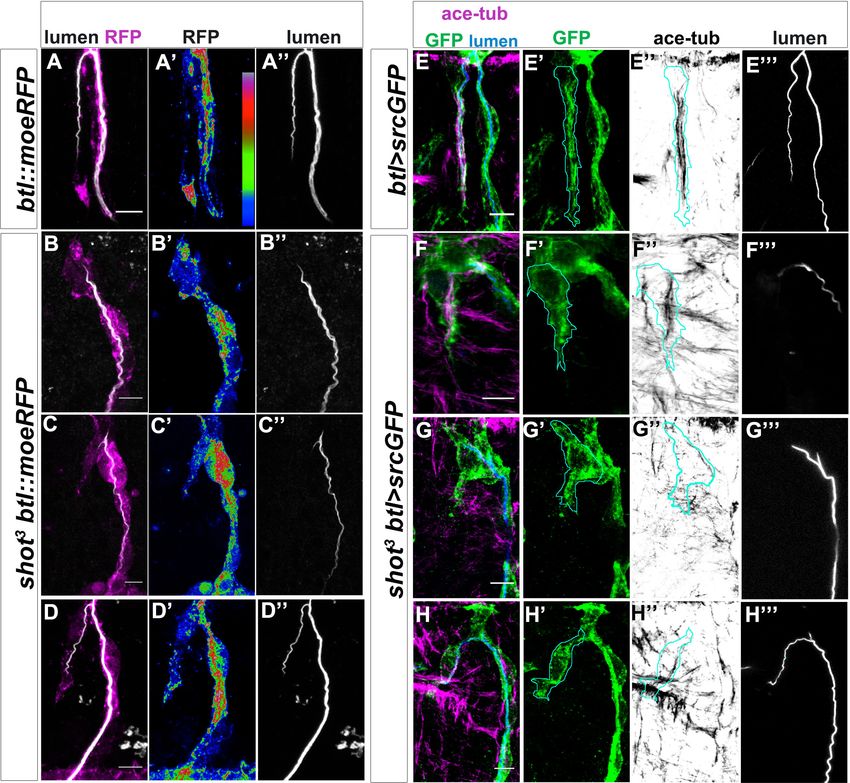
Research article                                                                               Cell Biology Developmental Biology

                                      While migrating and elongating, the TC invaginates a subcellular tube from its apical membrane,
                                   at the contact site with the stalk cell (Gervais and Casanova, 2010). The generation of this de novo
                                   subcellular lumen can be considered the beginning of the single-cell branching morphogenesis of
                                   this cell, which continues throughout larval stages to generate an elaborate single-cell branched
                                   structure with many subcellular lumina (Best, 2019).
                                      We have previously shown that centrosomes are key players in the initiation of subcellular branch-
                                   ing events during embryogenesis. Here, they act as microtubule organising centres (MTOCs) mediat-
                                   ing the formation of single or multiple-branched structures depending on their numbers in the TC
                                   (Ricolo et al., 2016). Centrosomes organise the growth of MT-bundles toward the elongating baso-
                                   lateral edge of the TC. These MTs have been suggested to serve both as trafficking mediators, guid-
                                   ing vesicles for delivery of membrane material, and as mechanical and structural stabilisers for the
                                   new subcellular lumen (Best, 2019). Actin filaments are present at the growing tip, the basolateral
                                   and the luminal membrane of the TC, and actin-regulating factors such as DSRF, Enabled (Ena) and
                                   Moesin (Moe) have been shown to contribute to TC morphogenesis (Gervais and Casanova, 2010;
                                   Guillemin et al., 1996; Schottenfeld-Roames et al., 2014). During TC elongation, the lumen
                                   extends along with the cell, stabilizing the elongating cell body and maintaining a more or less con-
                                   stant distance between its own tip and the migrating tip of the cell (Gervais and Casanova, 2010).
                                   At the TC basolateral side, a dynamic actin pool integrates the filopodia and aligns the growing sub-
                                   cellular tube with the elongation axis (JayaNandanan et al., 2014; Okenve-Ramos and Llimargas,
                                   2014; Oshima et al., 2006). Together, MT-bundles and the basolateral actin pool are necessary for
                                   subcellular lumen formation (Gervais and Casanova, 2010). However, not much is known on how
                                   these two cytoskeletal structures are coordinated within the TC.
                                      By the time the larva hatches, TCs have elongated and grown a full-length lumen, which becomes
                                   gas-filled along with the rest of the tracheal system. In the larva, terminal cells ramify extensively and
                                   form many new cytoplasmatic extensions each with a membrane-bound lumen creating tiny subcellu-
                                   lar tubes that supply the targets with oxygen (Baer et al., 2007; Ghabrial et al., 2011; Whit-
                                   ten, 1957). At larval stages, sprouting and extension of new branches in response to local hypoxia is
                                   generally considered to occur by essentially the same molecular mechanisms as the initial tube
                                   invagination and cell extension in the embryo (Jarecki et al., 1999; Sigurbjörnsdóttir et al., 2014).
                                   However, not much is known about how hypoxic signalling is transduced into cytoskeletal modula-
                                   tion to achieve the single-cell branching morphogenesis of the TC. Also, what coordinates the cross-
                                   talk between microtubules and actin at the basolateral growing tip, how cell elongation is stabilised
                                   by lumen formation and how both processes remain coordinated is still poorly understood in both
                                   embryonic and larval TCs.
                                      Spectraplakins are giant conserved cytoskeletal proteins with a complex multidomain architecture
                                   capable of binding MTs and actin. They have been reported to crosslink MT minus-ends to actin-net-
                                   works, making MT-bundles more stable and resistant to catastrophe (Dogterom and Koenderink,
                                   2019). Loss of spectraplakins has been shown in vivo to have remarkable effects on microtubule
                                   organisation, cell polarity, cell morphology, and cell adhesion (Röper et al., 2002; Suozzi et al.,
                                   2012). Drosophila has a single spectraplakin, encoded by short-stop (shot) (Gregory and Brown,
                                   1998; Lee et al., 2000; Röper et al., 2002). shot mutants display pleiotropic phenotypes in wing
                                   adhesion, axon and dendrite outgrowth, tracheal fusion, muscle-tendon junction, dorsal closure,
                                   oocyte specification and patterning, photoreceptor polarity and perinuclear microtubule network
                                   formation (Gregory and Brown, 1998; Khanal et al., 2016; Lee and Kolodziej, 2002a, Lee and
                                   Kolodziej, 2002b; Mui et al., 2011; Subramanian et al., 2003; Sun et al., 2019). Shot has been
                                   shown to bind both the microtubule plus-end-binding EB1 and the microtubule minus-end-binding
                                   protein Patronin, required for the establishment of acentrosomal microtubule networks
                                   (Khanal et al., 2016; Nashchekin et al., 2016; Subramanian et al., 2003). It also has been shown to
                                   bind actin and to crosslink MTs and actin contributing to cytoskeletal organisation and dynamics
                                   (Applewhite et al., 2010; Booth et al., 2014; Lee and Kolodziej, 2002b).
                                      In the present study, we uncover a novel role for the spectraplakin Shot in subcellular lumen for-
                                   mation and branching. Our results show that shot loss-of-function (LOF) leads to cells deficient in de
                                   novo subcellular lumen formation at embryonic stages. We show that Shot promotes the crosstalk
                                   between microtubules and actin, which leads to the extension and guidance of the subcellular lumen
                                   within the TC cytoplasm. We observe that Shot levels are enriched in cells undergoing the initial
                                   steps of subcellular branching as a direct response to FGF signalling. And an excess of Shot induces

Ricolo and Araujo. eLife 2020;9:e61111. DOI: https://doi.org/10.7554/eLife.61111                                                      2 of 29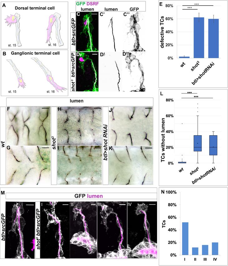
Research article                                                                           Cell Biology Developmental Biology

                                   ectopic acentrosomal branching points in the embryonic and larval tracheal TC leading to cells with
                                   extra-subcellular lumina. Furthermore, we find that Tau protein can functionally replace Shot in sub-
                                   cellular lumen formation and branching.

                                   Results
                                   Loss of shot causes defects in de novo subcellular lumen formation
                                   Shot is expressed during Drosophila development in several tissues such as the epidermis, the mid-
                                   gut primordia, the trachea and the nervous system (Lee and Kolodziej, 2002a; Röper and Brown,
                                   2003). We began by analysing the effect of shot LOF during TC subcellular lumen formation. To do
                                   so, we analysed dorsal (DB) and ganglionic branch (GB) TCs at late stages of embryogenesis (st.15/
                                   16) (Figure 1A,B).
                                       The shot3 null mutant TC phenotype consisted in subcellular lumen elongation defects with a pen-
                                   etrance of 100% per embryo (n = 40) and 62.5% per TC (n = 400) (Figure 1C,D and F–I and E). Of
                                   the total mutant TCs analysed, 25% did not develop a subcellular lumen (n = 600, Figure 1L). This
                                   phenotype resembled the previously reported for blistered (bs) mutants (Guillemin et al., 1996). bs
                                   encodes the transcription factor DSRF that regulates TC fate induction in response to Branchless-
                                   Breathless (Bnl-Btl) signalling (Gervais and Casanova, 2011; Guillemin et al., 1996). However, we
                                   observed that DSRF was properly accumulated in shot3 TC nuclei (Figure 1D), discarding a possible
                                   effect of Shot in TC fate induction.
                                       To analyse if the shot phenotype was tissue autonomous, we expressed shot-RNAi to knock-
                                   down Shot in all tracheal cells and found that, like in null mutant conditions, 60% of TCs analysed
                                   (n = 300) at the tip of the DBs (n = 150) or GBs (n = 150) were affected in subcellular lumen forma-
                                   tion (Figure 1E and J,K). Of these, 20% did not develop a terminal lumen at all (Figure 1L).
                                       shot3 embryonic TC lumen phenotypes range in expressivity from complete absence of subcellu-
                                   lar lumen to different lengths of shorter lumina (Figure 1M,N). When quantified in detail, out of the
                                   62.5% TCs that showed a luminal phenotype, 36% of TCs did not elongate a subcellular lumen at all
                                   (types III and IV) and 64% failed to accomplish a full-length lumen (types I and II) (n = 25)
                                   (Figure 1M,N).
                                       Recently, it has been reported that endocytosis is involved in subcellular lumen formation
                                   (Mathew et al., 2020). Bazooka (Baz), the Drosophila Par3, which is mainly associated with the api-
                                   cal membrane, has been shown to accumulate at the tip of the TC during lumen formation
                                   (Gervais and Casanova, 2010; Mathew et al., 2020). When we analysed this Baz accumulation in
                                   shot3 mutants, we could detect that it was very disrupted and no longer localised at the TC tip (Fig-
                                   ure 1—figure supplement 1).
                                       Taken together, these results indicated that Shot is involved in de novo subcellular lumen forma-
                                   tion and elongation.

                                   Shot overexpression induces extra-subcellular branching independently
                                   of the centrosome
                                   Having observed that Shot was necessary for subcellular lumen formation and extension, we hypoth-
                                   esised that Shot overexpression (ShotOE) would induce extra-subcellular branching events. Indeed,
                                   analysis of long-isoform ShotOE (shotA-GFP) in tracheal cells revealed that increasing Shot concen-
                                   trations induced extra-subcellular lumina (ESL) in GB and DB TCs (Figure 2A–C,J). Since MTs and
                                   actin are essential for subcellular lumen formation (Gervais and Casanova, 2010), we then asked
                                   whether supernumerary luminal branching was due to the MT- or the actin-binding domains present
                                   in the Shot molecule (Bottenberg et al., 2009; Voelzmann et al., 2017). To this end, we overex-
                                   pressed an isoform of Shot (ShotC-GFP) with a deletion of the first calponin domain (Figure 2K,L),
                                   resulting in a shorter actin-binding domain (ABD), which binds actin very weakly or not at all
                                   (Lee and Kolodziej, 2002a; Lee and Kolodziej, 2002a). The tracheal overexpression of shotA
                                   induced phenotypes in 95% of the embryos (n = 20), with an average of two TC bifurcations per
                                   embryo (n = 400). shotC overexpression induced phenotypes in 90% of the embryos (n = 20), with
                                   an average of two TC bifurcations per embryo (n = 400) (Figure 2D–F and G). In both cases approxi-
                                   mately 15% of all TCs analysed displayed an ESL phenotype (Figure 2J). In all cases, we could detect
                                   more MT-bundles in TCs, associated with the ESLs (Figure 2A–F and Figure 2—figure supplement

Ricolo and Araujo. eLife 2020;9:e61111. DOI: https://doi.org/10.7554/eLife.61111                                                 3 of 29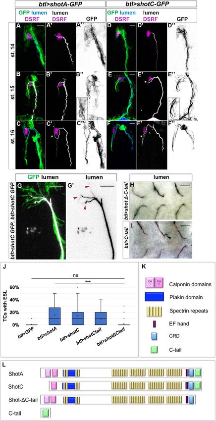
Research article                                                                                          Cell Biology Developmental Biology

Figure 1. shot loss-of-function induces defects in subcellular lumen formation. (A–B) Representation of dorsal and ganglionic TCs from embryonic st.15
to st.16 (DB and GB in grey, TC in pink). At st.15, the TC (cytoplasm in pink, nucleus in yellow, basal membrane in grey, apical membrane in blue and
lumen in white) emits filopodia in the direction of cell elongation; apical membrane grows in the same direction giving rise to the outline of the
subcellular lumen. At the same time the subcellular lumen is filled of chitin (white). At the end of st.16 the TC is elongated and the subcellular lumen is
Figure 1 continued on next page

Ricolo and Araujo. eLife 2020;9:e61111. DOI: https://doi.org/10.7554/eLife.61111                                                                    4 of 29

Research article                                                                                         Cell Biology Developmental Biology

Figure 1 continued
formed. (C–D) DBs at st.15 of btl >srcGFP (control) and shot3; btl >srcGFP fixed embryos stained with GFP to visualise tracheal cells, green in C and D,
grey in C’’ and D’’, CBP to visualise the lumen, white in C and D black in C’ and D’ and DSRF in magenta. Anterior side is on the left and dorsal is up,
scale bars 5 mm. (E) Quantification of total defective TCs in btl >shotRNAi (60%), shot3 (62.5%) and wt (2.25%) n = 20 embryos, 400TCs. Error bars
are ± SEM and asterisks represent a p-valueshotRNAi (J and K) (L) Quantification of total TCs
(genotype indicated) without subcellular lumen (wt 1.34% n = 400, shot3 25% n = 400, btl >shotRNAi 20%n = 300). *** p-valuesrcGFP control and shot3 embryos stained with GFP (grey) to visualise membrane and CBP (in
magenta) to visualise the lumen. Anterior side is on the left and dorsal side is up. Scale bars 5 mm. (I) TC partially elongated with formed lumen but with
wrong directionality (52%); (II) the elongation was stopped prematurely and a primordium of subcellular lumen was formed (12%); (III) the cell elongated
partially but the lumen was completely absent (16%); and (IV) the cell was not able to elongate and the lumen was completely absent (20%). Types III
and IV were quantified in L as TCs without lumen. (E) Detailed quantification, by confocal microscopy, of the different types of TC mutant phenotypes
reported as I-IV (n = 25 TCs).
The online version of this article includes the following source data and figure supplement(s) for figure 1:
Source data 1. Quantification of shot loss-of-function defects in subcellular lumen formation.
Figure supplement 1. Par3 vesicles are mislocalised in shot mutant TCs.

                                     1). ShotA-GFP and ShotC-GFP displayed different localisations within the TC. Full-length ShotA-GFP
                                     localisation can be detected at the cell-junctions, around the crescent lumen, in MT-bundles, and
                                     throughout the cytoplasm, whereas ShotC-GFP localised more to the MT/lumen region, in agree-
                                     ment with the lack of actin-binding capability of ShotC isoform (Figure 2—figure supplement 1).
                                     Interestingly, we observed a highly ramified subcellular lumen when higher amounts of ShotC were
                                     expressed in tracheal cells (Figure 2G) suggesting that the effect of ShotOE in subcellular lumen
                                     branching was dosage dependent.
                                         Tracheal overexpression of shotC phenocopied that of shotA in inducing ESLs (14.63% and 15.5%
                                     ESL respectively, Figure 2D–F and J), suggesting that the ABD is not necessary for the induction of
                                     additional luminal branching events. In order to clarify this, we used two other isoforms of Shot:
                                     shotDCtail, lacking the C-terminal MT-binding domain, and shotCtail, a truncated form containing
                                     only the C-terminal MT-binding domain (Alves-Silva et al., 2012; Figure 2K,L). Whereas overex-
                                     pressing shotD-Ctail in TCs we could only detect a branching phenotype in 1,5% of TCs analysed
                                     (n = 400), (Figure 2K), overexpression the C-tail domain alone induced TCs with extra branching in
                                     9.5% of TCs (n = 400) (Figure 2I), indicating that the C-tail alone was sufficient to induce ESLs in
                                     TCs. Taken together these results using different Shot isoforms, lead us to conclude that the Shot
                                     MT-binding domain alone is sufficient for the extra branching events observed in ShotOE TCs.
                                         ESLs were previously observed when higher numbers of centrosomes were present in TCs
                                     (Ricolo et al., 2016). We therefore asked if the observed extra branching phenotypes could be due
                                     to supernumerary centrosomes induced by ShotOE in TCs. Consequently, we quantified the number
                                     of centrosomes in the TCs of ShotOE embryos. In wt TCs we detected an average of 2.3 ± 0.5
                                     (n = 33) centrosomes per TC, and in ShotOE 2.2 ± 0.2 centrosomes per TC (n = 33) (Figure 3A–C).
                                     In both conditions, and as previously described (Ricolo et al., 2016), this centrosome pair was
                                     detected at the apical side of the TCs (Figure 3A,B). Besides, analysing ShotOE TCs at embryonic
                                     st.15, (n = 16) we could detect that the ESL arose from the pre-existing subcellular lumen, distally
                                     from the centrosome pair (Figure 3B’ arrow). These data indicate that ShotOE did not change TC
                                     centrosome number and induced ESL by a distinct mechanism from centrosome duplication.
                                         Regulator of cyclin A1 (Rca1) is the Drosophila ortholog of vertebrate Emi1 and a regulator of
                                     APC/C activity at various stages of the cell cycle (Grosskortenhaus and Sprenger, 2002). Rca1
                                     mutants have supernumerary centrosomes at the TCs and develop ESLs at embryonic stages
                                     (Ricolo et al., 2016). In contrast with ShotOE alone (Figure 3B’), in Rca1 mutants the bifurcated sub-
                                     cellular lumen arose from the apical junction and continued to extend during TC development
                                     (Ricolo et al., 2016). When we analysed the luminal origins in Rca1, ShotOE conditions, both types
                                     of ESL where detected in the same TC in 25% of the cases (n = 12). In the same TCs two types of
                                     ESL were generated, one from the apical junction and another sprouted from the pre-existing lumen
                                     distally from the junction (Figure 3D, asterisks). In addition, the effect of Rca1 LOF and ShotOE was
                                     additive in producing TCs with a multiple-branched subcellular lumen (Figure 3I). These

Ricolo and Araujo. eLife 2020;9:e61111. DOI: https://doi.org/10.7554/eLife.61111                                                                   5 of 29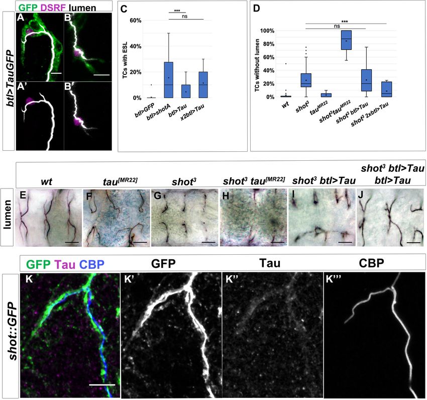
Research article                                                                                      Cell Biology Developmental Biology

Figure 2. ShotOE induces luminal branching through its microtubule-binding domain. Lateral view of DB tip cells from st.14 to st.16, of btl >shotA-GFP
embryos (A–C) and btl >shotCGFP (D–F). Embryos were stained with GFP (green in A-F and grey A’’-F’’) to visualise Shot-GFP, DSRF to mark the TC
nuclei (in magenta) and CBP to stain the chitinous lumen (blue in A-F and white A’-F’). Both overexpressing conditions induced ESLs (white stars). Note
the GFP was more diffuse in the cytoplasm of the TCs of embryos overexpressing shotA, and more organised in bundles in the TCs overexpressing
Figure 2 continued on next page

Ricolo and Araujo. eLife 2020;9:e61111. DOI: https://doi.org/10.7554/eLife.61111                                                                6 of 29
Research article                                                                                         Cell Biology Developmental Biology

Figure 2 continued
shotC. Anterior side of embryo is on the left and dorsal side up. Scale bars 5 mm. (G) ESL induction by ShotOE is dosage sensitive. Example of dorsal
TC of an embryo overexpressing two copies of btl >shotC-GFP, stained with anti-GFP (green) and CBP (white in G, black in G’). Red arrows indicate
extra-subcellular lumen branching. Note that the extra-subcellular lumina are very thin and they follow Shot positive bundles detected with GFP.
Anterior side is on the left, dorsal midline is on the top. Scale bars 5 mm. (H, I) Tips of GB TCs from btl >shotDCtail embryos with a single subcellular
lumen each (H) and btl >C-tail (I) in which one TC is bifurcated; stained with anti-Gasp (ventral view, anterior side of the embryo is on the left). Scale
bars 10 mm. (J) The C-tail domain is involved in ESL formation. Percentage of TCs displaying ESLs in embryos overexpressing GFP, shotA, shotC, C-tail,
and shotDCtail in the tracheal system (n = 400 TCs all genotypes except btl >shotC where n = 800). *** p-value0.1.
Statistics by two-tailed Student’s t-test. There was no significant difference between overexpression of shotDCtail and GFP alone. (K) Schematic
representation of spectraplakin protein domains and (L) the different Shot constructs used in this study.
The online version of this article includes the following source data and figure supplement(s) for figure 2:
Source data 1. Quantification of ShotOE phenotypes in subcellular lumen formation.
Figure supplement 1. ShotOE does not perturb the TC cytoskeletal organisation.

                                     morphological ESL differences suggested that Rca1 and shot operate in different ways in the de
                                     novo formation and branching of the subcellular lumen.

                                     Shot associates with stable microtubules and actin
                                     Spectraplakin expression is critical in cells that require extensive and dynamic cytoskeleton reorgan-
                                     isation, such as epithelial, neural, and migrating cells. Loss of spectraplakin function leads to a variety
                                     of cellular defects due to disorganised cytoskeletal networks (Hahn et al., 2016). In a plethora of tis-
                                     sues and in cultured S2 cells, Shot can physically interact with different cytoskeletal components
                                     (Applewhite et al., 2010; Lee and Kolodziej, 2002b; Sanchez-Soriano et al., 2009). Therefore, we
                                     investigated Shot localisation and its interaction with MTs and actin in control TCs.
                                         We analysed live embryos using time-lapse imaging and observed that Shot localisation was
                                     extremely dynamic throughout subcellular lumen formation. We could detect Shot in the apical TC
                                     junction as well as extending together with the growing subcellular lumen (Video 1 and Figure 4—
                                     figure supplement 1A). It was apparent that Shot localised dynamically with the growing luminal
                                     structures, showing a strong localisation at the middle/tip of the extending TC (Video 1 and Fig-
                                     ure 4—figure supplement 1A).
                                         In control conditions actin concentrated strongly at the tip of the TC, but was also detected in
                                     the TC cytoplasm, and these different actin populations have been shown to be important for sub-
                                     cellular lumen formation and extension (Gervais and Casanova, 2010; Oshima et al., 2006). During
                                     TC elongation, MTs polymerise from the centrosome pair at the apical junction toward the tip of the
                                     cell, reaching the area of high actin accumulation at the migrating tip of the TC (Gervais and Casa-
                                     nova, 2010; Ricolo et al., 2016). So, we next analysed ShotA and ShotC localisation in relation to
                                     the dynamically localised actin core present in the cytoplasm and at the tip of the migrating TC in
                                     live embryos (Videos 2, 3, and 4 and Figure 4—figure supplement 1). In both GB and DB TCs we
                                     could detect a dynamic interaction between the long Shot isoform (ShotA) and actin as detected by
                                     Moe::RFP (the ABD of moesin fused to RFP, thereby labelling the actin cytoskeleton of these cells,
                                     Video 4) or Life-actRFP (Video 3 and Figure 4—figure supplement 1C). ShotA dynamically inter-
                                     acted with different actin populations, namely the actin core and basal, filopodial actin (Videos 3
                                     and 4 and Figure 4—figure supplement 1C). However, as the lumen extended, ShotC fibres
                                     extended with the cell, surrounding the growing luminal area, like Shot A, but no strong interaction
                                     was detected with the dynamic actin core and with the basal, filopodial actin (Video 2 and Fig-
                                     ure 4—figure supplement 1B).
                                         We followed these analyses, observing endogenous Shot in fixed and antibody stained embryos.
                                     At early stages, when TCs started to elongate, we detected Shot co-localizing with actin at the tip of
                                     the TC (Figure 4A). The overlap between Shot and actin was maintained until late st.15 (Figure 4B).
                                     Then, we examined Shot localisation in relation to MTs. Shot was strongly detected in the TC from
                                     early stages of lumen extension and until the end of TC elongation (Figure 4D–E). At the beginning
                                     of de novo lumen formation, when MTs emanated from the junction/centrosome pair, Shot co-local-
                                     ised with the first sprouting stable MTs (Figure 4C–D). The overlap between Shot and stable MTs
                                     was strongly observed also at embryonic st.15 when a MT track preceded subcellular lumen

Ricolo and Araujo. eLife 2020;9:e61111. DOI: https://doi.org/10.7554/eLife.61111                                                                   7 of 29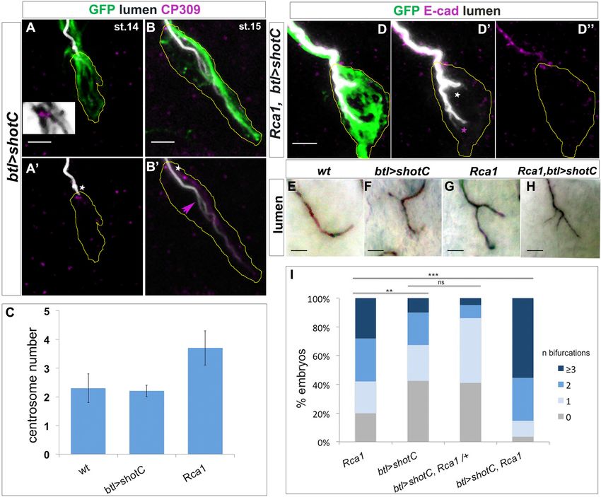
Research article                                                                                        Cell Biology Developmental Biology

Figure 3. ESL induction by ShotOE is not associated with centrosome amplification. (A,B) GB TC of a st.14 embryo (A), prior to lumen extension, and
st.15 when a bifurcated lumen can be detected in ShotOE conditions. (A,B) btl >shotC-GFP embryos stained with CBP to mark lumen (white), anti-GFP
to visualise Shot (green) and anti-CP309 antibody to mark centrosomes (magenta); the outline of TCs is drawn in yellow. The box in A is a magnification
of the apical side, showing the TC centrosome pair (in magenta) and GFP positive Shot bundles (in grey) emanating from centrosomes. White stars
indicate centrosomes apically localised. Note in B’ the subcellular lumen (magenta arrow) bifurcated in a point downstream from the centrosome pair.
Anti-CP309 antibody stains all centrosomes throughout the embryo and not just in TCs and cross-reacts with an unidentified antigen at the subcellular
lumen. (C) Quantification of centrosome number in wt, btl >shotC and Rca1 embryos ± SEM. (D) GB tips from Rca1; btl >shotC-GFP embryos at st.15,
stained with CBP (in white) to visualise the lumen and E-cadherin (magenta) to recognise the TC apical junction. Anterior side of the embryo is on the
left and ventral is down. Scale bar 2 mm. In these cases, it is possible to detect two types of luminal bifurcations: one from the apical junction (white
arrow), caused by Rca1 mutant supernumerary centrosomes, and another one arising from a pre-existing lumen, induced by ShotOE. (E–H) Details of
GB TCs at st.16 from embryos stained with anti-Gasp antibody to mark the lumen. (E) wt TCs with a single lumen each; (F) btl >shotC showing
subcellular lumen bifurcations; (G) Rca1 showing subcellular lumen bifurcations; (H) Rca1; btl >shotC showing a multi-branched subcellular lumen.
Anterior side of the embryo is on the left ventral midline is down. Scale bars 5 mm. (I) Quantification of the number of bifurcations (GB TCs) per embryo
of the indicated genotype. ‘n bifurcations’ is the number of ESL per TC.
The online version of this article includes the following source data for figure 3:
Source data 1. Quantification of centrosome number and luminal phenotypes.

Ricolo and Araujo. eLife 2020;9:e61111. DOI: https://doi.org/10.7554/eLife.61111                                                                  8 of 29
Research article                                                                                   Cell Biology Developmental Biology

                                                                                            Video 2. Shot localises with the extending subcellular
                                                                                            lumen in TCs during subcellular lumen extension. In
                                                                                            vivo Shot colocalisation with actin during lumen
                                                                                            formation in a wild-type dorsal branch (DB) TC. Time-
                                                                                            lapse images of a wt embryo expressing
                                                                                            btlGAL4UASShotC-GFP; btl::moeRFP visualised from a
                                                                                            dorsal view. Note the colocalisation of Shot with actin
                                                                                            the apical junction of the TC. As the cell extends,
                                                                                            ShotC localises to the growing lumen and there is no
                                   Video 1. Shot localisation within the TCs is dynamic     detectable strong association with the actin core and
                                   and accompanies the growing subcellular lumen. In        filopodial actin. Frames were taken every 2 min for 3.3
                                   vivo Shot localisation during lumen formation in two     hr.
                                   wild-type ganglionic branch (GB) TCs. Time-lapse
                                                                                            https://elifesciences.org/articles/61111#video2
                                   images of a wt embryo expressing btlGAL4UASShotC-
                                   GFP visualised from a dorsal view. Note the
                                                                                       detection (Figure 4D). At st.16, both Shot and
                                   accumulation of Shot at the apical junction of the TC
                                                                                       stable MTs localised to the apical side of the TCs
                                   and subsequently in association with subcellular lumen
                                   extension. Frames were taken every minute for 3.5 hr.
                                                                                       in the area surrounding the subcellular lumen
                                   https://elifesciences.org/articles/61111#video1     (Figure 4E).
                                                                                          Shot localisation within the TC suggested that
                                                                                       the spectraplakin localised with stable MTs all
                                                                                       around the nascent lumen and with the actin at
                                   the tip of the TC, during the time of cell elongation and subcellular lumen formation. This suggests
                                   that Shot mediates the crosstalk between these two cytoskeletal components, helping their stabilisa-
                                   tion and organisation during subcellular lumen formation and growth (Figure 4F).

                                   Absence of shot leads to disorganised microtubules and actin
                                   We then asked how actin and MTs were localised and organised in shot3 mutant embryos. We ana-
                                   lysed the different types of TC mutant phenotypes ranging from cases in which the TC did not elon-
                                   gate and the subcellular lumen was not formed, to cases in which the TC was able to elongate and
                                   form the lumen albeit not to the levels in control embryos (Figure 5). In all cases, we found defects
                                   in both MTs and actin accumulation in mutant TCs.
                                      Considering actin localisation, in control embryos at early st.16, Moe::RFP detecting actin was
                                   strongly localised at the tip of the TC, in front of the tip of the growing lumen (86% of TCs analysed,
                                   n = 21). Moreover, a few spots of actin were detectable in the cytoplasm, around the subcellular
                                   lumen (Figure 5A and Video 2). In shot3, we observed reduced actin accumulation at the TC-tip and
                                   an increase of scattered spots into the cytoplasm (86% of TCs analysed, n = 23) (Figure 5B–D), indi-
                                   cating that Shot contributed to TC actin organisation.
                                      Regarding MT-bundles, we observed stable MTs organised in longitudinal bundles around the
                                   subcellular lumen in control TCs (Figure 5E). In shot3 TCs (n = 20), we detected MT-bundle defects.
                                   In particular, we observed that when the TC was not elongated, MT-bundles no longer localised to
                                   the apical region and seemed to be fewer than in wt (Figure 5F). A general disorganisation in MT-

Ricolo and Araujo. eLife 2020;9:e61111. DOI: https://doi.org/10.7554/eLife.61111                                                             9 of 29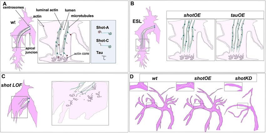
Research article Cell Biology Developmental Biology
Video 3. Shot localises with actin in TCs during the
early steps of subcellular lumen extension. In vivo Shot
long-isoform colocalisation with actin during lumen
formation in a wild-type dorsal branch (DB) TC. Time-
lapse images of a wt embryo expressing
btlGAL4UASShot-A-GFPUASlifeActRFP visualised from Video 4. Shot localises with actin in TCs during later
a dorsal view. Note the colocalisation of Shot with actin steps of subcellular lumen extension. In vivo Shot long-
at the apical junction of the TC and subsequent isoform colocalisation with actin during lumen
association with the actin core and filopodial actin formation in a wild-type dorsal branch (DB) TC. Time-
during subcellular lumen extension. Frames were taken lapse images of a wt embryo expressing
every 40 s for 32 min. btlGAL4UASShotA-GFP; btl::moeRFP visualised from a
https://elifesciences.org/articles/61111#video3 dorsal view. Note the colocalisation of Shot with
filopodial actin during subcellular lumen extension.
Frames were taken every 30 s for 36 min.
bundles in respect to the control was also https://elifesciences.org/articles/61111#video4
observed in TCs partially able to elongate a sub-
cellular lumen (Figure 5G,H).
These analyses, taken together with the previous analysis of Shot localisation in control TCs, sug-
gested a spectraplakin role in organizing/stabilizing both MTs and Actin accumulation in the TC.
Subcellular branching depends on both actin and microtubule-binding
domains of shot
In order to analyse how the different domains of Shot affected luminal development and branching,
we expressed different isoforms of Shot in shot3 mutant TCs. As described previously, shot3
embryos displayed a variable expressivity in TC phenotypes. To simplify the quantification of the res-
cue experiments, we took in consideration the most severe luminal phenotype: the complete
absence of a subcellular lumen. In shot3, we quantified that 22% of TCs (at the tip of GBs and DBs)
did not develop a subcellular lumen at all (Figure 1H,I and Figure 6B,J). Targeted expression of full-
length ShotA in the trachea of shot3 mutant embryos was able to rescue the subcellular lumen phe-
notype to the level of only 6% of the TCs analysed (n = 200) not developing a subcellular lumen
(Figure 6C).
We then proceeded to molecular dissect the function of Shot in TCs. To do so, we used the three
different constructs Shot: ShotC, ShotDCtail and ShotCtail (Figure 2L). When we expressed ShotC in
the tracheal TCs we found that 20% of TCs analysed (n = 200), had TCs with no lumen (Figure 6D
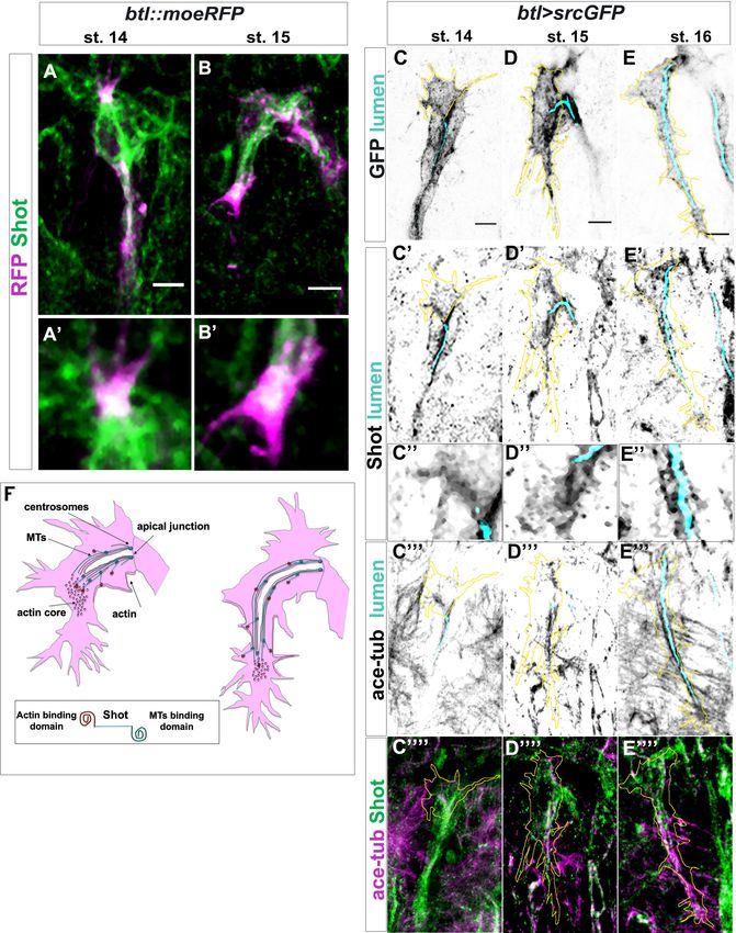
Ricolo and Araujo. eLife 2020;9:e61111. DOI: https://doi.org/10.7554/eLife.61111 10 of 29Research article Cell Biology Developmental Biology Figure 4. Shot colocalises with TC cytoskeletal components. (A–B) Endogenous Shot, detected by an antibody in btl::moeRFP embryos, colocalised with actin during TC development. Tip of dorsal branches from st. 14 to late st. 15 of btl::moeRFP embryos stained with RFP (magenta) and Shot (green). In the magnification of the tip of the TCs (A’–B’) note Shot and RFP co-localisation in both embryonic stages. (C–E) Endogenous Shot accumulated around stable microtubules during subcellular lumen formation. The whole TC morphology changes overtime. This is a developing Figure 4 continued on next page Ricolo and Araujo. eLife 2020;9:e61111. DOI: https://doi.org/10.7554/eLife.61111 11 of 29
Research article                                                                                                 Cell Biology Developmental Biology

Figure 4 continued
structure and both cell-shape and the cytoskeleton are changing throughout its development. Here we provide snapshots of different stages showing
the beginning, middle and final stages of TC lumen formation. Dorsal TCs from fixed embryos btl >srcGFP stained with Shot and acetylated-tubulin
antibodies and fluostain to detect chitin, from st.14 to st.16. GFP staining is showed in grey and cell contour in yellow (C–E), endogenous Shot is shown
in grey in panels C’- E ‘’ (C’’- E’’ are magnification of C’- E’) and green C’’’’- E’’’’. Acetylated tubulin is in grey in C’’’- E’’’ and magenta in C’’’’ -E’’’’. The
chitinous lumen was detected with fluostain, represented in cyan (C– E’’’). Acetylated tubulin and Shot are both accumulated ahead of the subcellular
lumen at earliest stages (st. 14–15) and around the subcellular lumen at later stages (st.16). Note that co-localisation between acetylated tubulin and
Shot is detected in the TCs. Anterior side is on the left, dorsal midline is up. Scale bar 5 mm. (F) Schematic representation of dorsal TC development
from st.15 to st.16. Basal membrane in grey, apical membrane in light blue, subcellular lumen in white, the actin network in red and MTs are in green.
Between st.14 and st.15 actin dots mature in an actin core in front of the tip of the subcellular lumen in formation that is surrounded by microtubules.
Shot (represented on the bottom of the figure) was detected both inside the actin core and surrounding the lumen where stable MTs are organised.
The online version of this article includes the following figure supplement(s) for figure 4:
Figure supplement 1. Shot is dynamically localised in TCs and interacts with actin.

                                        and J), suggesting that the ABD domain is necessary for the correct de novo luminal
                                        morphogenesis.
                                            We next expressed shotC-tail in order to address whether the Shot MT-binding domain alone
                                        could restore subcellular lumen formation. We observed that 24% of TCs analysed at the tip of GBs
                                        and DBs (n = 250) were still not able to form a subcellular lumen (Figure 6E and J), suggesting that
                                        the tracheal expression of shotC-tail was not enough to rescue the null phenotype. Finally, we
                                        expressed shot-DC-tail to test whether Shot without the MT-binding domain could restore subcellu-
                                        lar lumen formation. We observed that 16% of TCs analysed at the tip of GBs and DBs (n = 250)
                                        were still unable to form a subcellular lumen (Figure 6F and J). Taken together, these analyses sug-
                                        gested that full-length isoform A, allowing Actin-MT crosslinking is necessary for correct de novo
                                        subcellular lumen formation.
                                            In order to further test the hypothesis that full-length Shot is needed to correctly form a subcellu-
                                        lar lumen, we analysed shotkakP2 mutant phenotype. This allele carries an insertion of a transposable
                                        element into the intron between the second and the third transcriptional start site of shot abolishing
                                        all isoforms containing the first Calponin domain (CH1) and interfering with Shot actin-binding activ-
                                        ity (Bottenberg et al., 2009). The penetrance and expressivity of the phenotype observed in shot-
                                        kakP2
                                               TCs was very similar to shot3 null allele with 18% of these (n = 600; 300 ganglionic and 300
                                        dorsal TCs) not forming a subcellular lumen at all (Figure 6G,J). In addition, shotkakP2 TCs display
                                        the same MT and actin disorganisation phenotypes as shot3 TCs (Figure 6—figure supplement 1B–
                                        D). Phenotypic data from shotkakP2 together with data from transgenic rescues with the ShotC con-
                                        struct, lacking the CH1 domain, indicate that Shot full length is required for de novo subcellular
                                        lumen formation.
                                            Since the actin and MT-binding domains were shown to be necessary for the proper formation of
                                        a subcellular lumen, we asked whether it was necessary to have both domains in the same protein or
                                        if simply the independent presence of these domains was enough to generate a subcellular lumen.
                                        To do so, we generated transheterozygous flies expressing two different Shot isoforms, ShotkakP2
                                        and ShotDEGC. ShotDEGC is a truncated protein, lacking the EF-hand, the Gas2 and the C-tail domains
                                        of Shot, leading to complete loss of the MT-binding activity (Takács et al., 2017). The analysis of
                                        shotDEGC mutant TC phenotypes revealed that 18% of TCs (n = 400; 200 ganglionic and 200 dorsal
                                        TCs) did not develop a TC lumen at all (Figure 6H,L) and that shotDEGC mutant TCs display the same
                                        MT and actin disorganisation phenotypes as shot3 TCs (Figure 6—figure supplement 1E–G).
                                            In shotDEGC/shotkakP2 transheterozygous embryos, Shot molecules contained exclusively either the
                                        CH1 or the C-tail, but neither molecule had actin- and MT-binding activity simultaneously. These
                                        embryos displayed the same phenotype as either homozygous mutant (18% TCs with no lumen,
                                        n = 400) (Figure 6I), indicating that both the actin- and the MT-binding domains need to be present
                                        in the same Shot molecule for proper TC subcellular lumen formation.
                                            Taken together these results indicate that Shot is able to mediate the crosstalk between MTs and
                                        actin during subcellular lumen formation, via its MT and actin-binding domains and that these have
                                        to be present in the same molecule for proper subcellular lumen formation.

Ricolo and Araujo. eLife 2020;9:e61111. DOI: https://doi.org/10.7554/eLife.61111                                                                             12 of 29
Research article Cell Biology Developmental Biology Figure 5. Shot LOF lead to disorganised MT-bundles and actin. (A–D) Asymmetric actin accumulation in extended TCs was affected in shot3 mutant embryos. Dorsal TC from fixed shot3/+; btl::moeRFP controls (A) and shot3; btl::moeRFP mutant embryos (B–D), stained with RFP (Magenta in A-D or in a colour scale in which blue is low, green is middle and red high intensity in A’- D’) and CBP (in white). In shot mutant TCs actin appeared affected in its accumulation (B–D). (A) control; (B) when the cell was not elongated and the lumen was not formed; (C) when the cell was partially elongated and the lumen was not formed; (D) when the cell elongated and the lumen was partially formed (D). Note that actin was affected even when the cell was elongated and a lumen was partially formed. (E–H) TC MT-bundles in shot3. Dorsal TC from embryo at st.16 control (A) and shot3 mutant embryos (B– D) stained with GFP (green) acetylated tubulin (in magenta in E-H and in grey in E’’-H’’) and CBP (in blue in E-H and grey in E’’’-H’’’). The border of TC was drawn in cyan (E–H’’). In all cases, the organisation and the amount of stable MTs was strongly affected, in (F) MT-bundles were observed to be disorganised along the cytoplasmatic protrusion without subcellular lumen and in G and H only a thin track of MTs surrounds the subcellular lumen. Anterior side is on the left and dorsal midline is up. Scale bars 5 mm. Ricolo and Araujo. eLife 2020;9:e61111. DOI: https://doi.org/10.7554/eLife.61111 13 of 29
Research article Cell Biology Developmental Biology
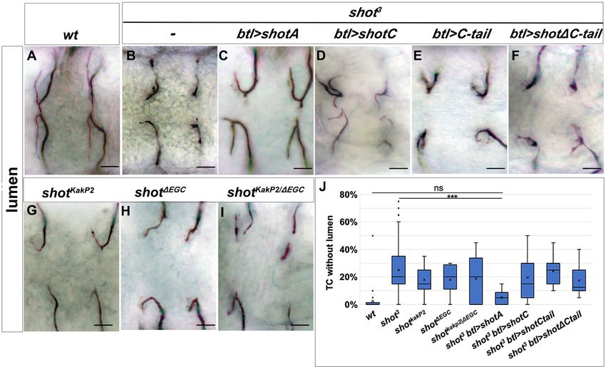
Figure 6. Shot Actin- and MT-binding domains are necessary for proper subcellular lumen formation. (A–I) Dorsal branches of st.16 embryos, stained
with anti-GASP to visualise the lumen. Genotype is indicated above each panel. Null allele, shot3, rescue experiments (B–F) indicate that both the actin-
binding domain (ABD) and the microtubule-binding domain are involved in subcellular lumen formation since the only construct able to rescue the null
allele phenotype is the UASShotA (B and J). Both functional domains are needed in the same molecule since mutants affected only in the ABD
(shotKakP2) or in the microtubule-binding domain (shotDEGD) and the transheterozygous shot KakP2/DEGD display the same phenotype as shot3. Scale bars
are 10 mm. (J) Quantification of TCs without lumen: wt (n = 820); shot3 (n = 600); shotKakP2 (n = 400); shotDEGD (n = 400); shot KakP2/DEGD (n = 320 TCs);
shot3, btl >shotA (n = 240); shot3; btl >shotC (n = 240); shot3; btl >shotCtail (n = 240); shot3; btl >shotDCtail (n = 240). *** p-value0.1. Statistics by two-tailed Student’s t-test. Only ShotA significantly rescued the shot3 TC luminal phenotype.
The online version of this article includes the following source data and figure supplement(s) for figure 6:
Source data 1. Quantification of rescue experiments and different shot mutant phentotypes and transheterozygous combinations.
Figure supplement 1. Asymmetric actin accumulation is affected in shot kakp2 and shot DEGC Dorsal TCs from btl::moeRFP.
Increased levels of shot are induced in TCs by DSRF
The TC-specific transcription factor bs/DSRF is important for TC specification and growth, and has
been suggested to regulate the transcription of genes that modify the cytoskeleton
(Guillemin et al., 1996; Olson and Nordheim, 2010). Considering the luminal phenotypes associ-
ated with bs LOF in TCs and the role of MTs in subcellular luminal formation, we asked whether shot
expression in TCs could be regulated by DSRF.
In order to test this, we searched in silico for DSRF binding sites in the promoter regions of all
shot isoforms using the Matscan software (Blanco et al., 2006) and the reported position weight
matrix (PWM) corresponding to SRF (Khan et al., 2018; Supplementary file 1). We found seven
regions with at least one putative binding site (binding score larger than 70% of maximum value)
within 2000 bases of the shot annotated TSS (Figure 7F and Supplementary file 1). These regions
mapped to the locations of known Shot promoters (Figure 7F; Hahn et al., 2016). We then asked if
lower Shot protein levels could be detected in bs mutant TCs. Indeed, when analysing bs in compari-
son to bs/+ TCs, we could detect lower levels of endogenous Shot protein and shot mRNA
Ricolo and Araujo. eLife 2020;9:e61111. DOI: https://doi.org/10.7554/eLife.61111 14 of 29Research article Cell Biology Developmental Biology Figure 7. Shot expression is regulated by DSRF in TCs GB TC at st.15 from bs heterozygous controls. (A) and homozygous (B) mutant embryos, stained with Shot (magenta in A and B, grey in A’ and B’), DSRF (green) antibodies and CBP (grey). In yellow, the outline of the TCs. Shot protein was less accumulated in TCs from homozygous bs embryos (B, B’ and E) n = 9 TCs. Scale bars are 5 mm. (C,D) DB TCs from bs heterozygous (C) control and homozygous (D) mutant embryos from wholemount FISH with a ribo-shot probe (magenta), stained with anti-betagal (green, to detect the DSRF Figure 7 continued on next page Ricolo and Araujo. eLife 2020;9:e61111. DOI: https://doi.org/10.7554/eLife.61111 15 of 29
Research article                                                                                         Cell Biology Developmental Biology

Figure 7 continued
enhancer trap lacZ expression) and CBP (blue) to mark the lumen. LacZ expression is higher in mutant embryos, homozygous for the lacZ P-element
insertion (D). The yellow line marks the TC outline. Lower levels of shot mRNA were detected in bs mutant TCs when compared to control TCs (n = 8).
Scale bars are 5 mm. (E) Quantification of Shot protein in control and mutant TCs and stalk cells (SC). Quantification of raw integrated pixel density in
arbitrary units measured in Fiji in the TC and attached SC in each embryo. **p0.1. Statistics by two-tailed Student’s t-test.
(F) P1, P2 and P3 transcription start sites of the shot locus together with the specific sequences recognised by the DSRF transcription factor (squares in
magenta) (adapted from Hahn et al., 2016). Dorsal and ventral TCs from control (G and J) bs (H and K) mutant embryos. The tracheal overexpression
of Shot is sufficient to restore the growth of TC subcellular lumina in bs mutant background (I, L). (M) Quantification of TCs with an extended lumen: bs/
+ (n = 350); bs/bs (n = 280) and bs/bs;btl >Shot (n = 210). *** p-value
Research article                                                                                          Cell Biology Developmental Biology

Figure 8. Shot and Tau functionally overlap during subcellular lumen formation. (A–B) DB (A) and GB (B) embryonic TCs expressing tauGFP in the
tracheal system, stained with GFP (green), CBP (white) and DSRF (magenta), showing the ESL phenotype induced by Tau overexpression. In A’ in B’
lumen and TC nuclei are shown, anterior side on the left, dorsal side is up; scale bar 5 mm. (C) Quantification of TCs with ESL in embryos
overexpressing GFP (n = 240); ShotA (n = 400); one copy of btl >tauGFP (n = 440) or two copies of btl >tauGFP (n = 300). ***p-value0.1. Statistics by two-tailed Student’s t-test. (D) Quantification of TCs without subcellular lumen in control (n = 820), shot3(n = 600), tau[MR22]
(n = 180), shot3; tau[MR22](n = 180), shot3; btl >Tau (n = 440) and shot3; btl >Tau btl >Tau (n = 260) embryos. ***p-value0.1. Statistics by two-tailed Student’s t-test. (E–J) Dorsal view of TCs from st. 16 embryos (genotype indicated) stained with anti-Gasp. tau
deletion mutant does not display a subcellular lumen phenotype (D and F) but enhances the effect of shot mutation in the double mutant shot3;
tau[MR22]. One copy of Tau is not sufficient to rescue shot3 (D and I, n = 400) but two copies rescues the shot LOF TC phenotype (D and J n = 260).
Scale bars 10 mm. (K) Tau is detected in embryonic TCs. Embryonic shot::GFP dorsal TC stained with GFP (green in K, grey in K’), anti-Tau antibody
(magenta in K, grey in K’) and CBP (blue in K grey in K’). Scale bar 5 mm.
The online version of this article includes the following source data and figure supplement(s) for figure 8:
Source data 1. Quantification of tau mutant and overrexpression phenotypes and rescues.
Figure 8 continued on next page

Ricolo and Araujo. eLife 2020;9:e61111. DOI: https://doi.org/10.7554/eLife.61111                                                                    17 of 29
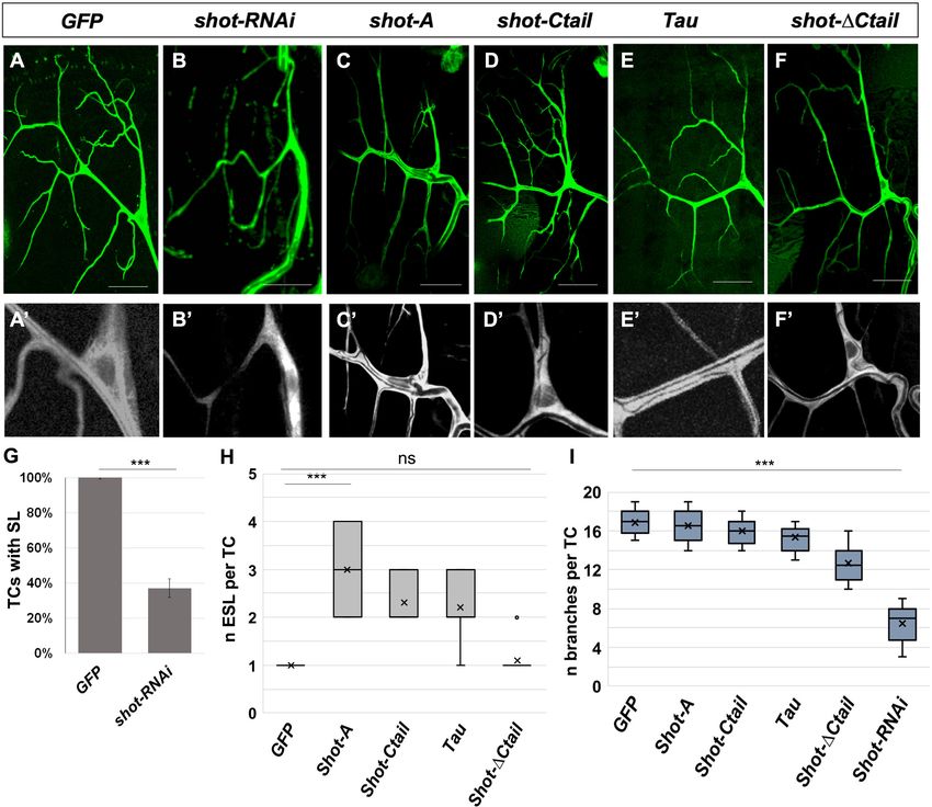
Research article                                                                                          Cell Biology Developmental Biology

Figure 8 continued
Figure supplement 1. MT disorganisation in shot mutant TCs is rescued by ShotA and Tau, but not ShotC.
Figure supplement 2. Actin disorganisation in shot mutant TCs is rescued by ShotA and Tau, but not ShotC.
Figure supplement 3. Double shot3;tauMR22 mutant embryos display defects in lumen formation but not in tracheal cell number or TC fate.

Figure 9. Shot and Tau modulate luminal branching in larval TCs. Wandering larval (L3) TCs expressing only GFP (A) and different Shot and Tau
constructs (B, C, D, E, F) under the control of a tracheal DSRFGAL4 driver (all except A and E where the driver used was btlGAL4). (A, A’) UASGFP
(n = 8) (B, B’) UASshotRNAi, UASGFP (n = 8); (C, C’) UASShotA-GFP (n = 10); (D, D’) UASshotCtail-GFP (n = 8); (E, E’) UASTauGFP (n = 8); (F,F’)
UASshotDCtail-GFP (n = 8). Scale bars 50 mm. (G) Quantification of the percentage of TCs with subcellular lumen; (H) quantification of the number of
ESL per TC; (I) quantification of the number of branches per larval TC. *** represent a p-value0.1. Statistics by two-tailed
Student’s t-test.
The online version of this article includes the following source data for figure 9:
Source data 1. Quantification of larval branching phenotypes.

Ricolo and Araujo. eLife 2020;9:e61111. DOI: https://doi.org/10.7554/eLife.61111                                                                   18 of 29
Research article                                                                                       Cell Biology Developmental Biology

                                     6.5 ± 0.6 branch points each (n = 8) (Figure 9B and I). We then overexpressed the long isoform of
                                     Shot (ShotA-GFP aka ShotOE condition) and could not detect extra branching points in TCs, sug-
                                     gesting that more than just an increased Actin-MT crosstalk is needed for the induction of TCs with
                                     supernumerary cytoplasmatic extensions (Figure 9C). Nonetheless, overexpression of ShotA, ShotC-
                                     tail and Tau induced ESL in TCs, with two or more lumina in all TCs analysed (n = 10) (Figure 9C–E
                                     and H). Like in embryos, targeted expression of Shot-DC-tail did not induce ESL in larval TCs
                                     (Figure 9F and H). Taken together, these results indicate that Shot is necessary for larval lumen for-
                                     mation and branching and that Actin-MT crosstalk by Shot or Tau is sufficient for ESL formation
                                     within each TC cytoplasmatic extension.

                                     Discussion
                                     In this study, we analysed the importance of MT-actin crosstalk through Shot and Tau in subcellular
                                     lumen formation in Drosophila embryonic and larval tracheal cells. Our work reveals novel insights
                                     into the formation of lumina by single-cells. First, that a spectraplakin in involved in the crosstalk
                                     between actin and MTs in tracheal TCs and that this crosstalk is necessary for de novo lumen forma-
                                     tion. Absence of Shot leads to defects in MT and actin organisation and a profound alteration of the
                                     cytoskeleton in TCs (Figure 10A,C). Consequently, membrane delivery is disrupted and a novel sub-
                                     cellular lumen cannot be formed. Second, that once a primary lumen is formed de novo in TCs, nei-
                                     ther actin-MT crosstalk, nor supernumerary centrosomes, are necessary for the formation of new

Figure 10. Shot and Tau dynamically modulate the cytoskeleton during subcellular lumen formation. Schematic representation of st.16 embryonic (A, B,
C) and third instar larval (D) TCs; cytoplasm is in pink and luminal space in white. (A) Cytoskeletal components in a wt embryo with the actin-network
(dark pink) and MTs (green). Shot and Tau are able to organise the cytoskeleton by crosslinking MTs and actin; Shot (represented with the actin domain
in red and the MT-binding domain in green) mediates the crosstalk between actin and MTs as the longer isoform (ShotA), but shorter isoforms lacking
part of the ABD were reported not to bind/or very weakly bind actin (ShotC). Tau is represented in blue. (B) ESLs are formed by overexpressing shot or
tau by an excess of MT stabilisation from the pre-existing lumen, which probably acts as a MTOC in this case. ESLs can be induced by Shot isoforms
with affected (ShotC) or without ABD (ShotC-tail). (C) In the absence of the longer isoform of Shot (ShotA) proper cytoskeletal organisation, is not
established, by defective MT-actin crosslinking, and cell elongation and lumen formation fail to occur. (D) Schematic representation of larval TCs in wt,
in shotOE (or tauOE) where ESLs are formed without concomitant single-cell branching and in shot KD, where both single-cell and luminal branching
are reduced.

Ricolo and Araujo. eLife 2020;9:e61111. DOI: https://doi.org/10.7554/eLife.61111                                                                19 of 29
Research article Cell Biology Developmental Biology
supernumerary lumina (ESLs). New lumina can arise from branching points along the length of the
pre-existing lumen, only by MT stabilisation by isoforms of Shot lacking entirely the ABD
(Figure 10B,D). In these cases, we can form ESLs acentrosomally, perhaps from the MTOC activity
provided by the gamma-tubulin present along the crescent lumen (Gervais and Casanova, 2010) or
by other types of MTOCs. Third, spectraplakin activity is necessary to organise MTs and actin in TCs;
without Shot TCs exhibit a disrupted MT and actin cytoskeleton, which can be restored by tissue
specific expression of this spectraplakin. Fourth, increased levels of Shot are induced in TCs by
DSRF, and Shot can rescue the subcellular lumen formation phenotypes in bs mutants. This agrees
with previous observations in other systems where bs and shot mutants display similar phenotypes
(Prout et al., 1997). And fifth, high-levels of Tau can replace Shot in subcellular lumen formation
and branching.
Shot promotes subcellular branching by organizing and mediating the
crosstalk between microtubules and actin
Previously, it was shown that Shot was involved in tracheal fusion cell anastomosis during embryonic
development (Lee and Kolodziej, 2002a). It was observed that Shot accumulates at E-cadherin-
dependent contacts between fusion cells and shot LOF disrupts this contact leading to cell-fusion
phenotypes. In these cells, interactions of Shot with F-actin and microtubules are functionally redun-
dant and both targeted expression of ShotC or ShotA is sufficient to rescue the cell-fusion pheno-
type (Lee and Kolodziej, 2002a). Our results are more akin to what has been reported in neuronal
growth cones, and both actin and MT-binding domains of Shot are required for TC extension and
subcellular lumen formation (Figure 10A). In neurons, like in tracheal cells, ShotC is unable to rescue
the phenotype caused by shot LOF, which is only rescued by expression of the full-length ShotA iso-
form (Lee and Kolodziej, 2002b). Shot has also been shown to be required for sealing epithelial
sheets during dorsal closure (Takács et al., 2017). In these epithelial cells, Shot acts as a MT-actin
crosslinker to regulate proper formation of the MT network. As in the case of tracheal TCs presented
here, the actin- and microtubule-binding activities of Shot are simultaneously required in the same
molecule, indicating that like in TCs Shot is engaged as a physical crosslinker also during dorsal clo-
sure (Takács et al., 2017).
MTs and the actin cytoskeleton perform many functions in tracheal TCs that are regulated by dif-
ferent actin- and MT-binding proteins. While mediators of actin function, such as Ena (Gervais and
Casanova, 2010), and of MT function, like D-Lissencephaly-1 (DLis-1), have been identified previ-
ously, we show here that Shot is able to mediate crosstalk between MTs and actin during subcellular
lumen formation. In Shot LOF conditions, MTs and actin are disorganised. Consequently, this Shot
crosslinking function is essential for de novo lumen formation and extension. It has been previously
described that in TCs of mutants affected in MT organisation, the actin-network is not perturbed
(Gervais and Casanova, 2010), so the ‘actin phenotype’ observed in shot LOF cannot be a conse-
quence of defects in the MT network. This observation indicates a possible spectraplakin function in
organizing TC actin in agreement with previous observations that Shot and ACF7 can promote filo-
podia formation (Lee et al., 2007; Sanchez-Soriano et al., 2009).
Shot expression is regulated by DSRF in TCs
Our results show that molecular levels of Shot are important for cytoskeletal rearrangements, indi-
cating that there is a dosage dependent effect in lumen formation and extension as well as in luminal
branching events. Shot is present in many cells during development but Shot level regulation is likely
to be more important in cells such as neurons and tracheal terminal cells, due to their morphology
(Voelzmann et al., 2017). bs/DSRF is a TC-specific transcription factor, whose expression is trig-
gered by Bnl signalling (Guillemin et al., 1996; Sutherland et al., 1996), and is required for TC cyto-
skeletal organisation (Gervais and Casanova, 2010). DSRF has also been shown to be necessary not
just for the establishment of TC fate, but to ensure the progression of TC elongation (Gervais and
Casanova, 2011). Cytoskeletal organisation and remodelling as well as TC elongation are tightly
coupled during subcellular lumen formation and in bs mutants actin accumulation was impaired at
the TC tip (Gervais and Casanova, 2010). We observe a similar actin phenotype in Shot mutants
(Figure 5A–D) suggesting that the actin defects observed in DSRF mutants may be due to a lower
expression of Shot in these cells.
Ricolo and Araujo. eLife 2020;9:e61111. DOI: https://doi.org/10.7554/eLife.61111 20 of 29You can also read