Human Cytomegalovirus Host Interactions: EGFR and Host Cell Signaling Is a Point of Convergence Between Viral Infection and Functional Changes in ...
←
→
Page content transcription
If your browser does not render page correctly, please read the page content below
REVIEW
                                                                                                                                                 published: 07 May 2021
                                                                                                                                        doi: 10.3389/fmicb.2021.660901

                                             Human Cytomegalovirus Host
                                             Interactions: EGFR and Host Cell
                                             Signaling Is a Point of Convergence
                                             Between Viral Infection and
                                             Functional Changes in Infected Cells
                                             Byeong-Jae Lee 1,2,3 , Chan-Ki Min 1,2,3 , Meaghan Hancock 4 , Daniel N. Streblow 4 ,
                                             Patrizia Caposio 4 , Felicia D. Goodrum 5 and Andrew D. Yurochko 1,6,7,8*
                                             1
                                               Department of Microbiology & Immunology, Center for Molecular and Tumor Virology, Louisiana State University Health
                                             Sciences Center Shreveport, Shreveport, LA, United States, 2 Center for Applied Immunology and Pathological Processes,
                                             Louisiana State University Health Sciences Center Shreveport, Shreveport, LA, United States, 3 Center of Excellence
                                             for Emerging Viral Threats, Louisiana State University Health Sciences Center Shreveport, Shreveport, LA, United States,
                                             4
                                               Vaccine and Gene Therapy Institute, Oregon Health & Science University, Beaverton, OR, United States, 5 BIO5 Institute,
                          Edited by:         University of Arizona, Tucson, AZ, United States, 6 Feist-Weiller Cancer Center, Louisiana State University Health Sciences
               Maria Isabel Colombo,         Center Shreveport, Shreveport, LA, United States, 7 Center for Cardiovascular Diseases and Sciences, Louisiana State
       Universidad Nacional de Cuyo,         University Health Sciences Center Shreveport, Shreveport, LA, United States, 8 Center of Excellence in Arthritis
                            Argentina        and Rheumatology, Louisiana State University Health Sciences Center Shreveport, Shreveport, LA, United States

                     Reviewed by:
                       John Sinclair,        Viruses have evolved diverse strategies to manipulate cellular signaling pathways
           University of Cambridge,
                                             in order to promote infection and/or persistence. Human cytomegalovirus (HCMV)
                    United Kingdom
                 Laura Ruth Delgui,          possesses a number of unique properties that allow the virus to alter cellular events
       CONICET Mendoza, Argentina            required for infection of a diverse array of host cell types and long-term persistence.
                  *Correspondence:           Of specific importance is infection of bone marrow derived and myeloid lineage cells,
                  Andrew D. Yurochko
                   ayuroc@lsuhsc.edu         such as peripheral blood monocytes and CD34+ hematopoietic progenitor cells (HPCs)
                                             because of their essential role in dissemination of the virus and for the establishment of
                   Specialty section:        latency. Viral induced signaling through the Epidermal Growth Factor Receptor (EGFR)
         This article was submitted to
               Microbial Immunology,         and other receptors such as integrins are key control points for viral-induced cellular
               a section of the journal      changes and productive and latent infection in host organ systems. This review will
             Frontiers in Microbiology
                                             explore the current understanding of HCMV strategies utilized to hijack cellular signaling
        Received: 01 February 2021
           Accepted: 07 April 2021
                                             pathways, such as EGFR, to promote the wide-spread dissemination and the classic
           Published: 07 May 2021            life-long herpesvirus persistence.
                              Citation:
                                             Keywords: human cytomegalovirus (HCMV), monocytes, progenitor cells, epidermal growth factor receptor
       Lee B-J, Min C-K, Hancock M,          (EGFR), glycoproteins, cell signaling, differentiation, latency
              Streblow DN, Caposio P,
Goodrum FD and Yurochko AD (2021)
        Human Cytomegalovirus Host
     Interactions: EGFR and Host Cell
                                            INTRODUCTION
  Signaling Is a Point of Convergence
Between Viral Infection and Functional
                                            Human cytomegalovirus (HCMV) infection is a significant public health threat worldwide (Stagno
            Changes in Infected Cells.      et al., 1986; Bentz et al., 2006; Arvin, 2007; Nogalski et al., 2014; Fulkerson et al., 2021). HCMV is
          Front. Microbiol. 12:660901.      a leading cause of morbidity and mortality in developing fetuses and the immunocompromised
    doi: 10.3389/fmicb.2021.660901          population (Stagno et al., 1986; Bentz et al., 2006; Arvin, 2007; Nogalski et al., 2014;

Frontiers in Microbiology | www.frontiersin.org                                     1                                             May 2021 | Volume 12 | Article 660901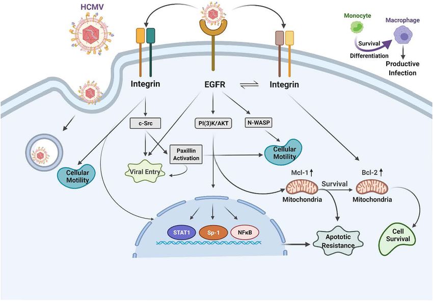
Lee et al.                                                                                                                   EGFR and HCMV Signaling

Fulkerson et al., 2021). HCMV infection in neonates is the leading           Stegmann et al., 2017a; Liu et al., 2018; Nguyen et al., 2018; Gerna
cause of congenital central nervous system damage and deafness               et al., 2019; Nishimura and Mori, 2019). There are some receptors
(Boppana et al., 2011). HCMV is also a leading infectious agent              that seem to be present on most infected cell types and these
negatively affecting the outcome of solid organ and bone marrow              include heparan sulfate proteoglycans, cellular integrins (α2β1,
transplants. In healthy individuals, HCMV infection can cause                α6β1, and αvβ3) (Feire et al., 2004; Wang et al., 2005) and Toll-
mononucleosis and is associated with some vascular diseases and              like receptors (Compton et al., 2003; Boehme et al., 2006), while
cancers (Soderberg-Naucler, 2008; Pass et al., 2009; Wang et al.,            other receptors such as EGFR (Wang et al., 2003, 2005), are only
2017; Styczynski, 2018; Gerna and Lilleri, 2020).                            found on some cell types and may help dictate tropism. Other
    A hallmark of HCMV infection is wide-spread dissemination                potential tropism receptors include the Platelet-Derived Growth
of the virus from the blood in monocytes to most organs                      Factor Receptor [PDGFR (Wu et al., 2017; Nguyen and Kamil,
and then the establishment of latency in the bone marrow in                  2018; Wu et al., 2018; Nishimura and Mori, 2019)], Neuropilin-
progenitor cells that in turn, during reactivation, differentiate            2 [Nrp-2 (Martinez-Martin et al., 2018)], and Olfactory Receptor
into monocytes to again disseminate the virus to host organ                  Family 14 Subfamily I Member 1 [OR14I1 (Xiaofei et al., 2019)].
tissue (Sinzger et al., 1995; Smith et al., 2004a; Stevenson                 In general, whether these receptors are specific for a specific cell
et al., 2014; Collins-McMillen et al., 2018b). Therefore, one of             type or are more conserved across a variety of cell types, they
the first essential steps for hematogenous dissemination is the              are important for viral attachment, internalization, and fusion
infection of monocytes (Smith et al., 2004b; Yurochko, 2008;                 and entry into the cytoplasm of the infected cell (Nowlin et al.,
Chan et al., 2010, 2012a; Nogalski et al., 2011). The virus, in              1991; Navarro et al., 1993; Boyle and Compton, 1998; Compton,
turn, hijacks infected monocyte signaling pathways in order to               2000; Isaacson and Compton, 2009). They also all seem to
promote efficient viral spread to most peripheral organs and                 initiate multiple downstream cellular signaling pathways during
the bone marrow (Smith et al., 2004b; Yurochko, 2008; Chan                   infection (Nogalski et al., 2007; Smith et al., 2007; Yurochko,
et al., 2010, 2012a; Nogalski et al., 2011). Furthermore, infiltration       2008; Reeves et al., 2012).
of infected monocytes into host organ systems allows for viral                   As an extended introduction, we would like to emphasize
spread to additional hosts through release of new virus in a                 why we chose to focus primarily on monocytes and CD34+
variety of body fluids (such as in saliva and urine) and spread              HPCs in this review. Both cell types are important targets for
to the bone marrow where latency is established in CD34+                     HCMV infection and are critical to life-long persistence and
hematopoietic progenitor cells (HPCs). The CD34+ HPCs during                 dissemination within an individual infected host and the general
reactivation differentiate along myeloid lineage development                 human population. Monocytes are essential for hematogenous
pathways to become monocytes allowing organ dissemination                    dissemination of HCMV to multiple organ systems following
and additional host to host spread (Goodrum et al., 2002; Kim                primary infection (Nogalski et al., 2011). That is, HCMV uses the
et al., 2017; Collins-McMillen et al., 2018a). This review examines          biological features of monocytes: motility, ability to migrate to
the processes of HCMV infection of these two critical cells and              all tissue types and differentiation into long-lived macrophages
specifically how viral signaling through cellular receptors is a key         (Nogalski et al., 2012; Stevenson et al., 2014; Collins-McMillen
molecular trigger for HCMV infection and persistence.                        et al., 2017b, 2018b) to promote initial dissemination to the
    Human cytomegalovirus must first attach to the cell via an               multiple host organs that serve as the source of viral spread
interaction between viral membrane glycoproteins and specific                from host to host and for the eventual establishment of the viral
cell surface receptor-binding molecules (Chan et al., 2009b,                 persistence/latency seen in the bone marrow in CD34+ HPCs.
2012a; Collins-McMillen et al., 2017a). Several viral glycoprotein           Monocytes also seem to play an essential role in the establishment
complexes are crucial for viral entry/internalization of monocytes           of viral latency through this initial spread and then in the
and other cell types, two of which (gB and the gH complexes)                 organ dissemination following reactivation in CD34+ HPCs
will be focused on extensively in this report. gB and the                    and the differentiation of these CD34+ HPCs into monocytes
gH/gL complexes help dictate infection tropism and serve as                  (Stevenson et al., 2014; Collins-McMillen et al., 2017b, 2018a;
central ligands for viral attachment to clinically relevant cell             Kim et al., 2017). Like infection of monocytes, initial infection of
types (Nogalski et al., 2013). The gB and the gH/gL complexes                CD34+ HPCs relies on receptor-ligand engagement with EGFR
engage the cell through the epidermal growth factor receptor                 engagement being key to early infection (Kim et al., 2016).
(EGFR) and cellular integrins, respectively (Chan et al., 2009b;             Although we did not focus on the receptor-ligand engagement
Nogalski et al., 2011, 2013; Kim et al., 2016). This receptor-ligand         for different cell types, distinct downstream signaling pathways,
engagement in turn activates cellular signaling and the functional           and functional consequences in this review, it also occurs during
changes in the target monocytes to promote both productive                   the infection of epithelial, endothelial cells, fibroblasts, and others
infection and persistence in host organ systems.                             (Sinzger et al., 1995; Chan et al., 2008b; Yurochko, 2008).
    To contact a target host cell, the viral ligands in the envelope,            For HCMV to initially infect monocytes, the virus must
gB (UL55), gH (UL75)/gL (UL115)/gO (ULl74), which make up                    deal with several biological barriers, and one such barrier is
the gH/gL/gO complex or the trimer, and the gH/gL/UL128-131                  the short lifespan of blood monocytes and the initial lack of
complex or the pentamer, and gM (UL100)/gN (UL73) interact                   viral gene expression and replication in these cells. HCMV-
with cellular receptors that in turn lead to the activation of various       infected monocytes are initially short lived and are non-
signal molecules in the infected host cells (Mach et al., 2005;              permissive for productive infection (Yurochko et al., 1989,
Shimamura et al., 2006; Shen et al., 2007; Fields et al., 2013;              1990, 1992, 1995, 1997a,b, 1999; Taylor-Wiedeman et al., 1991;

Frontiers in Microbiology | www.frontiersin.org                          2                                        May 2021 | Volume 12 | Article 660901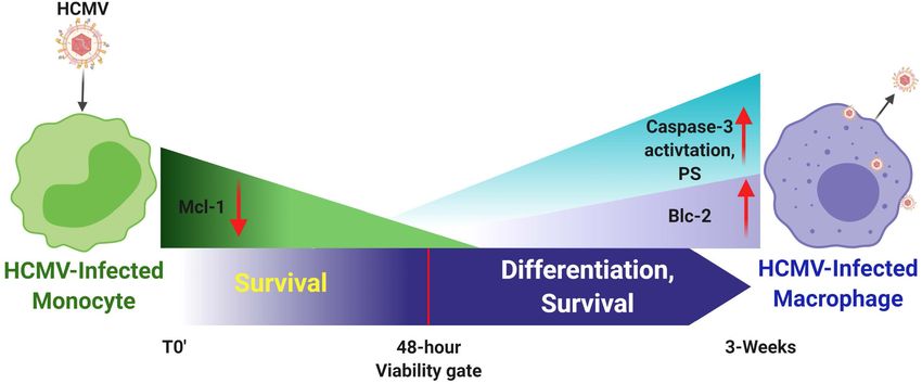
Lee et al.                                                                                                                   EGFR and HCMV Signaling

Mendelson et al., 1996; Hahn et al., 1998; Yurochko and Huang,                mature gB is what engages several cell-surface proteins, including
2000; Chan et al., 2010). To regulate these processes, HCMV                   the primary receptor, EGFR (Wang et al., 2003; Chan et al., 2012a;
utilizes a unique signalosome generated initially by the signaling            Fulkerson et al., 2020), as well as perhaps PDGFR-α (Soroceanu
downstream of gB/EGFR and pentamer/β1 and β3 integrin                         et al., 2008) and some integrins (Bentz and Yurochko, 2008; Feire
engagement at the cell surface and then activation of EGFR                    et al., 2010). In monocytes, this receptor likely helps determine
and integrin dependent events post entry (Collins-McMillen                    tropism for blood leukocytes, as B cells and T cells do not
et al., 2017b; Fulkerson et al., 2020). This signalosome, as                  express EGFR, correlating with the lack of infection of these
discussed in more detail below, alters key biological steps in                cells (Chan and Yurochko, 2014). In addition, PDGFR is not
infected monocyte motility (Smith et al., 2004b; Chan et al.,                 reported to be expressed on freshly-isolated blood monocytes
2009a) and survival through manipulation of Bcl-2 family                      (Chan et al., 2009b) or freshly-isolated CD34+ HPCs (Su et al.,
members (Chan et al., 2010; Collins-McMillen et al., 2016).                   2002) suggesting that the gB/EGFR interaction plays a key role in
In addition, this viral ligand-induced signalosome promotes                   the infection of these cell types. For this review and for infection
monocyte-to-macrophage differentiation and polarization of                    of monocytes and CD34+ HPCs, gB engagement of EGFR is
these differentiated macrophages (Yurochko et al., 1989;                      essential for entry and the post entry events related to nuclear
Stevenson et al., 2014; Collins-McMillen et al., 2017a,b, 2018b).             translocation as well as for the associated downstream signaling
Because macrophages are naturally long-lived cells and once                   (Chan et al., 2009b; Kim et al., 2017; Fulkerson et al., 2020).
differentiated can produce and shed virus for weeks to months                     There are multiple gH complexes, from the newly discovered
(Smith et al., 2004b; Stevenson et al., 2014), they can serve as a            gH interaction with UL116 [gH (UL75)/UL116] (Caló et al., 2016)
source of organ persistence for the virus and shedding in bodily              to the known gH complexes of gH/gL (UL115)/gO (UL174) or
fluids to other hosts. Although distinct, latent infection of CD34+           the trimer (Stegmann et al., 2017a) and gH/gL/UL128-131 or the
HPCs also produces a unique signalosome that is essential for the             pentamer (Nguyen and Kamil, 2018; Nishimura and Mori, 2019).
establishment of, maintenance of, and reactivation from latency               These unique complexes directly affect cell-type-specific entry.
and the associated differentiation of these progenitor cells into             For example, fibroblast entry processes are largely mediated
monocytes (Goodrum et al., 2002; Streblow and Nelson, 2003).                  by the glycoproteins/glycoprotein complexes gB, gH/gL/gO
We hope to shed light on HCMV signaling through key cellular                  (the trimer), and gM (UL100)/gN (UL73), whereas entry into
receptors, and how that signaling initiates productive infection              epithelial cells, endothelial cells, and monocytes/macrophages
and persistence in this review.                                               also require the gH/gL/UL128-131 pentameric complex (Hahn
                                                                              et al., 1998, 2004; Jarvis and Nelson, 2002; Ricciotti and
                                                                              Fitzgerald, 2011; Aoki and Narumiya, 2012; Stegmann et al.,
                                                                              2017b). The HCMV gM/gN complex (Mach et al., 2005) is
HCMV RECEPTORS AND
                                                                              the most abundant glycoprotein complex found on virions
LIGAND-INDUCED SIGNALING                                                      (Soroceanu et al., 2008; Feire et al., 2010) and has been shown
PATHWAY AND CELLS                                                             to interact with heparin sulfate proteoglycans on the cell surface
                                                                              (Kari and Gehrz, 1992), and may also have some intracellular
HCMV Glycoproteins                                                            roles during viral replication (Pignatelli et al., 2003; Mach et al.,
Human cytomegalovirus is a species-specific β-herpesvirus that                2005). HCMV UL116 has recently been shown to be required
infects an extensive range of cell types, including monocytes,                for the production of infectious virus and may help chaperone
macrophages, endothelial cells, epithelial cells, vascular smooth             gH complexes into virions (Caló et al., 2016; Nguyen and Kamil,
muscle cells, stromal cells, neuronal cells, fibroblasts, CD34+               2018; Gatault et al., 2021).
HPCs, and hepatocytes (Sinzger et al., 1995; Jarvis and Nelson,                   There are both shared and unique features of the gH/gL
2002). Although HCMV can infect many different cell and tissue                glycoprotein complexes among the different herpesviruses. For
types, the virus has a set number of viral entry complexes that               example, gH/gL (along with gB) comprise the central herpesvirus
can be used, which means the virus has adapted to utilize these               membrane fusion machinery (Weed and Nicola, 2017). However,
complexes in a way that promotes efficient transmission and                   other aspects of these complexes are unique to HCMV and/or
infection of different cell types in the new host to allow for lifelong       the other β-herpesviruses. gO (and homologs) are found only
persistence (see Figure 1 for a cartoon of viral glycoproteins and            in β-herpesviruses, and the gO-associated trimer is required
their cognate ligands on monocytes/CD34+ HPCs and other cell                  for cellular infectivity of cell-free HCMV virions (Wille et al.,
types). HCMV gB, like all herpesvirus gBs, is essential for viral             2010; Liu et al., 2018); however, its role in modulating signal
membrane fusion with cellular surface or vesicular membranes                  transduction pathways in virus-infected host cells is not resolved.
(Compton et al., 1992; Navarro et al., 1993; Tugizov et al., 1994;            The pentameric complex is unique to HCMV (Wang and
Vanarsdall et al., 2008; Isaacson and Compton, 2009; Zhou et al.,             Shenk, 2005b; Ryckman et al., 2008; Ciferri et al., 2015;
2015) and in an HCMV-specific manner, HCMV gB is a receptor-                  Chandramouli et al., 2017) and is required for infection of
ligand binding protein for EGFR (Wang et al., 2003, 2005; Chan                epithelial and endothelial cells, as well as leukocytes (Bentz
et al., 2012a; Fulkerson et al., 2020). HCMV gB is synthesized                et al., 2006; Kim et al., 2017). The complex is genetically
as a 160 kDa precursor that is cleaved by furin in the Golgi,                 unstable during HCMV passage in fibroblasts and many lab-
resulting in 116 and 55 kDa fragments that become disulfide-                  adapted stains have lost this complex through mutation in one
linked to make the mature gB (Britt and Auger, 1986). This                    or more of the pentamer complex-specific glycoprotein coding

Frontiers in Microbiology | www.frontiersin.org                           3                                       May 2021 | Volume 12 | Article 660901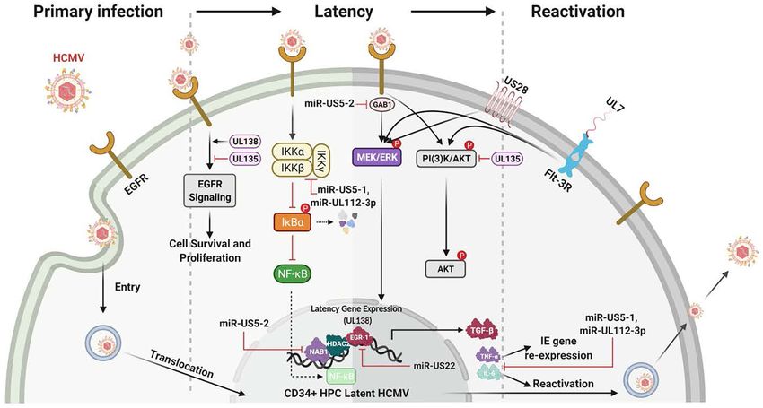
Lee et al. EGFR and HCMV Signaling
FIGURE 1 | Schematic of the HCMV glycoprotein complexes and their cognate receptors. HCMV attachment and entry into monocytes depends on several
envelope glycoprotein complexes including gM/gN, gB, and the gH complexes. gM/gN mediates initial attachment via glycosaminoglycans. gB is known to interact
with the epidermal growth factor receptor (EGFR) on monocytes. gB may also interact with integrins on monocytes, although this is unresolved. The trimeric
gH/gL/gO complex interacts with integrins and the platelet-derived growth factor receptor alpha (PDGFRα) on fibroblasts; at present the trimer only has been
reported to interact with integrins on monocytes. The pentameric gH/gL/UL128-131 complex interacts with β1- and β3-integrins on monocytes. Several other
receptors, such as OR14I1 and Nrp2, have been reported to bind to the pentamer; this engagement remains unresolved during infection of monocytes. The
membrane of fibroblasts is illustrated on the right and the membrane of epithelial cells is illustrated on the left and they include the multiple receptors that have been
reported to engage the HCMV glycoprotein complexes during infection of fibroblasts and epithelial cells, respectively.
sequences (Akter et al., 2003; Wille et al., 2010). Repairing the and the many receptors together or independently alters host
HCMV pentamer complex in a fibroblast-adapted laboratory signaling pathways in the many cells that HCMV infects.
strain restores infectivity of epithelial and endothelial cells, and
monocytes (Wang and Shenk, 2005a,b; Nogalski et al., 2013). EGFR and Integrins Are Essential Entry
The pentamer binds to different cellular integrins and seems to
Receptors Required for Initiating Cellular
have different integrin binding partners on different cell types
(Nogalski et al., 2014; Collins-McMillen et al., 2017b; Vanarsdall Signal Pathway During HCMV Infection
et al., 2018). In addition, the pentamer was recently reported to of Monocytes
bind to Nrp2 (Martinez-Martin et al., 2018) and OR14I1 (Xiaofei Epidermal Growth Factor Receptor (EGFR) (See
et al., 2019) on epithelial cells, perhaps providing epithelial cell Figure 2)
tropism. The pentamer has also been documented to interact The HCMV glycoproteins regulate the key intracellular signaling
with thrombomodulin (Hsu et al., 2015; van den Boomen and pathways in human primary monocytes required for productive
Lehner, 2015). CD147 (Vanarsdall et al., 2018) and CD151 infection and the establishment of persistence. Following the
(tetraspanin) and THY-1 (CD90) have also been found to play engagement of heparan sulfate proteoglycans, HCMV infection
a role during HCMV infection of some cell types (Hochdorfer of monocytes begins via the interaction of gB and the gH/gL
et al., 2016). However, the biological relevance and importance of complexes with host cell surface EGFR and β1 and β3
these receptors in infection monocytes or CD34+ HPCs and in integrins (Chan et al., 2009b, 2012a; Nogalski et al., 2011, 2013;
cell signaling remain unresolved. Overall, viral glycoproteins are Kim et al., 2016; Fulkerson et al., 2020). Epidermal growth
an indispensable cell type specific strategic weapon for successful factor receptor (EGFR or ErbB-1) is a member of the ErbB
viral infection via their receptor binding capabilities, their ability receptor tyrosine kinase family, which can bind epidermal
to induce cellular signaling in host cells and to allow viral entry growth factor (EGF) or transforming growth factor-alpha (TGF-
into that target cell. Further studies are needed to determine the α) (Wieduwilt and Moasser, 2008). Studies have revealed that
precise mechanisms by which the many HCMV glycoproteins downstream EGFR signaling can be unique and functionally
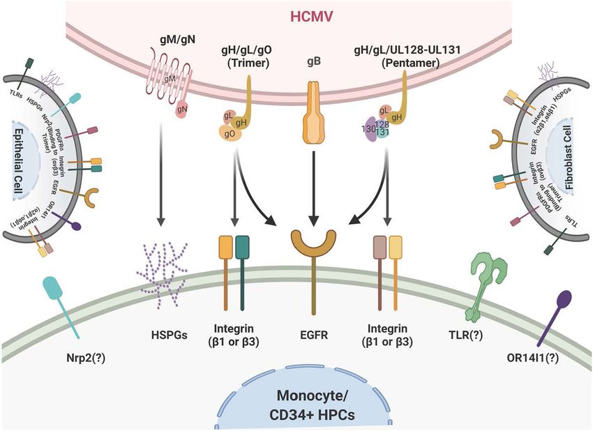
Frontiers in Microbiology | www.frontiersin.org 4 May 2021 | Volume 12 | Article 660901Lee et al. EGFR and HCMV Signaling FIGURE 2 | Human cytomegalovirus (HCMV) glycoprotein binding to cognate receptors controls viral signaling, trafficking and the key cellular functions required for productive infection of monocytes. The HCMV glycoproteins bind several cellular receptors, including EGFR and the β1- and β3-integrins. Following binding, these receptor engagements trigger a variety of signal transduction pathways in the infected monocyte. For example, gB binding to EGFR results in activation of the intrinsic EGFR tyrosine kinase and down stream signaling that is required for entry, for the unique multi-vesicular nuclear translocation process seen in monocytes, as well as for motility, and the survival of the monocytes and their differentiation into macrophages. The engagement of the pentameric complex, gH/gL/UL128-131 with β1- and β3-integrins on the surface of monocytes activates c-Src to drive the required signaling pathways needed for entry, nuclear translocation, motility, survival and differentiation. More specifically, this downstream signaling following EGFR and integrin engagement acts in concert to activate numerous down stream signaling pathways, including the NF-κB pathway, PI(3)K, and the MAPK pathway, which culminates in enhanced transactivation of many host cell promoters via increases in transcriptional regulators, altercations in actin regulatory proteins that modulate motility (such as N-WASP and paxillin) and changes in Mcl-1 and Bcl-2 to promote survival of short-lived monocytes. distinct based on the ligand and the cell type expressing EGFR mechanism to promote heightened motility (Chan et al., 2009b), (Wieduwilt and Moasser, 2008). In support of this cell type as opposed to the use of the standard Wiskott–Aldrich Syndrome specific EGFR biology during HCMV infection, one can note the protein (WASP) that controls chemokine-induced motility in differences between EGFR-dependent signaling seen in infected monocytes (Snapper and Rosen, 1999; Tsuboi, 2006; Chan et al., monocytes (Chan et al., 2009b, 2012a; Nogalski et al., 2011, 2013; 2009b; Lee et al., 2017). That is, under normal circumstances, Kim et al., 2016; Fulkerson et al., 2020) vs. that seen in infected WASP usually helps modulate monocyte motility (Snapper and endothelial cells (ECs) (Bentz and Yurochko, 2008) or infected Rosen, 1999; Tsuboi, 2006; Chan et al., 2009b; Lee et al., 2017). trophoblasts (LaMarca et al., 2006). There are also reported WASP and N-WASP are actin nucleators that mechanistically differences in EGFR signaling in CD34+ HPCs (Kim et al., 2017) drive changes in the actin cytoskeleton associated with cell and that will be discussed in more detail below. movement. This use of N-WASP showcases some of the distinct In monocytes, HCMV induces the phosphorylation of properties of the gB/EGFR receptor-ligand engagement in EGFR following viral binding and that engagement initiates the monocytes. N-WASP is responsible for highly motile cancer activation of PI(3)K and Akt. When considering other peripheral cell movement (Yamaguchi and Condeelis, 2007; Kurisu and blood cell types, such as T cell and B cells, EGFR expression Takenawa, 2010; Frugtniet et al., 2015) and biochemically is seems to be involved in the select myeloid tropism of the virus more efficient at nucleating actin than WASP. Thus, we propose (Xiaofei et al., 2019). This HCMV-activated EGFR in monocytes this switch to N-WASP during HCMV infection of monocytes promotes motility and transendothelial migration to allow promotes migration in the absence of a chemotactic gradient into virus-infected cells to gain entry into peripheral organ tissues organ tissues (Chan et al., 2009b). Rapid activation of the EGFR (Smith et al., 2004a,b, 2007; Bentz et al., 2006; Chan et al., 2009b; signaling pathway following HCMV infection also turns out to Collins-McMillen et al., 2017a,b). Mechanistically, gB/EGFR be essential for viral entry into monocytes [Figure 2 (Chan et al., engagement results in the upregulation and select usage of the 2009b; Fulkerson et al., 2020)]. Thus, EGFR plays an essential Neural Wiskott–Aldrich Syndrome protein (N-WASP) as a role in the pathobiology of HCMV by mediating viral entry into Frontiers in Microbiology | www.frontiersin.org 5 May 2021 | Volume 12 | Article 660901
Lee et al.                                                                                                             EGFR and HCMV Signaling

monocytes and stimulating the cellular signaling activity that          infection, including the requirement for multiple integrins, that
promotes hematogenous dissemination of the virus.                       was not observed in fibroblasts (Nogalski et al., 2013; Campadelli-
   The HCMV gB engages EGFR and then rapidly and directly               Fiume et al., 2016). That is, engagement of both β1 and
activates downstream signaling via its intrinsic tyrosine kinase        β3 integrins are required during viral attachment to mediate
(Walker et al., 1998). gB mainly interacts with EGFR on                 entry and outside-in signaling in monocytes (Campadelli-Fiume
monocytes, although in some cell types, it may also interact with       et al., 2016). HCMV binding to integrins on monocytes rapidly
cellular integrins (Yurochko et al., 1992; Bentz and Yurochko,          activates the integrin/c-Src signaling pathway to induce PI(3)K
2008; Nogalski et al., 2011, 2013; Collins-McMillen et al., 2016;       and other essential signaling pathways.
Kim et al., 2016). This receptor-ligand engagement stimulates              The HCMV binding to host cell receptors induces signals
the activation of transcription factors, NF-κB (discussed in more       in a distinct manner for each cell type. In monocytes, this
detail below) and Sp-1 (Yurochko et al., 1995, 1997a,b) and             receptor-ligand engagement results in cellular differentiation
numerous signaling pathways (Chan et al., 2012a; Campadelli-            and long-term cellular survival (Smith et al., 2004b; Chan
Fiume et al., 2016; Eberhardt et al., 2016; Collins-McMillen            et al., 2008a; Yurochko, 2008; Campadelli-Fiume et al., 2016);
et al., 2017b; Yurochko, 2017). In addition to activating               but how are integrins involved? Integrins are a family of
signaling molecules and transcription factors, these functional         heterodimeric receptors composed of a single α-chain and a
changes initiated by HCMV gB binding to EGFR serve as a                 single β-chain that when engaged via their various cognate
molecular convergence point linking the changes in molecular            ligands leads to the induction of specific signaling pathways
and biochemical pathways with the biological changes required           via activation of c-Src and other tyrosine kinases (Yamada
for productive infection. One example is the viral activation           and Miyamoto, 1995; Hynes, 2002). Based on transcriptome
of STAT1 (Collins-McMillen et al., 2017a,b). Not only is                analysis, integrin-mediated signaling plays a central role as a
STAT-1 activated within minutes of viral binding, as well as            convergent point linking the specific cellular molecules that
chronically (3 plus weeks after infection) as documented by             have been activated by HCMV viral binding on monocytes. For
changes in the patterns of appropriate activating phosphorylated        example, the integrin-mediated rapid phosphorylation of paxillin
residues, its specific activation is required for the survival          identified that paxillin is both required for HCMV entry and
and differentiation of monocytes that ultimately is needed for          for the hematogenous dissemination of the virus in monocytes
productive infection and viral persistence in organ macrophages         through altered motility (Nogalski et al., 2011). Paxillin is an
(Collins-McMillen et al., 2017b).                                       adaptor protein and an important signal transducer in the
   The HCMV infection elicits a robust immune response                  regulation of actin rearrangement during cellular adhesion and
(Gerna et al., 2005; Sinzger et al., 2006; Mason et al.,                movement. HCMV-infected human primary monocytes showed
2012). However, symptomatic infections largely only occur               heightened levels of paxillin mRNA and protein expression in
in immunocompromised patients (Stagno et al., 1986; Bentz               an integrin-dependent manner (Nogalski et al., 2011). The use
et al., 2006; Arvin, 2007; Nogalski et al., 2014; Fulkerson             of siRNA knockdown in monocytes revealed that paxillin is
et al., 2021). Thus, the immune response keeps the virus                essential for virus-induced motility and viral entry (Nogalski
under tight control in healthy individuals. How viral infection         et al., 2011). Thus, the activation of the integrin/c-Src/paxillin
and the immune response interacts during life-long persistence          signaling pathway is essential for triggering the cellular changes
remains unresolved? With monocytes playing a central role               that allow for HCMV entry and hematogenous dissemination.
during infection, understanding how the virus manipulates EGFR             It is important to note that both the gB/EGFR and the
signaling pathways in monocytes (Yurochko, 2008; Stevenson              pentamer/integrin signaling axes drive independent events, as
et al., 2014), promotes reactivation (Streblow and Nelson,              well as cooperate to modify the infected cell. For example, as
2003; Nogalski et al., 2014; Collins-McMillen et al., 2018a,            discussed briefly in this review, integrins activate paxillin through
2019), enhances production of suppressive cytokines (Chan               integrin/c-Src activation (Nogalski et al., 2011), and EGFR
et al., 2009a; Wu et al., 2017; Mikell et al., 2019) and alters         separately regulates the expression of the N-WASP (Chan et al.,
differentiation and phenotypic polarization of monocytes and            2009b). Together, both are required for activation of motility,
macrophages (Chan et al., 2008a, 2012a; Collins-McMillen et al.,        since inhibition of either pathway block infected monocyte
2017a,b) could pave the way for a better understanding of the           motility and migration. From this data, HCMV-cell interactions
pathobiology of the virus.                                              emerge as an essential trigger for the cellular alterations that allow
                                                                        for HCMV entry and hematogenous dissemination.
Integrins
We previously described that viral engagement of β1 and
β3 integrins are essential for infection of human peripheral
                                                                        Extended Cellular Signaling and
monocytes via the interaction of the HCMV gH/gL/UL128-131               Intracellular Trafficking During HCMV
complex with these two integrin families [Figure 2 (Nogalski            Infection of Monocytes
et al., 2013; Kim et al., 2016)]. It is also known from other           An investigation into receptor-ligand mediated signaling and
groups that HCMV binds to integrins and facilitates viral               the associated regulation of survival and differentiation led us
entry into human fibroblasts (Compton, 2000; Wang et al.,               to a finding that cellular signaling during viral binding also
2005; Campadelli-Fiume et al., 2016). However, we have noted            controls unique post entry events; these include the intracellular
that there are functional changes that occur during monocytes           trafficking and nuclear translocation of the virus during infection

Frontiers in Microbiology | www.frontiersin.org                     6                                       May 2021 | Volume 12 | Article 660901
Lee et al. EGFR and HCMV Signaling
of monocytes (Figure 2). All herpesviruses replicate their DNA viral gene expression initiate along with all of the other steps
in the nucleus, and thus the steps of intracellular trafficking associated with productive infection. This process is essential for
and nuclear translocation are essential steps following viral entry organ infection following primary infection (Min et al., 2020)
for productive infection. As this brief discussion will show, and following reactivation of virus from latency. Figures 2, 3
the virus has evolved to utilize the same signaling pathways show the signaling associated with these discussed biological
required for entry to modulate, in a cell specific manner, these changes. Mcl-1, a member of the Bcl-2 gene family, serves to
key post entry events. Briefly, we observed in monocytes that regulate monocyte survival (Yang et al., 1996; Zhou et al., 1997).
the gH/gL/UL128-131 complex through binding to β1 and β3 HCMV uses the EGFR/PI3K signaling pathway to upregulate
integrins and activation of c-Src signaling drives a pattern this anti-apoptotic protein after infection; thus, overcoming
of intracellular trafficking and nuclear translocation not seen the intrinsic apoptotic fate of monocytes (Chan et al., 2010).
in other cell types. That is, HCMV nuclear translocation in Mcl-1 was upregulated/maintained early in HCMV-infected
monocytes is distinct from that seen in fibroblasts, endothelial monocytes (Chan et al., 2010) and then begins to diminish to
cells, and CD34+ cells (Kim et al., 2016; Fulkerson et al., 2020). near background levels around 48 h post infection (hpi). Reeves
Nuclear translocation occurs roughly 30 min post infection in et al. (2012) have also shown the induced expression of Mcl-
fibroblasts and endothelial cells (Kim et al., 2016). In contrast, 1 during monocyte infection. siRNA-mediated knockdown of
post entry of viral DNA into the nucleus of monocytes is extended Mcl-1 revealed the strong anti-apoptotic role that Mcl-1 plays
until 3 days post infection (dpi) (Kim et al., 2016; Fulkerson et al., in HCMV-infected monocytes during the first 48 hpi (Chan
2020). Furthermore, we showed that mature HCMV particles are et al., 2010). Additional studies showed that Bcl-2 then takes
initially retained in early endosomes, they then move to the trans- over for Mcl-1 in monocytes after the first 48 hpi (Chan et al.,
Golgi network (TGN) and then finally to recycling endosomes in 2010; Stevenson et al., 2014; Collins-McMillen et al., 2016). Bcl-
monocytes, where de-envelopment occurs (Kim et al., 2016). We 2 is known to be an anti-apoptotic factor in the survival of
recently added to this work and showed that gB/EGFR binding myeloid cells (Lin et al., 1996; Lagasse and Weissman, 1997; Sanz
continues past the entry process and that this chronic gB/EGFR et al., 1997; Adams and Cory, 1998; Perlman et al., 2000; Liu
engagement was essential for nuclear translocation following the et al., 2001; Zhang et al., 2003; Reeves et al., 2012; Zhu et al.,
integrin/pentamer/c-Src decision point (Fulkerson et al., 2020). 2018; Shnayder et al., 2020). Molecularly, known to be anti-
That is, viral engagement of EGFR continues from the attachment apoptotic, their regulation and linear regulation (Mcl-1 and then
step through the post entry phase in order to promote correct Bcl-2) is not seen in mock infected cells and monocytes treated
viral tracking and nuclear translocation in monocytes (Fulkerson with other stimuli (Zhou et al., 1997; Collins-McMillen et al.,
et al., 2020). Although studied in less detail, it seems to be a 2016). Both of these factors are induced by cellular signaling
similar, although expedited, process occurs in CD34+ HPCs (Kim with Mcl-1 early regulation being controlled by the gB/EGFR
et al., 2017). This unique signaling pathway suggests that HCMV- axis and the later Bcl-2 upregulation being largely controlled by
infected monocytes use a distinctive mechanism in infected the pentamer/integrin axis (Zhou et al., 1997; Chan et al., 2010;
monocytes to promote productive infection and that this process Reeves et al., 2012; Collins-McMillen et al., 2016). To add to
is controlled by pentamer/integrin and gB/EGFR engagement. the potential complexities of the control of monocyte survival,
HCMV seems to block full caspase-3 activation, while inducing
partial activation of caspase-3, which in turn is important for
MONOCYTE-TO-MACROPHAGE monocyte-to-macrophage differentiation (Chan et al., 2012b).
Thus, it seems that HCMV modulation of monocyte survival
DIFFERENTIATION REQUIRES is related to monocyte-to-macrophage differentiation (discussed
REGULATION OF MONOCYTE AND below) and these combined major biological changes, usurped
MACROPHAGE APOPTOTIC during infection of monocytes, are essential for productive organ
PROGRAMS DURING HCMV INFECTION infection and long-term persistence of the virus in a variety of
organ macrophages (Min et al., 2020).
Monocyte-to-macrophage differentiation in HCMV-infected
monocytes is essential for long-term viral persistence. Monocytes
have a short lifespan of 1–3 days in circulation and are not UNIQUE CELLULAR SIGNALING
initially permissive for viral gene expression and replication (Min PATHWAYS INDUCE
et al., 2020). Thus, HCMV must extend the life span of the
infected monocytes and promote differentiation of these cells into
MONOCYTE-TO-MACROPHAGE
long lived macrophages that are permissive for viral replication DIFFERENTIATION LIKELY REQUIRED
(Min et al., 2020). Briefly, as described in the mentioned review FOR PRODUCTIVE INFECTION IN
and associated references (Min et al., 2020), HCMV binds PERIPHERAL ORGANS
to and enters monocytes and initiates the process of nuclear
translocation; simultaneously to the viral manipulated events Because infected monocyte-to-macrophage differentiation is
that are essential to getting the genome into the nucleus, the required for viral replication and long-term viral persistence,
virus must promote survival and differentiation of that infected we have focused on the control of differentiation of monocytes
monocyte. Only in the differentiated cell (the macrophage) does into macrophages and how cellular signaling via gB-EGFR
Frontiers in Microbiology | www.frontiersin.org 7 May 2021 | Volume 12 | Article 660901Lee et al. EGFR and HCMV Signaling FIGURE 3 | Human cytomegalovirus infection of monocytes drives a number of key molecular mechanisms required to enhance survival of normally short-lived monocytes and for the promotion of the differentiation of these infected monocytes into long-lived productively infected macrophaqes. It is important to note that monocytes, although they can be infected, they are not permissive for viral replication; only upon differentiation is de novo viral gene expression observed. HCMV-infected monocytes show enhanced expression of Mcl-1 early after infection (
Lee et al.                                                                                                                EGFR and HCMV Signaling

the nature of the differentiation and the M1- and M2-polarized              HOST CELL SIGNALING AND LATENCY
phenotypes (Stevenson et al., 2014), and that this end polarization
phenotype does not fall into a strict M1- or M2-like phenotype              The HCMV latency and reactivation are crucial for long-term
or perhaps does not fall into a strict proinflammatory or                   persistence and stable spread within the human population
immunosuppressive state.                                                    (Jarvis and Nelson, 2002; Streblow and Nelson, 2003; Petrucelli
                                                                            et al., 2009; Buehler et al., 2016; Collins-McMillen and Goodrum,
                                                                            2017; Kim et al., 2017; Collins-McMillen et al., 2018a, 2019;
HCMV MODULATES NF-κB CELLULAR                                               Hancock et al., 2020a). HCMV latency also exploits cellular
SIGNALING                                                                   signaling pathways to help establish and maintain latency and
                                                                            then upon recognition of appropriate signals, to manipulate
NF-κB is essential for initial HCMV gene expression since                   the cellular environment and allow successful viral reactivation
the HCMV major immediate early promoter requires NF-                        and differentiation of the infected CD34+ HPCs to blood
κB (Fields and Knipe, 2013; Fields et al., 2013). This early                monocytes and then to tissue macrophages (Jarvis and Nelson,
identification of the impact of NF-κB on the initiation of                  2002; Petrucelli et al., 2009; Collins-McMillen et al., 2018a, 2019;
HCMV gene expression served as the impetus to drive early                   Hancock et al., 2020a). As with primary infection, EGFR signaling
studies into the regulation of NF-κB. It has been shown that                is also an important cellular receptor controlling this aspect of
HCMV activates both the canonical and non-canonical NF-κB                   the viral infection process (Figure 4). HCMV activation of EGFR
signaling pathways (Figure 2) to aid in viral gene expression,              and downstream PI3K signaling is important for viral entry into
viral replication, and productive infection (Andreoni et al.,               CD34+ HPCs (Kim et al., 2017). Furthermore, through the use
2002; Chan et al., 2008b, 2012a; Yurochko, 2008; Collins-                   of the EGFR inhibitor, AG1478, it was shown that inhibition of
McMillen et al., 2017b). From a regulatory standpoint, it was               EGFR signaling post entry resulted in increased transcription of
shown in both fibroblasts and monocytes that gB- and gH-                    IE1/IE2, while curbing the transcription of the latency-associated
mediated signaling induces rapid activation of NF-κB signaling              UL138 transcript (Kim et al., 2017). EGFR is also essential for the
pathways (and degradation of IκBα); only in monocytes,                      maintenance of latency, with the virus controlling a low level of
however, did this rapid activation of NF-κB lead to increased               constant signaling to maintain stable latency (Buehler et al., 2019;
inflammatory cytokine production, motility, endothelial cell                Mikell et al., 2019); this will be discussed in more detail below.
adhesion, and transendothelial migration (Yurochko et al.,                  Thus, viral signaling during attachment and entry is important
1997a, 1999; Smith et al., 2004b, 2007; Yurochko, 2008; Chan                in a distinct manner in numerous cell types essential for viral
et al., 2009b), suggesting cell type-specific effects. Because viral-       persistence; while long-term regulation of EGFR signaling and
induced changes in NF-κB seem to control cellular function in               possibly integrin signaling post entry is critical for latency and
monocytes separate from the direct role on viral gene expression,           persistence. Cellular differentiation (Min et al., 2020) is also
NF-κB activation and its role in changes in infected monocyte               closely related to both latency and reactivation and one expects
function remain a topic of interest. Furthermore, because NF-               multiple aspects of altered signaling during reactivation and
κB controls multiple aspects of hematopoiesis and cellular                  latency to be central to the process in much the same way that
development (Hancock et al., 2017; Zhang et al., 2017), it is               signaling is essential for the establishment of persistence during
likely the receptor-ligand changes in this transcription factor             primary infection.
may help explain some of the broad biological changes described
in the sections above. ‘Omics profiling is consistent with this
idea, where inhibition of NF-κB signaling blocked or down-                  The EGFR and pUL135 and pUL138
regulated over 500 genes at 4 hpi, including multiple gene                  Connection
products associated with apoptosis, motility, and differentiation           UL135 and UL138 are two key viral genes in the ULb’ region
(Chan et al., 2008b). These observations imply that modulation              important in latency and reactivation. They have been shown
of NF-κB signaling serves as a cell-specific convergence point              to have opposing roles in regulating EGFR’s function in viral
where viral activation mediated by receptor-ligand engagement               replication/reactivation (Umashankar et al., 2014; Buehler et al.,
through EGFR and integrins stimulates a cell in a specific manner           2016). The ULb’ region encodes the UL133-UL138 locus, which
to promote the required changes for infection of that cell. It              is required for infection and persistence in the host. The UL133,
is important to point out that the viral miRNAs miR-US5-1                   UL135, UL136, and UL138 genes have been shown to have
and miR-UL112-3p target the IκB kinases IKKa and IKKb for                   important functions in vascular endothelial cells (Bughio et al.,
downregulation and can negatively influence proinflammatory                 2013, 2015) and to differentially regulate latency and reactivation
cytokine expression suggesting additional mechanisms by which               in CD34+ HPCs (Petrucelli et al., 2009, 2012; Umashankar
HCMV may specifically control this key pathway or specific parts            et al., 2011, 2014). pUL135 appears to promote replication, via
of this pathway during lytic and/or latent infection (Hancock               inhibition of EGFR signaling through the turnover of EGFR
et al., 2017). Other transcription factors have also been reported          from the cell surface. UL135 promotes endocytic trafficking of
to be regulated by receptor-ligand signaling and during key points          EGFR for lysosomal degradation through its interaction with the
of latent or lytic infection cycle (Fields and Knipe, 2013; Fields          adapter proteins, Abelson interactor 1 (ABI-1) and SH3 domain-
et al., 2013). The entirety of this process is beyond the scope of          containing kinase-binding protein 1 (CIN85) (Rak et al., 2018).
the current review.                                                         Loss of UL135 or disruption of its interaction with Abi-1 or

Frontiers in Microbiology | www.frontiersin.org                         9                                      May 2021 | Volume 12 | Article 660901
Lee et al. EGFR and HCMV Signaling
FIGURE 4 | Many HCMV-expressed proteins and miRNAs regulate the signaling pathways known to be important for the establishment, maintanence and
reactivation from latency. The initial infection of CD34+ HPCs lays the foundation for the establishment of latency. Initially, gB engagement of EGFR and the ensuing
signaling is required for HCMV entry into CD34+ HPCs. This initial ligand-dependent event prepares the cell for the establishment of latency through the manipulation
of host signaling pathways and regulation of viral latent gene expression. These latency gene products in turn are critical for the establishment, the maintenance of
and/or reactivation from latency. For example, gene products such as UL138 and UL135 regulate viral latency and host interactions by regulating various signaling
pathways [such as Egr-1 activity or PI(3)K signaling] during the establishment of and exit from latency. Other products such as the viral miRNAs miR-US5-1 and
miR-UL112-3p target and down regulate IKKα and IKKβ to control inflammatory cytokine regulation (such as IL-6 and TNFα) and likely to mitigate NF-KB dependent
signaling that might interfere with latency programming. Other products such as soluble UL7 are released during reactivation and through binding to the Flt-3R
induces activation of the downstream PI3K/AKT and MAPK/ERK signaling pathways that in turn triggers the HPC and monocyte differentiation required for complete
viral reactivation. US28, another critical latency modulator, is involved in the MEK/ERK and EGFR/PI3K signaling required for HCMV reactivation from latency, as well
as playing a key role in monocyte to macrophage differentiation. In addition, a variety of viral miRNAs (such as miR-US5-1, miR-US5-2, miR-US22, and
miR-UL112-3p) target cellular processes and regulate virus-mediated signaling. As examples, miR-US5-2 blocks NAB1 activity, a represser of TGFβ resulting in
increased production of the TGFβ that suppresses hematopoietic events; miR-US5-2 also down regulates GAB1, thus lowering MEK/ERK signaling, as well as
regulates EGR1 and UL138 expression supporting a role for miR US5-2 during reactivation. Overall these viral miRNAs, UL7, US28, UL138/135 control vital signaling
pathways during the establishment, maintenance of and during reactivation from latency and are intimately involved in the differentiation of the infected CD34+ HPCs
into monocytes and then into productive tissue macrophages.
CIN85 results in increased cellular and surface levels of EGFR herpesvirus miRNAs target broadly similar cellular functions,
and a failure to reactivate from latency (Buehler et al., 2016; including secretory pathways, immune evasion, survival, and
Rak et al., 2018). In contrast, pUL138 activity is required for proliferation of infected cells. A number of the HCMV
continued surface expression of EGFR (Buehler et al., 2016). miRNAs control the signaling pathways associated with long-
Thus, in this model, pUL135 and pUL138, along with EGFR term viral persistence; some of these key miRNAs are briefly
signaling, comprise a molecular convergence point to control discussed below.
latency and reactivation. HCMV miR-US5-1, miR-US5-2, and miR-UL112-3p down-
modulate secretion of the inflammatory cytokines tumor necrosis
Viral MicroRNAs (miRNAs) and Host factor-alpha (TNF-α) and interleukin-6 (IL-6), by targeting
Signaling multiple members of the endocytic secretory pathway (Hook
Viruses encode miRNAs that regulate viral and host gene et al., 2014). In addition, miR-US5-1 and miR-UL112-3p
expression to generate a more favorable cellular environment block NF-κB-dependent induction of proinflammatory cytokines
and/or inhibit host immune responses. Several herpesvirus family through direct targeting of the IκB kinase (IKK) complex
members express miRNAs that regulate host cell signaling components IKKα and IKKβ (Hancock et al., 2017). It has also
pathways (Gottwein and Cullen, 2008; Hook et al., 2014; been determined that miR-UL112-3p can modulate the Toll-Like
Hancock et al., 2020a). The 14 HCMV miRNAs (Stark et al., Receptor (TLR) innate immune pathway through direct targeting
2012) are unique among human herpesviruses because they of TLR2. Because TLRs play a critical role in controlling viral
are not clustered within defined latency-associated genomic infections (Akira et al., 2006), this altered signaling could also be
regions and thus are differentially expressed during lytic involved in viral persistence. Specifically, it was shown that miR-
and latent infection (Dunn et al., 2005; Pfeffer et al., UL112-3p efficiently down-regulated the TLR2/IRAK1/NF-κB
2005; Buck et al., 2007; Dolken et al., 2007). Overall, signaling axis (Landais et al., 2015).
Frontiers in Microbiology | www.frontiersin.org 10 May 2021 | Volume 12 | Article 660901Lee et al. EGFR and HCMV Signaling
The HCMV reactivation from latency can cause significant progenitor cells (Beaudin et al., 2014). The binding of HCMV
disease in transplant recipients (Safdar et al., 2002; Ljungman UL7 to the Flt-3R triggers HPC/monocyte differentiation via
et al., 2011; Hancock et al., 2020a). Clinical manifestations PI3K and MAPK/ERK1/2 signaling (MacManiman et al., 2014;
include myelosuppression and graft rejection as a result of Crawford et al., 2018). UL7 has also been shown to promote
infection of CD34+ HPCs (Fries et al., 1993). Mechanistically, angiogenesis (MacManiman et al., 2014) and alter cytokine
little had been uncovered to account for the global suppression production (Engel et al., 2011). The differences in UL7’s effect
caused by infection of only a small number of CD34+ HPCs, on monocytes vs. other cell types suggest its effects on signaling
until a recent study examining the regulation of TGF-β. TGF-β may vary from cell type to cell type. Deletion of UL7 from the
is a member of a multifunctional cytokine family responsible for virus results in loss of reactivation from latency in vitro and
many cellular processes (Marshall et al., 2000; Capron et al., 2010; in humanized mice, further highlighting the functional role of
Blank and Karlsson, 2015), including potent inhibition of early UL7 during viral reactivation (Crawford et al., 2018; Hancock
progenitor cell proliferation (Sing et al., 1988). Recently it was et al., 2020a). Finally, it was recently published that HCMV
shown that latently expressed HCMV miR-US5-2 downregulates utilizes secreted protein pUL7, miR-US5-1, and miR-UL112-3p
NAB1, a repressor of the transcriptional factor NGFI-A (Egr-1), to reduce the proapoptotic transcription factor FOXO3a, which
which in turn results in increased TGF-β production that causes in turn reduces expression of proapoptotic gene BCL2L11 and
myelosuppression in CD34+ HPCs. These results implicate prevents virus-induced apoptosis after infection of CD34+ HPCs
viral miRNAs in regulating HCMV-induced myelosuppression (Hancock et al., 2021).
during solid organ and hematopoietic stem cell transplantation
(Hancock et al., 2020a). Viral miRNAs can also influence viral US28 HCMV Signaling Molecules
gene expression. For example, miR-US5-1 and miR-US5-2 can US28, one of the 4 HCMV encoded G-protein coupled receptors
both regulate HCMV US7 expression (Tirabassi et al., 2011) while (GPCRs), has been documented to signal through a number
miR-UL112-3p targets the immediate early protein IE1 (IE72) of pathways and this chemokine receptor has a number of
(Grey et al., 2007; Murphy et al., 2008), which has implications biological functions (Streblow et al., 1999; Miller et al., 2003, 2012;
in latency establishment and immune cell recognition (Lau et al., Vischer et al., 2006; Elder and Sinclair, 2019; Elder et al., 2019).
2016). Together, these examples show how the viral miRNAs US28 binds to both members of the CC and CX3 C chemokine
can influence cell signaling and cytokine secretion that in turn families making it unique among GPCRs; and furthermore, the
promotes key elements of viral persistence. receptor elicits ligand-specific signaling and functions that are
So far, several studies have addressed how HCMV miRNAs also cell type specific (Vomaske et al., 2009). US28 can also
influence EGFR and EGFR-dependent downstream signaling. signal in a ligand-dependent and -independent manner (Streblow
HCMV miR-US5-2 targeting of the EGFR adaptor protein GAB1 et al., 2003; Miller et al., 2012). US28 has been shown to alter
results in a block in MEK/ERK signaling downstream of EGFR smooth muscle cell migration (Streblow et al., 1999) and to
activation that affects both cell proliferation and expression of be important in vascular disease, as well as play a role during
pUL138, thus interfering with the feed-forward loop of EGFR latency where US28 is argued to be key in both maintenance
activation of pUL138 expression (Hancock et al., 2020b). pUL138 of and reactivation from latency (Crawford et al., 2019). US28
expression is also regulated by the HCMV miRNA miR-US22, is expressed in naturally infected human peripheral blood cells
which directly targets the transcription factor Egr-1 downstream during periods of latency (Krishna et al., 2017), reactivation
of EGF-mediated MEK/ERK signaling. Through targeting Egr- in lung transplant recipients (Boomker et al., 2006), and in
1, miR-US22 regulates pUL138 expression and CD34+ HPC the model of HCMV latency using CD34+ HPCs, monocytes,
proliferation and plays an important role in viral reactivation and in monocyte-derived macrophages during active infection
(Mikell et al., 2019). With the role chronic EGFR signaling plays (Zipeto et al., 1999; Beisser et al., 2001; Goodrum et al., 2002;
in the maintenance of latency (Buehler et al., 2016), it seems Cheung et al., 2006; Humby and O’Connor, 2015; Crawford
probable that these miRNAs will strongly tune the EGFR response et al., 2019). Overall, US28 regulates host cell signaling, which
by modulating signaling pathways to promote latency or, when in many cases is through changes in PI3K/Akt and MAPK
appropriate, viral reactivation. signaling and alterations in EGFR and downstream processes.
Together these varied mechanisms briefly discussed along with
UL7 as an HCMV Signaling Molecule others not discussed (Fields et al., 1996, 2013; Fields and Knipe,
UL7 is a secreted HCMV-encoded signaling molecule known 2013) demonstrate the complex mechanisms controlling HCMV
to play a robust role during lytic and latent infection (Engel reactivation from latency and the intimate linking of these
et al., 2011; Crawford et al., 2018; Perez-Carmona et al., 2018; various viral gene products in controlling the cellular signaling
Min et al., 2020). UL7 belongs to the HCMV RL11 gene family required for reactivation.
(RL11-13, UL1, UL4-11, RL6, and RL5A) and signals through
Fms like tyrosine kinase 3 receptor (Flt-3R), a Class III receptor
tyrosine kinase predominantly expressed in HCMV infection CONCLUSION
of myeloid lineage cells, such as CD34+ HPCs or monocytes
(Beaudin et al., 2014). The HCMV has evolved a complicated strategy to disrupt
Flk2/Flt3 promotes both myeloid and lymphoid development biological pathways in infected host cells. The understanding of
by expanding non-self-renewing multipotent hematopoietic the relationship between viral infection and cellular signaling
Frontiers in Microbiology | www.frontiersin.org 11 May 2021 | Volume 12 | Article 660901You can also read