The Lon protease temporally restricts polar cell differentiation events during the Caulobacter cell cycle
←
→
Page content transcription
If your browser does not render page correctly, please read the page content below
RESEARCH ARTICLE
The Lon protease temporally restricts
polar cell differentiation events during
the Caulobacter cell cycle
Deike J Omnus†, Matthias J Fink†, Klaudia Szwedo, Kristina Jonas*
Science for Life Laboratory and Department of Molecular Biosciences, The Wenner-
Gren Institute, Stockholm University, Stockholm, Sweden
Abstract The highly conserved protease Lon has important regulatory and protein quality control
functions in cells from the three domains of life. Despite many years of research on Lon, only a few
specific protein substrates are known in most organisms. Here, we used a quantitative proteomics
approach to identify novel substrates of Lon in the dimorphic bacterium Caulobacter crescentus.
We focused our study on proteins involved in polar cell differentiation and investigated the devel-
opmental regulator StaR and the flagella hook length regulator FliK as specific Lon substrates in
detail. We show that Lon recognizes these proteins at their C-termini, and that Lon-dependent
degradation ensures their temporally restricted accumulation in the cell cycle phase when their
function is needed. Disruption of this precise temporal regulation of StaR and FliK levels in a Δlon
mutant contributes to defects in stalk biogenesis and motility, respectively, revealing a critical role
of Lon in coordinating developmental processes with cell cycle progression. Our work underscores
the importance of Lon in the regulation of complex temporally controlled processes by adjusting the
concentrations of critical regulatory proteins. Furthermore, this study includes the first characteriza-
tion of FliK in C. crescentus and uncovers a dual role of the C-terminal amino acids of FliK in protein
function and degradation.
*For correspondence:
kristina.jonas@su.se
†
These authors contributed
equally to this work
Introduction
Intracellular proteolysis is a critical process in all cell types that is carried out by dedicated proteases.
Competing interest: The authors By removing damaged and non-functional proteins, proteases are necessary for maintaining protein
declare that no competing
homeostasis, in particular under stress conditions that threaten the proteome. Additionally, proteases
interests exist.
have important regulatory roles in precisely adjusting the amounts of specific functional proteins,
Funding: See page 20 thus complementing transcriptional and post-transcriptional control mechanisms. Because of their
Received: 14 September 2021 important cellular functions, human proteases are considered as promising therapeutic targets (Bota
Accepted: 22 September 2021 and Davies, 2016) and their bacterial counterparts as potential antimicrobial drug targets (Culp and
Published: 25 October 2021 Wright, 2016). Hence, extending the knowledge of the substrate pools of specific proteases and the
mechanisms underlying substrate selection is vital.
Reviewing Editor: Sonja V
Albers, University of Freiburg,
In prokaryotes and in the mitochondria and chloroplasts of eukaryotes, the majority of proteins
Germany is degraded by ATP-dependent proteases of the AAA+ (ATPases associated with various cellular
activities) protein family (Sauer and Baker, 2011). The protease Lon was the first ATP-dependent
Copyright Omnus et al. This
protease to be identified and is widely conserved across the three domains of life (Gur, 2013). Lon
article is distributed under the
terms of the Creative Commons
forms a hexameric protease complex, of which each monomer contains three functional domains:
Attribution License, which an N-terminal domain, an ATP-dependent unfoldase domain, and a peptidase domain forming the
permits unrestricted use and proteolytic chamber (Gur, 2013). As a heat shock protein (Phillips et al., 1984), Lon is upregulated
redistribution provided that the in response to protein unfolding stress, such as thermal stress, and contributes to the degradation of
original author and source are unfolded and misfolded proteins that accumulate under these conditions (Gur, 2013; Gur and Sauer,
credited. 2008; Tomoyasu et al., 2001). In addition to its role in protein quality control, Lon exerts important
Omnus, Fink, et al. eLife 2021;10:e73875. DOI: https://doi.org/10.7554/eLife.73875 1 of 24Research article Cell Biology | Microbiology and Infectious Disease
regulatory functions that can be traced to the degradation of specific sets of native substrate proteins
involved in stress responses, metabolism, pathogenicity, and cell cycle progression (Tsilibaris et al.,
2006).
Despite many years of research on Lon proteases, the number of validated Lon substrates is small
in most organisms. Most knowledge about Lon has been obtained by studying bacterial Lon ortho-
logs. In Escherichia coli, Lon specifically degrades several stress-induced regulators (Griffith et al.,
2004; Langklotz and Narberhaus, 2011; Mizusawa and Gottesman, 1983), metabolic enzymes
(Arends et al., 2018) as well as antitoxins of toxin-antitoxin systems (Muthuramalingam et al., 2016),
and in several pathogenic bacteria, Lon was shown to degrade regulators of pathogenicity (Joshi
et al., 2020; Puri and Karzai, 2017), thus playing important roles in the regulation of virulence path-
ways. In the alpha-proteobacterium Caulobacter crescentus, the number of identified Lon substrates
to date is small, however, Lon is known to impact cell cycle progression by degrading essential cell
cycle regulators.
The C. crescentus cell cycle is characterized by an asymmetric cell division event and morpho-
logically distinct cell cycle phases (Curtis and Brun, 2010). Each division yields a flagellated and
piliated swarmer cell and a sessile stalked cell. While the daughter stalked cell initiates DNA replica-
tion immediately after cell division, the daughter swarmer cell must differentiate into a stalked cell
before entering S-phase. Faithful progression through the C. crescentus cell cycle relies on precise
coordination of the polar differentiation events that trigger flagella, pili, and stalk biosynthesis with
core cell cycle events, such as DNA replication and cell division (Curtis and Brun, 2010). Previous
work established that around one-third of all genes in C. crescentus show cell cycle-dependent fluc-
tuations in their expression levels (Fang et al., 2013; Laub et al., 2000). Many of the corresponding
proteins have important developmental functions and peak in abundance in the cell cycle phase
in which their function is most needed (Laub et al., 2000). In addition to transcriptional regula-
tory mechanisms, active proteolysis must occur to rapidly adjust protein concentrations to enforce
these transcriptional changes (Grünenfelder et al., 2001). However, only a relatively small subset
of cell cycle-regulated factors with developmental functions has so far been found to be subject
to proteolysis in C. crescentus and the contributions of distinct proteases in this process remain
incompletely defined.
Previous work established that the ClpP protease with its unfoldases ClpA and ClpX has key
roles in C. crescentus development by mediating the temporally and spatially controlled degra-
dation of several important cell cycle regulators (Joshi and Chien, 2016; Schroeder and Jonas,
2021). In addition to ClpXP, Lon plays an important role in C. crescentus cell cycle regulation
(Joshi and Chien, 2016; Schroeder and Jonas, 2021). It degrades the swarmer cell-specific
transcription factor SciP (Gora et al., 2013), the methyltransferase and transcriptional regulator
CcrM (Wright et al., 1996), and the conserved replication initiator DnaA (Jonas et al., 2013).
Lon-dependent degradation of SciP and CcrM contributes to their cell cycle-dependent regu-
lation (Gora et al., 2013; Wright et al., 1996), while DnaA degradation by Lon ensures rapid
clearance of the protein at the onset of nutritional and proteotoxic stress, preventing cell cycle
progression under these conditions (Felletti et al., 2019; Jonas et al., 2013; Leslie et al., 2015).
Although C. crescentus cells lacking Lon are viable, they grow more slowly and show aberrant
chromosome content and division defects (Leslie et al., 2015; Wright et al., 1996), which can
in part be attributed to the stabilization of DnaA, SciP, and CcrM. Interestingly, Δlon cells exhibit
also characteristic developmental defects, that is, elongated stalks and motility defects (Wright
et al., 1996; Yang et al., 2018), suggesting that Lon degrades additional substrates involved in
cell differentiation.
Here, using a quantitative proteomics approach, we identified several proteins involved in the
dimorphic life cycle of C. crescentus as novel Lon substrates. We studied in detail the transcrip-
tional regulator of stalk biogenesis, StaR, and the flagella hook length regulator FliK as specific Lon
substrates. We show that Lon is required to establish cell cycle-dependent fluctuations of these
regulatory proteins, thereby contributing to their precise temporal accumulation during the cell
cycle phase in which their function is needed. Furthermore, we demonstrate that the increased
abundance of these proteins results in aberrant stalk length and motility defects. Taken together, our
work revealed a critical role of Lon in coordinating cell differentiation with core cell cycle events in
C. crescentus.
Omnus, Fink, et al. eLife 2021;10:e73875. DOI: https://doi.org/10.7554/eLife.73875 2 of 24Research article Cell Biology | Microbiology and Infectious Disease
Results
A quantitative proteomics approach identifies novel putative Lon
substrates involved in Caulobacter development
Previous work established that Lon degrades the cell cycle regulators DnaA, CcrM, and SciP in
C. crescentus (Gora et al., 2013; Jonas et al., 2013; Wright et al., 1996). Absence of Lon, either in
a Δlon strain or following Lon depletion, results in increased stability and abundance of these proteins
(Figure 1A–B). Conversely, lon overexpression leads to lower protein abundance of DnaA, CcrM, and
SciP (Figure 1C). Based on these results, we reasoned that it should be possible to identify novel
Lon substrates by monitoring proteome-wide differences in protein stability and protein abundance
in strains lacking or overexpressing lon in comparison to wild-type (WT) cells. Thus, we sampled cells
from the following strain backgrounds and conditions for quantitative proteomics analysis: (1) 0, 15,
and 30 min following protein synthesis shut down in the WT to assess protein stability in the presence
of Lon; (2) 0, 15, and 30 min following protein synthesis shut down in Δlon cells to examine protein
stability in the absence of Lon; (3) before and after 4.5 hr of Lon depletion in a Pvan-dependent Lon
depletion strain; and (4) before and after 1 hr of inducing lon overexpression from a medium-copy
plasmid. Tandem mass tag (TMT) labeling and mass spectrometrical (MS) analysis were used to detect
proteome-wide differences in protein levels across these samples. We obtained signals for 2270 or
2261 proteins, respectively, in our two biological replicates and sorted the proteins with respect to
four criteria (see Materials and methods for details): (1) to be more stable in Δlon cells than in the
WT after 30 min of translation inhibition, (2) to be present in higher abundance at t=0 in Δlon cells
compared to the WT, (3) to be upregulated after Lon depletion compared to non-depleting condi-
tions, and (4) to be downregulated following xylose-induced lon overexpression compared to non-
inducing conditions (Figure 1D). We identified 26 proteins that fulfilled all four criteria, one of them
being DnaA. 120 proteins fulfilled three criteria, and because CcrM and SciP were in this group of
proteins (Figure 1D), we considered proteins satisfying either three or four criteria as putative Lon
substrates. The 146 proteins that fell into this group contained proteins from all major functional
categories (Figure 1E). In comparison to all detected proteins, the group of putative Lon substrates
contained a lower proportion of metabolic proteins and proteins involved in core genetic information
processing, whereas proteins involved in cell cycle and cell differentiation processes, signal trans-
duction and stress responses as well as unclassified proteins showed a higher relative abundance
(Figure 1E). Notably, we did not detect FixT and HipB2 in our proteomics experiment, two other
recently reported Lon substrates in C. crescentus (Stein et al., 2020; Zhou et al., 2021).
Since Δlon cells have previously been shown to exhibit developmental defects (Wright et al.,
1996; Yang et al., 2018), we focused this study on the group of potential Lon substrates annotated
to have functions in cell cycle and cell differentiation processes. This group included proteins involved
in central cell cycle regulation, chromosome partitioning, stalk morphogenesis as well as motility and
chemotaxis (Figure 1F). Interestingly, according to previously published RNA-sequencing data, a
large subset of the genes encoding these proteins are subject to cell cycle-dependent regulation and
peak in their expression during a specific cell cycle phase (Figure 1F; Lasker et al., 2016; Schrader
et al., 2016).
The developmental regulator StaR is a specific Lon substrate
One of the proteins that satisfied the four criteria in our proteomics experiment most clearly was the
transcriptional regulator StaR, a protein previously reported to be involved in the regulation of stalk
biogenesis and holdfast production (Figure 1D and F; Biondi et al., 2006; Fiebig et al., 2014). Like
the three known substrates DnaA, CcrM, and SciP, StaR was more stable in the Δlon strain compared
to the WT and showed increased steady-state levels in Δlon and Lon-depleted cells, but reduced
protein levels in cells overexpressing lon (Figure 2A). To validate these proteomics data, we moni-
tored the stability of StaR in WT and Δlon cells using a StaR-specific antibody (Fiebig et al., 2014).
In the WT, the levels of StaR were below the limit of detection, however, we observed a strong and
stable band corresponding to StaR in the Δlon strain (Figure 2B), indicating that StaR is upregulated
in Δlon cells, a result that is consistent with the proteomics data (Figure 2A). To analyze the rate of
StaR degradation in the presence of Lon, we expressed staR from a medium-copy vector to elevate
its levels. In this strain, StaR was degraded with a half-life of approximately 12 min, when Lon was
present (Figure 2C). Absence of Lon resulted again in complete stabilization of StaR and increased
Omnus, Fink, et al. eLife 2021;10:e73875. DOI: https://doi.org/10.7554/eLife.73875 3 of 24Research article Cell Biology | Microbiology and Infectious Disease
$ % & :7
:7 6ORQ 6ORQS3YDQORQ S3[\OORQ
PLQDIWHU KYDQ K[\O
_/RQ V\QWKHVLV _/RQ _/RQ
VKXWRII
_'QD$ _'QD$ _'QD$
_&FU0 _&FU0 _&FU0
_6FL3 _6FL3 _6FL3
' +LJKHUOHYHOVDIWHU /RZHUOHYHOVDIWHU (
/RQGHSOHWLRQ ORQRYHUH[SU Q Q
0HWDEROLVP
+LJKHUOHYHOV 6WDELOL]HG
&RUHJHQHWLFLQIRUPDWLRQ
SURFHVVLQJ
LQ6ORQ LQ6ORQ
&HOOF\FOHDQGGLIIHUHQWLDWLRQ
'QD$ 6HQVLQJDQG
6WD5 6FL3 VLJQDOWUDQVGXFWLRQ
0RW'
6WUHVVUHVSRQVH
UHSDLUDQGPDLQWHQDQFH
&FU0
SXWDWLYH/RQ &&1$B
VXEVWUDWHV
8QFODVVLILHG
$OO 3XWDWLYH/RQ
1XPEHURIVDWLVILHGFULWHULD SURWHLQV VXEVWUDWHV
LQ LQ
) 7UDQVFULSW
&HOOF\FOHDQGSRODULW\ DFWLYDWLRQ
&&1$B 'QD$ &KURPRVRPDOUHSOLFDWLRQLQLWLDWRUSURWHLQ &HOOF\FOHUHJXODWLRQDQGSRODULW\GHWHUPLQDWLRQ 6:WR67
&&1$B
6FL3 &WU$LQKLELWRU\SURWHLQ6FL3 &HOOF\FOHUHJXODWLRQDQGSRODULW\GHWHUPLQDWLRQ 6:3'
&&1$B &FU0 0RGLILFDWLRQPHWK\ODVH&FU0, &HOOF\FOHUHJXODWLRQDQGSRODULW\GHWHUPLQDWLRQ 3'
&&1$B *FU$ &HOOF\FOHUHJXODWRU\SURWHLQ*FU$ &HOOF\FOHUHJXODWLRQDQGSRODULW\GHWHUPLQDWLRQ 67
&&1$B 'LY- +LVWLGLQHSURWHLQNLQDVH'LY- &HOOF\FOHUHJXODWLRQDQGSRODULW\GHWHUPLQDWLRQ 6:WR67
&&1$B &KS7 +LVWLGLQHSKRVSKWUDQVIHUDVH&KS7 &HOOF\FOHUHJXODWLRQDQGSRODULW\GHWHUPLQDWLRQ 67
&&1$B 6FS% 6HJUHJDWLRQDQGFRQGHQVDWLRQSURWHLQ% &KURPRVRPHSDUWLWLRQLQJ
&&1$B 3DU% &KURPRVRPHSDUWLWLRQLQJSURWHLQ3DU% &KURPRVRPHSDUWLWLRQLQJ
&&1$B
;HU& 7\URVLQHUHFRPELQDVH;HU& &KURPRVRPHSDUWLWLRQLQJ
&&1$B 6WD5 7UDQVFULSWLRQDOUHJXODWRURIVWDONELRJHQHVLV 6WDONPRUSKRJHQHVLV 6:
&&1$B 6KS$ +LVWLGLQHSKRVSKWUDQVIHUDVH6KS$ 6WDONPRUSKRJHQHVLV
&&1$B 3RO\VDFFKDULGHELRV\QWKHVLVSURWHLQ&HO' &HOOHQYHORSH 6:
&&1$B /LSRSRO\VDFFKDULGHELRV\QWKHVLVSURWHLQ &HOOHQYHORSH
0RWLOLW\DQG&KHPRWD[LV
&&1$B
0RW' &KHPRWD[LVSURWHLQ0RW' )ODJHOOXP 3'
&&1$B
)ODJHOODUKRRNOHQJWKGHWHUPLQDWLRQSURWHLQ )ODJHOOXP 3'
&&1$B )OL1IDPLO\SURWHLQ )ODJHOOXP 3'
&&1$B )OD< 5HJXODWRU\SURWHLQ)OD< )ODJHOOXP 6:3'
&&1$B &KH' 3UREDEOHFKHPRUHFHSWRUJOXWDPLQHGHDPLGDVH &KHPRWD[LV 3'
&&1$B
0&3VLJQDODVVRFLDWHGGRPDLQSURWHLQ &KHPRWD[LV 6:3'
&&1$B 0HWK\ODFFHSWLQJFKHPRWD[LVSURWHLQ &KHPRWD[LV 3'
&&1$B &KHPRWD[LVUHFHLYHUGRPDLQSURWHLQ&KHResearch article Cell Biology | Microbiology and Infectious Disease
Figure 1 continued
after 1 hr of induced lon overexpression compared to non-inducing conditions (red circle), and (4) to be stabilized in Δlon cells compared to the WT
30 min after translation shut off (yellow circle). Previously confirmed Lon substrates (shown in red) and the proteins investigated in this study (shown in
black) are highlighted. The bar graph below the Venn chart indicates the number of proteins satisfying four, three, two, or one criteria. Proteins satisfying
either three or four criteria were considered as putative Lon substrates. (E) Putative Lon substrates identified in (D) sorted by functional category (right
bar graph). For comparison, the sorting of all detected proteins into functional categories is shown (left bar graph). (F) Tables listing the putative Lon
substrates with functions in cell cycle and polarity as well as motility and chemotaxis. Previously known Lon substrates are marked with an asterisk.
Proteins satisfying four criteria are highlighted in green. The cell cycle phase in which the expression of the listed proteins is transcriptionally induced is
indicated (SW: swarmer cell, ST: stalked cell, PD: predivisional cell, SW to ST: swarmer to stalked cell transition).
The online version of this article includes the following figure supplement(s) for figure 1:
Source data 1. Proteomics data.
Source data 2. Unprocessed Western blot images.
steady-state levels, which is in line with our proteomics data, and confirms that StaR degradation
depends on Lon. To directly test if StaR is a Lon substrate, we performed an in vitro degradation assay
with purified StaR and Lon. This assay showed that Lon readily degrades StaR in an ATP-dependent
manner (Figure 2D). Hence, StaR is a Lon substrate and no additional factors are required for recog-
nizing and degrading StaR, at least in vitro.
We also investigated if Lon recognizes StaR via a degron sequence at one of the termini and moni-
tored the degradation of FLAG-tagged StaR variants. Addition of the 3xFLAG tag to the C-terminus
of StaR (StaR-F) completely abolished degradation (Figure 2E), indicating that a freely accessible
C-terminus of StaR is required for degradation by Lon. Addition of the tag to the N-terminus of
StaR (F-StaR) still enabled notable degradation within 60 min after shutting down protein synthesis
(Figure 2E). However, degradation of this N-terminally tagged StaR was also observed in the Δlon
strain, suggesting that another protease degrades this StaR variant, probably because of changes in
StaR folding that result from the addition of the tag. These data show that native N-termini and C-ter-
mini of the protein are required for Lon-dependent degradation.
Lon ensures cell cycle-dependent regulation of StaR abundance
As a transcriptional regulator of stalk biogenesis and holdfast production, StaR function is expected
to be particularly needed at the beginning of the cell cycle, when the swarmer cell differentiates into
a stalked cell (Figure 3A). Indeed, previously published RNA sequencing data show that staR mRNA
levels fluctuate during the cell cycle, peaking in the swarmer and early stalked cell before declining
during S-phase and remaining low until cell division (Figure 3B; Lasker et al., 2016; Schrader et al.,
2016). Furthermore, existing ribosome profiling data show that StaR is translated predominantly in the
swarmer and early stalked cells, but only at low levels during the remaining cell cycle (Lasker et al.,
2016; Schrader et al., 2016). Quantification of StaR protein levels by Western blot analysis in synchro-
nized cultures showed that protein levels follow this expression pattern in WT cultures (Figure 3C).
StaR was detectable within 30 min after synchronization before it was strongly downregulated and
remained below the limit of detection for the rest of the cell cycle. Strikingly, the cell cycle-dependent
changes in protein abundance were absent in the Δlon strain, in which StaR levels remained high until
75 min after synchronization (Figure 3C). Thus, Lon-dependent degradation of StaR is required for
establishing oscillations of StaR levels during the cell cycle.
Lon-mediated StaR proteolysis is required for proper stalk biogenesis
Next, we assessed the importance of Lon-mediated StaR proteolysis for correct stalk biogenesis.
Consistent with a previous study (Wright et al., 1996), we observed that the stalks of Δlon cells
are significantly elongated compared to the WT (Figure 3D–E). Because StaR overexpression was
shown to lead to an increase of stalk length (Biondi et al., 2006), we reasoned that the abnormal
stalk length of Δlon cells might be caused by the higher abundance of StaR in these cells. To address
this hypothesis, we introduced the ΔstaR deletion into the Δlon strain background and assessed stalk
length of this ΔstaR Δlon double mutant (Figure 3D–E, Figure 3—figure supplement 1). The ΔstaR
Δlon mutant phenocopied the ΔstaR single mutant, in which stalks are shortened compared to Δlon
cells, demonstrating that Lon affects stalk length through StaR (Figure 3D–E). To further investigate
the relationship between Lon, StaR, and stalk lengths, we also analyzed stalk morphology under
Omnus, Fink, et al. eLife 2021;10:e73875. DOI: https://doi.org/10.7554/eLife.73875 5 of 24Research article Cell Biology | Microbiology and Infectious Disease
$ ' /RQ6WD5í$73 /RQ6WD5$73
6WD5UHPDLQLQJ
6ORQ PLQ
LQFXEDWLRQ
/RQ
:7
FUHDWLQH
NLQDVH
PLQDIWHUV\QWKHVLVVKXWRII
6WD5DEXQGDQFH
6WD5
6WD5UHPDLQLQJ
í$73
:7 6ORQ YDQ YDQ [\O [\O í/RQ
6ORQ :7
S3YDQORQ S3[\OORQ
$73
% :7 6ORQ
PLQDIWHU
WLPH>PLQ@
_6WD5 VKXWRII
( 3[\OVWD5[)/$* 3[\O[)/$*VWD5
:7 6ORQ :7 6ORQ
& 3[\OVWD5 [\O [\O [\O [\O
:7 6ORQ PLQDIWHU
PLQDIWHU VKXWRII
_)/$*
_6WD5 VKXWRII
6ORQ6WD5)
:76WD5)
UHPDLQLQJ
UHPDLQLQJ
6ORQ
6ORQ)6WD5
:7 :7)6WD5
PLQDIWHUV\QWKHVLVVKXWRII PLQDIWHUV\QWKHVLVVKXWRII
Figure 2. The developmental regulator StaR is a Lon substrate. (A) Proteomics data obtained for StaR. The upper graph shows StaR stability in wild-
type (WT) and Δlon (LS2382) cells and the lower graph StaR abundance in the different strain backgrounds and conditions as determined by mass
spectrometry. Each data point represents the mean protein abundance of the two experimental replicates, error bars show standard deviations. (B) In
vivo degradation assay of StaR at native expression levels in WT and Δlon (KJ546) cells. (C) In vivo degradation assay of StaR after xylose-induced staR
overexpression in WT and Δlon (KJ546) backgrounds. The graph shows mean values and standard deviations of relative protein levels after protein
synthesis shut off determined from three independent experiments. (D) In vitro assay showing degradation of StaR by Lon. 4 µM StaR and 0.125 µM
Lon hexamer was incubated in the presence (+ATP) or absence (−ATP, control) of an ATP regeneration system. The graph shows the relative StaR levels
normalized to Lon or CK (in case of the −Lon sample) levels of three independent experiments and are represented as means with standard deviations.
(E) N-terminally and C-terminally tagged StaR (3xFLAG-StaR and StaR-3xFLAG, respectively) were expressed ectopically by xylose induction and their
protein levels were assessed in WT and ∆lon (KJ546) cells prior to induction (first lanes of each set of three) and after induction either before (t=0 min) or
after shutting off protein synthesis (t=60 min). The graph shows the mean values of relative protein levels and standard deviations of three independent
experiments.
The online version of this article includes the following figure supplement(s) for figure 2:
Source data 1. Unprocessed Western blot and protein gel images.
phosphate-limiting conditions, in which stalks are drastically elongated in C. crescentus (Schmidt and
Stanier, 1966). Stalk length in the different strain backgrounds followed the same trend as under
optimal conditions (Figure 3—figure supplement 1). However, all four strains were able to strongly
elongate their stalks under phosphate starvation, suggesting that StaR and Lon are not required
Omnus, Fink, et al. eLife 2021;10:e73875. DOI: https://doi.org/10.7554/eLife.73875 6 of 24Research article Cell Biology | Microbiology and Infectious Disease
$ * 6 * %
ULERVRPHSURILOLQJ
P51$OHYHO
VZDUPHU VWDONHG SUHGLYLVLRQDO
PLQ3Research article Cell Biology | Microbiology and Infectious Disease
$ %
REVHUYHG DQQRWDWHG
0RW'UHPDLQLQJ
UHPDLQLQJ
DEXQGDQFH
&&1$
PRW'
*
IOL.
0RW'DEXQGDQFH
)OL. 1 &
)OJKRRN
& :7 6ORQ
PLQDIWHU
:7 6ORQ YDQ YDQ [\O [\O _)OL.& VKXWRII
PLQDIWHUV\QWKHVLVVKXWRII 6ORQ :7
S3YDQORQ S3[\OORQ
' 3[\O[)/$*IOL. ( 3[\O[)/$*IOL.& * 3[\O[)/$*
:7 6ORQ :7 6ORQ IOL.&:7 IOL.&6
PLQDIWHU PLQDIWHU PLQDIWHU
_)/$* VKXWRII _)/$* VKXWRII _)/$* VKXWRII
6ORQ 6ORQ 6
UHPDLQLQJ
UHPDLQLQJ
UHPDLQLQJ
:7 :7
:7
PLQDIWHUV\QWKHVLVVKXWRII PLQDIWHUV\QWKHVLVVKXWRII PLQDIWHUV\QWKHVLVVKXWRII
) /RQ$73
PLQ
1 & &.
SURWHLQ UHPDLQLQJ
)OL.&
)OL.& & 6&
61
)OL.&61 &
)OL.&6
& 6
)OL.&
61
)OL.&6& & 6
&.
6&
PLQLQFXEDWLRQ
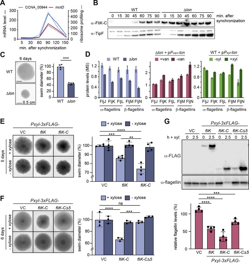
Figure 4. The flagella hook length regulator FliK is a Lon substrate with a C-terminal degradation tag. (A) Proteomics data obtained for CCNA_00944
and CCNA_00945 (MotD). The graphs on the left show CCNA_00944 and MotD stability in wild-type (WT) and Δlon (LS2382) cells, and graphs on the
right show CCNA_00944 and MotD abundance in the different strain backgrounds and conditions as determined by mass spectrometry. Each data
point for CCNA_00944 represents the mean protein abundance of the two experimental replicates, error bars show standard deviations. MotD was only
detected in one of the replicates. (B) Schematic representation of the CCNA_00944 and motD (CCNA_00945) genes as annotated in the Caulobacter
crescentus NA1000 genome, and how the presence of an additional guanosine (highlighted in red) merges the two genes to form one continuous gene
that was named fliK. The corresponding FliK protein contains a Flg hook domain. (C) In vivo degradation assay of full-length FliK in WT and ∆lon (KJ546)
cells. Samples were taken 0, 5, 10, and 15 min after shutting off protein synthesis. (D) In vivo degradation assay of ectopically expressed N-terminally
3xFLAG-tagged full-length FliK in WT and ∆lon (KJ546) cells. The graph shows mean values and standard deviation of relative protein levels 0, 30, and
60 min after protein synthesis shut off determined from three independent experiments. (E) In vivo degradation assay of the C-terminal part of FliK
(FliK-C) in WT and ∆lon (KJ546) cells. N-terminally 3xFLAG tagged FliK-C was ectopically expressed and samples were taken at indicated time points
after shut off of protein synthesis. Quantifications show mean values obtained from three independent biological replicates and error bars represent
standard deviation. (F) In vitro degradation assays showing Lon-dependent degradation of FliK-C and truncated FliK-C proteins lacking N-terminal,
internal, or C-terminal regions, graphically illustrated on the left side of the panel. Degradation assays were carried out in Lon reaction buffer with 4 µM
of one of the FliK-C variants, 0.125 µM of Lon hexamer in the presence of the ATP regeneration system (ATP, creatine phosphate, and creatine kinase
[CK]). Asterisks (*) mark a low molecular weight protein, that co-purifies with FliK-C (and some variants) but is unaffected by Lon. The band intensities
Figure 4 continued on next page
Omnus, Fink, et al. eLife 2021;10:e73875. DOI: https://doi.org/10.7554/eLife.73875 8 of 24Research article Cell Biology | Microbiology and Infectious Disease
Figure 4 continued
of the FliK-C variants from three independent experiments are represented as means with standard deviations on the right side of the panel. (G) In vivo
degradation assay of N-terminally 3xFLAG tagged FliK-C (WT) and a variant lacking the C-terminal five amino acids (FliK-C∆5) after xylose induction
in WT cells. Samples were taken at indicated time points after shut off of protein synthesis. Quantifications show mean values of relative protein levels
obtained from three biological replicates and error bars represent standard deviation.
The online version of this article includes the following figure supplement(s) for figure 4:
Source data 1. Unprocessed Western blot and protein gel images.
Figure supplement 1. Western blots of native FliK and 3xFLAG tagged FliK.
Figure supplement 1—source data 1. Unprocessed Western blot images.
Figure supplement 2. In vitro FliK-C degradation by Lon is ATP-dependent.
Figure supplement 2—source data 1. Unprocessed protein gel image.
A0A0H3C6R3), MotD contains a conserved Flg hook domain that is commonly present in the C-ter-
minal portion of FliK proteins that control flagella hook length in many bacteria (Waters et al., 2007).
When we attempted to clone CCNA_00944, we repeatedly observed one additional guanosine in
the cloned gene sequence that was not present in the reference genome sequence of C. crescentus
NA1000, the strain that we use. This insertion, which is also present in the sequence reads of previ-
ously published RNA-sequencing data (Schrader et al., 2014), generates a frameshift that merges the
CCNA_00944 gene with the downstream located motD gene, thus forming one single open reading
frame, of which the 3′ portion corresponds to motD (Figure 4B). This resulting gene corresponds
to a single open reading frame (CC_0900) in C. crescentus CB15, the isolate from which NA1000
is derived. These observations indicate that CCNA_00944 and motD are incorrectly annotated in
the reference genome of NA1000 and instead form one continuous open reading frame. Consis-
tently, when we performed Western blot analysis with antiserum raised against the protein portion
corresponding to MotD, we detected one single protein band that runs at high molecular weight
(Figure 4—figure supplement 1), confirming that CCNA_00944 and motD form together one single
open reading frame. Because the C-terminal portion of this new gene encodes a Flg hook domain
that is a characteristic of FliK proteins in other bacteria and because no other gene has so far been
annotated as fliK in C. crescentus, we named the new gene fliK (Figure 4B). This annotation of fliK is
also in line with a recent study in Sinorhizobium meliloti, which suggested that the motD gene from
alpha-proteobacteria should be renamed fliK (Eggenhofer et al., 2006).
FliK is a Lon substrate that is recognized at its C-terminus
Having established that CCNA_00944 and MotD are part of the same FliK protein, we next investi-
gated its regulation by Lon. Consistent with our proteomics data, we found that FliK abundance and
stability were notably increased in Δlon cells, consistent with FliK being a Lon substrate (Figure 4C). To
investigate the sequence determinants required for Lon to interact with FliK, we expressed an N-ter-
minally FLAG-tagged version of FliK and monitored its stability in vivo. Similar to the non-tagged FliK,
3xFLAG-FliK was efficiently degraded in WT cells but stable in cells lacking Lon (Figure 4D). This result
led us to hypothesize that the recognition by Lon occurs via the C-terminal domain of FliK. Therefore,
we monitored the stability of the C-terminal portion of FliK containing the Flg hook domain, which
corresponds to the formerly annotated MotD protein. Like the full-length protein, degradation of
this truncated FliK protein (FliK-C), with a FLAG-tag at the N-terminus, depended strongly on Lon
(Figure 4E). Furthermore, in vitro degradation assays showed that Lon degrades non-tagged FliK-C in
an ATP-dependent manner (Figure 4F, Figure 4—figure supplement 2). Based on these results, we
conclude that FliK is a Lon substrate, and that its C-terminal portion containing the Flg hook domain
is sufficient for Lon-dependent degradation.
To further pinpoint the regions within FliK-C that are required for Lon-dependent turnover, we
analyzed the degradation of a set of additional truncation mutants. According to the MobiDB data-
base (Piovesan et al., 2021), the C-terminal portion of FliK lists two unordered regions. We engi-
neered FliK-C variants that lack either of these unordered regions (FliK-C∆N44 and FliK-C∆59–91) or
the C-terminal part (FliK-C∆C44). In vitro degradation assays showed that deletion of the unordered
regions did not influence degradation, whereas removal of the 44 C-terminal amino acids completely
stabilized the protein (Figure 4F). This result, and the fact that some of the degrons recognized by
Omnus, Fink, et al. eLife 2021;10:e73875. DOI: https://doi.org/10.7554/eLife.73875 9 of 24Research article Cell Biology | Microbiology and Infectious Disease
Lon are located at the very C-terminus of Lon substrates (Burgos et al., 2020; Ishii et al., 2000; Puri
and Karzai, 2017; Zhou et al., 2019), prompted us to determine the in vivo stability of a FliK-C variant
lacking only the C-terminal five amino acids with the sequence LDIRI (3xFLAG-FliK-C∆5) (Figure 4G).
This deletion abolished FliK-C degradation (Figure 4G), demonstrating that the interaction between
Lon and FliK depends on these C-terminal amino acids.
Lon ensures temporally restricted accumulation of FliK during the cell
cycle
Like many other proteins involved in flagella biogenesis in C. crescentus, the transcription of the two
annotated genes CCNA_00944 and motD that together form the fliK gene is cell cycle regulated
(Lasker et al., 2016; Schrader et al., 2016), with mRNA levels and ribosome occupancy peaking in
late S-phase when a new flagellum at the pole opposite the stalk is being assembled (Figure 5A).
Consistently, Western blot analysis with synchronized WT C. crescentus cultures showed that FliK
protein was not detectable in the beginning of the cell cycle, but began to accumulate in late stalked
cells before reaching a maximum in abundance in predivisional cells shortly before cell division
(Figure 5B). This cell cycle-dependent pattern of FliK abundance was completely absent in the Δlon
strain, in which FliK was already detectable in swarmer cells and remained at high levels throughout
the cell cycle (Figure 5B). This result shows that, as in the case of StaR, Lon is absolutely necessary
to ensure that the protein is eliminated in the cell cycle phase when its function is no longer needed.
Precise regulation of FliK abundance is required for proper flagellin
expression and flagella function
Next, we wanted to investigate if the Lon-dependent regulation of FliK abundance is required for
proper motility in C. crescentus. Consistent with a previous study (Yang et al., 2018), we observed
that Δlon cells show reduced motility in soft agar compared to the WT (Figure 5C), which might be
caused by reduced flagella function in addition to growth and cell division defects. Additionally, our
proteomics data revealed that the levels of α-flagellins FljJ, FljK, and FljL and β-flagellins FljM and
FljN, which compose the structural components of flagella, are strongly downregulated in the Δlon
mutant (Figure 5D). Although a Lon-dependent effect on flagellin levels was not apparent after
4.5 hr of Lon depletion and only to a lesser extent upon lon overexpression (Figure 5D), these data
point to an indirect involvement of Lon in the regulation of flagella biosynthesis. One explanation for
the motility defect and the reduced flagellin levels in the Δlon mutant might be the stabilization and
higher levels of SciP in this strain (Figure 1A; Gora et al., 2013), which is known to negatively affect
flagellin gene expression through CtrA (Gora et al., 2010). Since correct regulation of FliK levels
was shown to be critical for proper flagella biosynthesis in other species (Muramoto et al., 1998;
Waters et al., 2007), we thought that the stabilization and thus increased abundance of FliK in the
absence of Lon might contribute to the motility defect of Δlon cells as well. To specifically study the
consequences of increased FliK abundance, we overexpressed FLAG-tagged FliK, FliK-C, and FliK-
CΔ5 from a medium copy vector in otherwise WT cells and assessed soft agar motility and flagellin
levels. Overexpression of FliK led indeed to a reproducible reduction in swim diameter to 85%
compared to the vector control strain (Figure 5E), indicating that elevated levels of FliK impair
motility. Interestingly, while this effect was clearly exacerbated in the strain overexpressing FliK-C
(Figure 5E–F), it was absent in the strain overexpressing FliK-CΔ5, demonstrating that the C-ter-
minus of FliK is critical for the FliK-dependent effect on motility. When analyzing flagellin protein
levels in the different overexpression strains, we found that the motility defects of the FliK and FliK-C
overexpression strains correlated with a significant downregulation of flagellin levels to 55% and
30%, respectively (Figure 5G). Conversely, overexpression of the FliK-CΔ5 affected flagellin levels
only mildly (Figure 5G).
Taken together, our data indicate that an oversupply of FliK, which can either be caused by overex-
pression or absence of Lon-dependent degradation, leads to reduced motility and flagellin levels. This
suggests that FliK likely contributes to the motility defects of Δlon cells along with SciP and potentially
other Lon substrates affecting flagella function that are stabilized and upregulated in the absence
of Lon. Furthermore, our data revealed that the C-terminal portion of FliK is critical not only for FliK
degradation, but also for its effects on motility and flagellin protein levels.
Omnus, Fink, et al. eLife 2021;10:e73875. DOI: https://doi.org/10.7554/eLife.73875 10 of 24Research article Cell Biology | Microbiology and Infectious Disease Figure 5. Lon-dependent degradation ensures temporal regulation of FliK levels during the cell cycle, which is needed for normal flagellin expression and motility. (A) RNA-sequencing and ribosome profiling data for CCNA_00944 and motD (CCNA_00945), as previously published (Lasker et al., 2016; Schrader et al., 2016). (B) Protein levels of FliK and TipF in synchronized wild-type (WT) and ∆lon (KJ546) cultures over 90 min following release of swarmer cells into PYE medium. TipF was included as a control. (C) Motility assay of Caulobacter crescentus WT and the ∆lon (KJ546) mutant in PYE soft agar after 6 days. The graph shows the relative swim diameters from four biological replicates, means (mean WT value was set to 100%), and standard deviations are indicated. Statistical significance was determined by paired two-tailed t-test: WT vs. ∆lon p=0.0002, ***. (D) Flagellin protein levels as determined by mass spectrometry in WT and ∆lon (LS2382) mutant cells as well as in the other strain backgrounds and conditions, see Figure 1. (E) Motility assay in soft agar of strains overexpressing 3xFLAG-tagged FliK and FliK-C by xylose induction (+xylose) in comparison to the vector control (VC) and non-inducing conditions (−xylose). The graph shows the relative swim diameters from four biological replicates, means (mean of VC was set to 100%) and standard deviations are indicated. Statistical significance was determined by ordinary one-way ANOVA (Šidák’s multiple comparisons test) for the following comparisons: VC +xyl vs. Pxyl-3xFLAG-fliK +xyl p=0.0004, ***; VC +xyl vs. Pxyl-3xFLAG-fliK-C +xyl
Research article Cell Biology | Microbiology and Infectious Disease Figure 5 continued Pxyl-3xFLAG-fliK-C +xyl p=0.0016, **. (F) Motility assay in soft agar of strains overexpressing 3xFLAG-tagged FliK-C and FliK-C∆5 by xylose induction (+xylose) in comparison to the VC and non-inducing conditions (−xylose). The graph shows the relative swim diameters from three biological replicates, means (relative to VC) and standard deviations are indicated. Statistical significance was determined by ordinary one-way ANOVA (Šidák’s multiple comparisons test) for the following comparisons: VC +xyl vs. Pxyl-3xFLAG-fliK-C +xyl p
Research article Cell Biology | Microbiology and Infectious Disease
flagellum synthesis
stalk biogenesis
accumulation clearance accumulation clearance
StaR Lon
hook
OM
IM
FliK Lon
stalk elongation / flagella hook synthesis /
holdfast production flagellin secretion
Figure 6. Lon ensures temporal regulation of stalk and flagella biogenesis during the Caulobacter crescentus cell
cycle. Lon specifically degrades StaR, a transcriptional regulator of stalk biogenesis and holdfast production, and
FliK, a protein involved in regulating flagella hook synthesis. The expression of staR peaks in swarmer cells, while
the expression of fliK peaks in late stalked and predivisional cells (Lasker et al., 2016; Laub et al., 2000; Schrader
et al., 2016). Our study shows that Lon-dependent proteolysis is required to rapidly eliminate these proteins when
their expression levels drop, thus outpacing synthesis. The combination of proteolysis and regulated transcription
ensures that StaR abundance is temporally restricted to the swarmer-to-stalked cell transition (shown in blue) and
FliK abundance to the late stalked and predivisional cell (shown in green) when their functions are needed for stalk
biogenesis or flagella synthesis, respectively.
The online version of this article includes the following figure supplement(s) for figure 6:
Figure supplement 1. FlgE degradation is partially dependent on Lon.
Figure supplement 1—source data 1. Unprocessed Western blot images.
In addition to proteins involved in stalk biogenesis and surface attachment, we identified several
proteins required for flagella-mediated motility and chemotaxis as potential Lon substrates (Figure 1F),
and investigated FliK in detail. FliK proteins are thought to function as molecular rulers that, while
being exported themselves, precisely measure flagellar hook length via the N-terminal domain and
mediate via the C-terminal domain a switch from export of hook protein to filament protein, once the
hook has reached a certain length (Erhardt et al., 2011; Minamino, 2018; Minamino et al., 1999;
Moriya et al., 2006; Shibata et al., 2007). Although previous work made significant progress in under-
standing the molecular function of this important protein in enterobacteria, proteolytic control mech-
anisms contributing to FliK regulation have so far not been described. Moreover, in bacteria outside
the gamma-proteobacteria FliK remains poorly studied. In C. crescentus, a fliK gene has previously
not been annotated, and our new data revealed that fliK corresponds to a gene that was previously
annotated as two separate but overlapping genes. We demonstrate that Lon-mediated proteolysis
ensures cell cycle-dependent accumulation of FliK during late S-phase, when the cell prepares for cell
Omnus, Fink, et al. eLife 2021;10:e73875. DOI: https://doi.org/10.7554/eLife.73875 13 of 24Research article Cell Biology | Microbiology and Infectious Disease
division by building a new flagellum and chemotaxis apparatus at the swarmer pole (Figure 6). The
results that overexpression of FliK results in reduced flagellin levels and impaired motility (Figure 5E
and G) indicate that this precise regulation of FliK abundance is critical for correct flagella biosyn-
thesis. Based on studies in other bacteria (Muramoto et al., 1998), we consider it possible that an
excess of FliK protein causes a premature termination of hook synthesis, leading to shorter hooks,
which may cause downstream effects, namely reduced flagellin levels and motility. Our observation
that overexpression of FliK-C, a truncated version of FliK lacking the N-terminal domain, which is
required for export in other bacteria (Hirano et al., 2005), exhibits an even stronger effect on flagellin
levels and motility may be explained by a dominant-negative effect that the truncated version of FliK
has over the WT. In this scenario, overexpressed FliK-C would block the normal function of native FliK,
possibly by occupying binding sites on interacting proteins, for example, the export apparatus protein
FlhB (Kinoshita et al., 2017; Minamino et al., 2009). Our result that deletion of the C-terminal five
amino acids of Caulobacter FliK-C abolishes the negative overexpression effects (Figure 5F and G) is
consistent with the finding that FliK’s export switch-inducing function depends on its C-terminal five
amino acids in other bacteria (Kinoshita et al., 2017; Minamino et al., 2006; Williams et al., 1996).
Importantly, we also uncovered a critical role of the C-terminal five amino acids of FliK for proteolytic
control (Figure 4G), thus, our data indicate a tight coupling between FliK function and degradation.
Interestingly, in addition to the flagella hook length regulator FliK, our data indicate that the
flagella hook protein FlgE itself is regulated by Lon in C. crescentus. FlgE was among the proteins that
satisfied two criteria in our proteomics approach, and we verified by Western blot analysis that FlgE
degradation partly depends on Lon (Figure 6—figure supplement 1). This finding is also consistent
with previous work showing that Lon degrades FlgE in E. coli (Arends et al., 2018), and reinforces the
previously made notion that precise regulation of the cellular concentrations of FlgE as well as FliK
is an important requirement for the correct temporal order of flagella assembly (Inoue et al., 2018).
In several other bacteria, including important human pathogens, Lon has been linked to flagella-
mediated motility (Ching et al., 2019; Clemmer and Rather, 2008; Fuchs et al., 2001; Rogers
et al., 2016). In many of these cases the nature of substrates mediating the observed Lon-dependent
effects on motility remains unknown. However, in Bacillus subtilis, it was shown that Lon specifically
degrades the master regulator of flagellar biosynthesis SwrA by a mechanism requiring the adaptor
SmiA (Mukherjee et al., 2015). In contrast to SwrA, both StaR and FliK are robustly degraded by
Lon in vitro (Figures 2D and 4F, Figure 4—figure supplement 2). While these data suggest that no
adaptors or other accessory proteins are required to mediate the interaction between these proteins
and Lon, it is possible that additional factors exist that modulate the rate of Lon-mediated proteolysis
of these proteins in response to specific conditions.
In conclusion, our work provides new insights into the cellular roles of Lon and emphasizes the
importance of proteolysis in adjusting the amounts of regulatory proteins involved in critical cellular
processes, including cell differentiation and cell cycle progression. Importantly, in addition to StaR and
FliK our work identified many other proteins as putative Lon substrates and the precise role of Lon in
the regulation of these proteins will be worthwhile to investigate in detail in future studies. Further-
more, our work highlights quantitative proteomics using isobaric mass tags as a powerful approach for
the identification of novel protease substrates that could be exploited to identify candidate substrates
under diverse growth conditions, in different species or of other proteases.
Materials and methods
Key resources table
Reagent type
(species) or
resource Designation Source or reference Identifiers Additional information
Gene (Caulobacter
crescentus) fliK-C; motD; CCNA_00945 GeneBank GeneBank:CCNA_00945
Gene
(C. crescentus) staR; CCNA_02334 GeneBank GeneBank:CCNA_02334
Continued on next page
Omnus, Fink, et al. eLife 2021;10:e73875. DOI: https://doi.org/10.7554/eLife.73875 14 of 24Research article Cell Biology | Microbiology and Infectious Disease
Continued
Reagent type
(species) or
resource Designation Source or reference Identifiers Additional information
Strain, strain Michael Laub, Massachusetts
background Institute of Technology;
(Escherichia coli) DH5α Other Chemical competent cells
Strain, strain Claes Andréasson, Stockholm
background (E. University;
coli) BL21-SI/pCodonPlus Other Electrocompetent cells
Strain, strain Michael Laub, Massachusetts
background Institute of Technology;
(C. crescentus) NA1000 Other Electrocompetent cells
Genetic reagent Martin Thanbichler, MPI
(plasmid) pBX-MCS-4 (plasmid) Thanbichler et al., 2007 Marburg
Genetic reagent
(plasmid) pSUMO-YHRC Holmberg et al., 2014 RRID:Addgene_54336
Goat anti-mouse IgG (H+L)
Antibody Secondary Antibody, HRP Thermo Fisher Scientific Cat# 32430; RRID:AB_1185566 (1:5000)
Goat anti-Rabbit IgG (H+L)
Antibody Secondary Antibody, HRP Thermo Fisher Scientific Cat# 32460; RRID:AB_1185567 (1:5000)
ANTI-FLAG M2 antibody
Antibody (Mouse monoclonal) Sigma-Aldrich Cat# F1804; RRID:AB_262044 (1:5000)
Antibody Anti-DnaA (Rabbit polyclonal) Jonas et al., 2011 (1:5000)
(1:10,000) kindly provided by
Antibody Anti-Lon (Rabbit polyclonal) Other R.T. Sauer
Antibody Anti-CcrM (Rabbit polyclonal) Stephens et al., 1996 (1:5000)
Antibody Anti-SciP (Rabbit polyclonal) Gora et al., 2010 (1:2000)
(1:5000) kindly provided by P.
Antibody Anti-TipF (Rabbit polyclonal) Davis et al., 2013 Viollier
Antibody Anti-StaR (Rabbit polyclonal) Fiebig et al., 2014 (1:500)
Antibody Anti-FliK-C (Rabbit polyclonal) This paper (1:500)
Anti-flagellin (Rabbit (1:2000) kindly provided by Y.
Antibody polyclonal) Brun and Shapiro, 1992 Brun
Source vector and purification
Peptide, protocol kindly provided by
recombinant Claes Andréasson (Stockholm
protein Ulp1-6xHis Other University)
Commercial assay SuperSignal West Femto
or kit Maximum Sensitivity Substrate Thermo Fisher Scientific Cat # 34095
Bio-Rad https://www.bio-rad.
Software, com/en-ca/product/image-lab-
algorithm Image Lab software RRID:SCR_014210 Version 6.0
Software,
algorithm GraphPad Prism https://www.graphpad.com RRID:SCR_002798 Version 7.0
Software, Schindelin et al., 2012
algorithm Fiji (ImageJ) https://fiji.sc/ RRID:SCR_002285
4–20% Mini-PROTEAN TGX
Stain-Free Protein Gels, 15
Other well, 15 µl Bio-Rad Cat # 4568096
Other Trans-Blot Turbo System Bio-Rad Cat # 1704150EDU
Other LI-COR Odyssey Fc Imaging LI-COR https://www.licor.com/bio/
System odyssey-fc/
Omnus, Fink, et al. eLife 2021;10:e73875. DOI: https://doi.org/10.7554/eLife.73875 15 of 24Research article Cell Biology | Microbiology and Infectious Disease
Strains and plasmids
All bacterial strains, plasmids, and primers used in this study are listed in Supplementary file 1.
Plasmid construction
Expression plasmids for protein purification
Plasmids used for protein expression are based on the pSUMO-YHRC backbone and were constructed
as follows: the coding sequences of the staR (pMF56-c88) and fliK-C (formerly motD; pMF61) genes
were amplified from C. crescentus NA1000 genomic DNA with the primers listed in Supplemen-
tary file 1 (see sheets listing primers and vector fragments for sequences and primer combina-
tions, respectively). The backbone vector pSUMO-YHRC was amplified in two parts disrupting the
kanamycin resistance gene in order to reduce background (using primer pairs oMJF34/oMJF36 and
oMJF37/oMJF38, Supplementary file 1). Following the PCR, the template was digested with DpnI
(10 U) and the remaining PCR fragments were subsequently purified by gel extraction. Fragments
were then assembled using Gibson assembly (Gibson et al., 2009). Vectors containing deletions of
an annotated gene (FliK-C truncations: pMF66, pMF67-A, and pMF68-A) were derived from vectors
harboring the full-length coding sequence in a similar manner using the primer pairs specified in
Supplementary file 1.
Replicating plasmids
pDJO145 (pBX-MCS-4 containing NdeI-3xFLAG-KpnI): plasmid pBX- MCS-4 (Thanbichler et al.,
2007) was amplified using primers oDJO13 and oDJO41. The sequence encoding the triple FLAG
tag (3xFLAG; GAC TAC AAA GAC CAT GAC GGT GAT TAT AAA GAT CAT GAC ATC GAC TAC AAG
GAC GAC GAC GAC AAG) was amplified from a plasmid using primers oDJO42 and oDJO43 adding
a KpnI-site followed by a stop codon to the 3′ end of 3xFLAG. The two amplified fragments were then
joined by Gibson assembly (Gibson et al., 2009).
pDJO151 (pBX-MCS-4 containing Pxyl-staR-3xFLAG): staR was amplified with primers oDJO44
and oDJO45 using chromosomal C. crescentus NA1000 DNA as template and cloned into NdeI-cut
pDJO145 using Gibson assembly.
pDJO157 (pBX-MCS-4 containing Pxyl-3xFLAG-staR): staR was amplified with primers oDJO46
and oDJO47 using chromosomal C. crescentus NA1000 DNA as template and cloned into KpnI-cut
pDJO145 using Gibson assembly.
pDJO173 (pBX-MCS-4 containing Pxyl-flgE-3xFLAG): flgE was amplified with primers oDJO65
and oDJO66 using chromosomal C. crescentus NA1000 DNA as template and cloned into NdeI-cut
pDJO145 using Gibson assembly.
pDJO200 (pBX-MCS-4 containing Pxyl-3xFLAG-fliK-C): fliK-C (CCNA_00945) was amplified with
primers oDJO87 and oDJO88 using chromosomal C. crescentus NA1000 DNA as template and
cloned into KpnI-cut pDJO145 using Gibson assembly.
pDJO410 (pBX-MCS-4 containing Pxyl-3xFLAG-fliK-CΔ5): fliK-CΔ5 was amplified with primers
oDJO87 and oDJO179 using C. crescentus NA1000 DNA as template and cloned into KpnI-cut
pDJO145 using Gibson assembly.
pDJO487 (pBX-MCS-4 containing Pxyl-3xFLAG-fliK): fliK was amplified with primers oDJO75 and
oDJO88 using chromosomal C. crescentus NA1000 DNA as template and cloned into KpnI- cut
pDJO145 using Gibson assembly.
Strain construction
To generate the ΔstaR Δlon strain (KJ1037), the ΔstaR deletion was introduced into the Δlon strain
(KJ546) by two-step recombination (Skerker et al., 2005) after transformation with plasmid pNTPS138-
ΔstaR (pAF491; Fiebig et al., 2014). Briefly, transformants were selected on kanamycin plates, single
colonies were grown overnight in PYE and plated on PYE containing sucrose. Single sucrose-resistant
colonies were subsequently screened for kanamycin sensitivity and the staR knockout was confirmed
by colony PCR using primers oDJO40 and oDJO38.
C. crescentus strains carrying replicating plasmids were created by transforming the plasmids into
the respective strain backgrounds by electroporation.
Omnus, Fink, et al. eLife 2021;10:e73875. DOI: https://doi.org/10.7554/eLife.73875 16 of 24You can also read