Acute and chronic neurological disorders in COVID-19
←
→
Page content transcription
If your browser does not render page correctly, please read the page content below
Page 1 of 30                                                             Brain

               REVIEW ARTICLE

                 Acute and chronic neurological disorders in COVID-19:
                                           potential mechanisms of disease
                                       Erin F. Balcom,1 Avindra Nath2 and Christopher Power1

                                                                                                                                              Downloaded from https://academic.oup.com/brain/advance-article/doi/10.1093/brain/awab302/6353020 by guest on 20 December 2021
               Abstract
               COVID-19 is a global pandemic caused by SARS-CoV-2 infection and is associated with
               both acute and chronic disorders affecting the nervous system. Acute neurological disorders
               affecting patients with COVID-19 range widely from anosmia, stroke,
               encephalopathy/encephalitis, and seizures to Guillain-Barre Syndrome. Chronic neurological
               sequelae are less well defined although exercise intolerance, dysautonomia, pain, as well as
               neurocognitive and psychiatric dysfunctions are commonly reported. Molecular analyses of
               cerebrospinal fluid and neuropathological studies highlight both vascular and immunologic
               perturbations. Low levels of viral RNA have been detected in the brains of few acutely ill
               individuals. Potential pathogenic mechanisms in the acute phase include coagulopathies with
               associated cerebral hypoxic-ischemic injury, blood-brain barrier abnormalities with
               endotheliopathy and possibly viral neuroinvasion accompanied by neuro-immune responses.
               Established diagnostic tools are limited by a lack of clearly defined COVID-19 specific
               neurological syndromes. Future interventions will require delineation of specific neurological
               syndromes, diagnostic algorithm development, and uncovering the underlying disease
               mechanisms that will guide effective therapies.

               Author affiliations:
               1 Division of Neurology, University of Alberta, Edmonton AB Canada
               2 Section of Infections of the Nervous System, NINDS-NIH, Bethesda, MD, USA

               Correspondence to: C. Power
               Department of Medicine (Neurology)
               University of Alberta, 6-11 Heritage Medical Research Centre
               Edmonton, AB Canada
               E-mail: chris.power@ualberta.ca

               Running title: Neurological disorders in COVID-19
               © The Author(s) (2021). Published by Oxford University Press on behalf of the Guarantors of Brain.
               This is an Open Access article distributed under the terms of the Creative Commons Attribution Non-Commercial License
               (http://creativecommons.org/licenses/by-nc/4.0/), which permits non-commercial re-use, distribution, and reproduction in any
               medium, provided the original work is properly cited. For commercial re-use, please contact journals.permissions@oup.com
Brain                                               Page 2 of 30

Keywords: SARS-CoV-2; COVID-19; nervous system; encephalopathy; stroke

Introduction

Since its discovery in Wuhan, China in late 2019, COVID-19, the illness caused by severe

                                                                                                            Downloaded from https://academic.oup.com/brain/advance-article/doi/10.1093/brain/awab302/6353020 by guest on 20 December 2021
acute respiratory syndrome coronavirus 2 (SARS-CoV-2) has resulted in over 3 million
deaths (https://covid19.who.int/) and placed unprecedented pressure on social, economic, and
health care systems worldwide. Many survivors of the acute infection experience persistent
and incapacitating neurological symptoms, which can have socio-economic and personal
consequences.1 It is thus imperative that there is a thorough understanding of the evolving
clinical syndromes and the underlying pathophysiological mechanisms, enabling rational
therapeutic interventions to be expeditiously deployed.

Retrospective cohort studies from around the world report neurologic signs and symptoms
such as headache, altered mental status, seizures, and stroke, in over a third of patients during
the acute phase of the illness,2-4 positioning SARS-CoV-2 as an emerging neuro-pathogen. A
similar proportion of infected persons develop a post-infectious viral syndrome with diverse
neuropsychiatric manifestations. Viral infections cause neurologic impairments through
multiple mechanisms,5 including direct infection of neurons, glia, or endothelial cells within
the nervous system resulting in acute cell death, as observed in herpes simplex virus type-1
(HSV-1) encephalitis.6 Alternatively, viruses e.g., the human immunodeficiency virus type-1
(HIV-1) can persist in cellular reservoirs within the central (CNS) and perhaps peripheral
(PNS) nervous system resulting in chronic inflammation and insidious progressive neurologic
damage.7 Among non-neurotropic viruses such as influenza and other respiratory viruses,
systemic infection is associated with inflammation, metabolic and hormonal derangements,
with vascular injury resulting in neurologic disease.8 The host immune responses triggered
during or following viral infections can also result in autoimmune damage of neural tissues,
as observed in the PNS (e.g., Guillain-Barre Syndrome/GBS) and in the CNS (e.g., Acute
Disseminated Encephalomyelitis/ADEM or Acute Transverse Myelitis/ATM). Each of these
mechanisms are implicated in SARS-CoV-2 infection and are addressed below. Of note, a
multisystem inflammatory syndrome in children (MIS-C) has been described in paediatric
cohorts with COVID-19; several cohort studies of children infected with SARS-CoV-2 report
neurological disorders resembling those observed in adults including headache,

          ScholarOne, 375 Greenbrier Drive, Charlottesville, VA, 22901 Support (434) 964 4100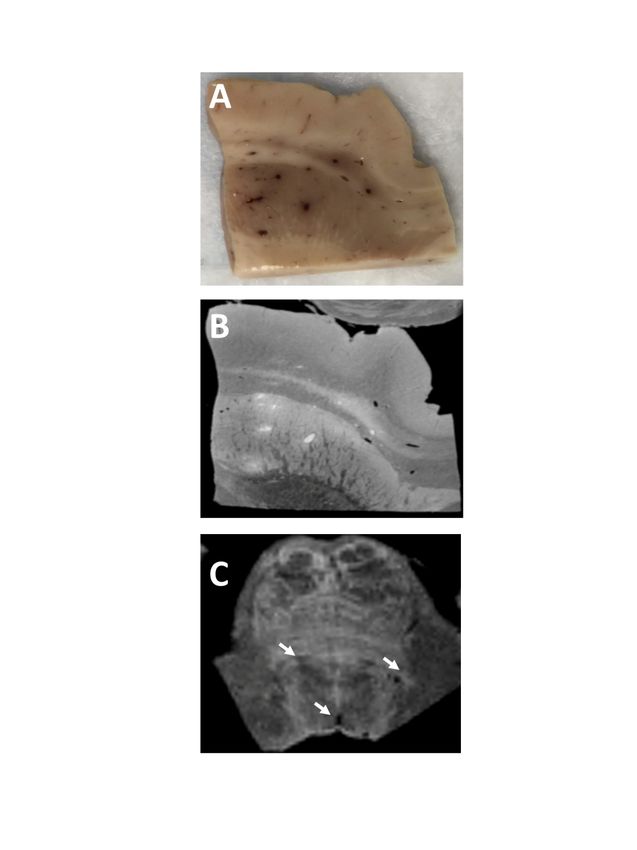
Page 3 of 30                                                    Brain

               encephalopathy, demyelinating disorders and stroke.9-13 This review provides an update on
               neurologic manifestations of COVID-19 that concentrates on adults while also examining
               contemporary evidence for the neuropathogenic mechanisms implicated in SARS-CoV-2
               infection (Figure 1) and their relationship to current and potential therapies (Table 1).
               Acute neurological syndromes
               Anosmia and ageusia were among the first focal neurologic symptoms described in COVID-

                                                                                                                    Downloaded from https://academic.oup.com/brain/advance-article/doi/10.1093/brain/awab302/6353020 by guest on 20 December 2021
               19, and generated interest in SARS-CoV-2’s potential neurotropism.14 Anosmia was reported
               in 5-35% of hospitalized patients, and may be higher among non-hospitalized patients with
               COVID-19.15 In some cases, it is the sole reported symptom, or persists far beyond the acute
               respiratory symptoms, negatively influencing the quality of life of survivors. Infection of the
               nasal mucosa and sustentacular cells with dissemination throughout olfactory nerve
               projections is one proposed mechanism of neuropathogenesis in COVID-19.16 Viral RNA has
               also been found in the olfactory bulb at post-mortem of some patients, though its association
               with neurologic injury is not established.16

               Altered mental status is a commonly reported neurological finding associated with COVID-
               19 hospitalization. Abnormalities in electroencephalography (EEG) in COVID-19 related
               encephalopathy correlate with disease severity.17 Frontal slowing is the most common pattern
               observed and has been proposed as a biomarker for COVID-19 encephalopathy.17 Seizures
               are uncommon among COVID-19 patients; in a retrospective cohort of 1043 patients, 0.7%
               developed seizures in hospital, and an even smaller proportion of those seizures occurred
               outside the context of pre-existing epilepsy18 although a recent retrospective study indicated
               that epileptiform abnormalities are frequently detected (48.7%) of hospitalized patients with
               COVID-19.19 Altered mental status, coma, and seizures in COVID-19 are almost certainly
               multi-factorial but can be stratified into metabolic/non-inflammatory versus inflammatory
               (e.g., encephalitis) categories.

               Non-inflammatory encephalopathy. Many patients with COVID-19 and altered mental status
               (e.g., lethargy, confusion, coma) have clinical courses complicated by hypoxia, renal failure,
               electrolyte disturbances, sedating medications, and underlying comorbidities. A third of
               critically ill patients with COVID-19 present with encephalopathy, often with frontal lobe-
               associated features,20, 21 either at the onset of illness or during the course of hospitalization,
               and encephalopathy is associated with increased mortality and poor functional outcome.22-24
               Although encephalopathy has been reported in COVID-19 patients at all ages, patients

                          ScholarOne, 375 Greenbrier Drive, Charlottesville, VA, 22901 Support (434) 964 4100
Brain Page 4 of 30
beyond the sixth decade of life and those with pre-existing neurologic conditions (stroke,
dementia, Parkinson’s disease) are most affected, particularly in the context of severe
respiratory illness.22, 25 While encephalopathy in the aforementioned circumstances is likely
multifactorial, reports of patients with encephalopathy in the absence of severe respiratory
illness suggest other possible mechanisms including bioenergetic failure and vascular
dysfunction in SARS-CoV-2 infection.26 The association between encephalopathy and
Downloaded from https://academic.oup.com/brain/advance-article/doi/10.1093/brain/awab302/6353020 by guest on 20 December 2021
morbidity exists independently of respiratory disease, similar to that observed in sepsis-
associated encephalopathy.27 Encephalopathy in non-COVID-19 patients is attributed to
mitochondrial dysfunction, excitotoxicity, and macro- or micro-ischemic injury.27 One recent
post-mortem analysis in New York revealed cerebral microthrombi in a subset of 67
hospitalized, deceased COVID-19 patients.28 Small, sub-cortical ischemic events may result
in confusion and cognitive dysfunction.28 Patients can also manifest multifocal cerebral
microhemorrhages or vascular leakage (Figure 2) due to compromise of the cerebral
endothelial cells.29, 30 Radiographic findings in COVID-associated encephalopathy include
non-specific white matter hyperintensities, diffusion restriction, micro-hemorrhage, and
leptomeningeal enhancement29, 31. In one study of ICU patients with COVID-19 and
encephalopathy, bilateral frontotemporal hypoperfusion was evident in all patients who
underwent perfusion imaging for altered mental status,32 though this finding was disputed and
has not been replicated.33 Surprisingly, brain MRI was normal in up to 46% of patients with
COVID-19 and associated encephalopathy.31 In another study, patients with COVID-19 and
cognitive impairment showed decreased metabolism in the fronto-parietal regions on FDG-
PET scans.24 Indeed, correlations between neuropathology and brain MRI findings (including
normal imaging) have yet to be established.
Inflammatory encephalitis. A minority of COVID-19 patients encompass established
diagnostic criteria for infectious encephalitis.34 There are convincing reports of encephalitis-
like presentations associated with elevated levels of soluble IL-6, IL-18, TNF-, CXCL10
and markers of glial and astrocyte activation in CSF.35-38 Radiologic findings associated with
COVID-19 meningoencephalitis include mesial temporal lobe T2/FLAIR hyperintensities,
varying from punctate to diffuse in subcortical white matter, the brainstem, and claustrum,
often accompanied by cerebral edema.39-41 Case reports indicate that COVID-19 can present
with an acute disseminated encephalomyelitis (ADEM) phenotype,31, 42 including oculomotor
dysfunction, seizures, and coma. Others have reported cases of acute necrotizing hemorrhagic
encephalopathy that present initially with symmetric lesions in the thalami and are thought to
ScholarOne, 375 Greenbrier Drive, Charlottesville, VA, 22901 Support (434) 964 4100Page 5 of 30 Brain
be cytokine mediated.43 Some patients may present with isolated pseudotumor cerebri/benign
intracranial hypertension presumably from meningitis.44, 45Opsoclonus-myoclonus syndrome
(OMS), which has been observed in association with infections such as Epstein-Barr virus,
Chikungunya, and Mycoplasma pneumoniae, has also been reported in patients with COVID-
19, including those with mild respiratory disease.46 Most patients with COVID-19 and OMS
had partial recovery at 4 weeks after treatments including pulse steroids, IVIg and
Downloaded from https://academic.oup.com/brain/advance-article/doi/10.1093/brain/awab302/6353020 by guest on 20 December 2021
antiepileptic medications.46-48
For presumed COVID-19 associated encephalitis, favourable therapeutic responses to
corticosteroids and plasma exchange (PLEX) were observed in a subset of patients although
factors predicting a beneficial therapeutic response remain to be defined.49, 50 Whether the
neuroinflammation observed clinically, neuroradiologically, and neuropathologically is due
to direct viral invasion, para-infectious or autoimmune processes remains unknown. In most
COVID-19 cases with encephalitis, SARS-CoV-2 RNA is not detectable in CSF via RT-
PCR, favouring an immune-mediated mechanism of disease.37, 49, 51, 52
Cerebrovascular disease. Patients with COVID-19 have an increased rate of stroke compared
to other disease cohorts, with higher NIHSS scores compared to non-COVID-19 associated
stroke.53 Over half of strokes among patients with COVID-19 are cryptogenic, with a higher
proportion of large vessel occlusions.54 Some series have reported higher than expected rates
of posterior circulation strokes (35.3%).53, 54 Hypercoagulability induced by systemic and
focal inflammation has been implicated in COVID-19 associated strokes that include both
arterial and venous thromboembolic events (VTE).55 Cerebral venous sinus thrombosis
(CVST) among patients with COVID-19 can also occur with an abnormally activated
prothromboplastin time (aPTT) and elevated D-dimer levels.56, 57 COVID-19 associated
CVST has an estimated in-hospital mortality of 40% in a cohort that included non-ventilated
patients.56 In fact, CVST represents 4% of cerebrovascular complications in COVID-19 with
an estimated frequency of 0.08% among hospitalized patients.56 In comparison, CVST
accounts for 0.5-1% of all strokes among non-COVID-19 patients and occurs in
approximately 2-5 per million people each year (0.0002-0.0005%).58 Elevated D-dimer levels
are more common among COVID-19 patients presenting with both ischemic and
hemorrhagic stroke and are associated with higher all-cause mortality.54 As there is an
increased risk of thromboembolism during COVID-19, multiple studies have compared
standard dose thromboprophylaxis to high and intermediate dose prophylactic anticoagulation
ScholarOne, 375 Greenbrier Drive, Charlottesville, VA, 22901 Support (434) 964 4100Brain Page 6 of 30
in hospitalized patients with COVID-19. The largest of these trials, INSPIRATION
randomized clinical trial found no difference in all-cause mortality or VTE in patients treated
with standard versus intermediate dose thromboprophylaxis in critically ill patients with
COVID-1959, in contrast to earlier retrospective studies showing potential benefit in ICU
patients.60 Interim unpublished results of the multiplatform merged randomized control trial
(mpRCT) ATTACC/REMAP-CAP/ACTIV-4A found similar results in the critically ill
Downloaded from https://academic.oup.com/brain/advance-article/doi/10.1093/brain/awab302/6353020 by guest on 20 December 2021
cohort as well as a signal toward harm with therapeutic anticoagulation.61 Interestingly, this
study found improved survival in moderately ill patients with COVID-19 treated with
intermediate-dose anticoagulation, suggesting severity of disease may be important in
determining appropriate thromboprophylaxis in hospitalized patients.62, 63 Further studies are
required to determine if prophylactic anticoagulation specifically reduces the risk of stroke in
COVID-19, particularly because of reports of hemorrhagic stroke in hospitalized patients
while receiving therapeutic anticoagulation.64 Indeed, the risk of hemorrhagic stroke is higher
than predicted among COVID-19 patients65 and is associated with elevated serum ferritin.53
Similarly, microhemorrhages29 and acute hemorrhagic necrotizing encephalitis (AHNE) have
been reported in patients with COVID-19.43 Outcomes including risk of death and duration of
hospitalization following intracerebral or subarachnoid haemorrhages are worse among
patients with COVID-19.66
Recent reports of vaccine-induced thrombotic thrombocytopenia (VITT) following
administration of adenovirus vector-based COVID-19 vaccines have raised concern.67-69 As
of April 4th, 2021, there had been 169 cases of VITT-CVST reported to the European
Medicines Agency out of 34 million doses administered of the ChAdOx1 nCoV-19
(AstraZeneca) vaccine, with an incidence of VITT estimated at 1 per 100,000 exposures.67
The reported rate of VITT-CVST after administration of 6.86 million doses of the
Ad26.COV2.S adenoviral vector vaccine (Johnson & Johnson/Janssen) was 0.87 cases per
million (0.000087%).70, 71 The majority of cases occurred in women less than 50 years old,
and the majority of patients displayed high levels of antibodies to platelet factor 4 (PF4)-
polyanion complexes, prompting comparisons to heparin-induced thrombotic
thrombocytopenia.72 The precise pathophysiology of VITT is unknown to date; simultaneous
thrombosis and thrombocytopenia in VITT has only been reported for adenoviral vector
vaccines although there have been 5 possible reports of CVST with normal platelet counts
among 4 million doses of the Moderna mRNA vaccine.73 Among 54 million doses of the
Pfizer-BioNTech mRNA vaccine, there have been 35 reports of central nervous system
ScholarOne, 375 Greenbrier Drive, Charlottesville, VA, 22901 Support (434) 964 4100Page 7 of 30 Brain
thrombosis without thrombocytopenia (0.00006%).73 CSVT is a serious but rare condition
associated with SARS-CoV-2 vaccination,74 but there remains a consensus among health
authorities that the benefits of widespread vaccination outweigh the potential risks,
particularly when one considers the rate of thrombosis in COVID-19 infection.
Acute PNS disorders. Autoimmune polyradiculoneuropathies such as GBS or Miller Fisher
Downloaded from https://academic.oup.com/brain/advance-article/doi/10.1093/brain/awab302/6353020 by guest on 20 December 2021
Syndrome have been reported in patients with SARS-CoV-2 infection, with and without
respiratory symptoms.75 These disorders can be triggered by systemic infections and have
been reported in patients with other coronavirus infections such as MERS and SARS-CoV-
1.76 The majority of case reports of GBS in COVID-19 describe the common syndrome of
ascending weakness, areflexia with supporting CSF and nerve conduction studies, and are of
the acute inflammatory demyelinating polyneuropathy (AIDP) type. Disease onset is between
5-10 days after acute COVID-19 symptoms (including anosmia, respiratory and
gastrointestinal symptoms), which in the intensive care unit (ICU) settings helps distinguish
GBS from critical illness neuropathy that appears later in disease course.36, 76, 77 Patients with
COVID-19-associated GBS respond to standard treatments (e.g., IVIg, PLEX) although how
COVID-19 impacts treatment responsiveness remains uncertain.76 Of note, a UK
epidemiological cohort study showed rates of GBS have fallen during the current pandemic,78
likely resulting from increased public health efforts that have reduced transmission of more
common infectious triggers.
Myalgia and weakness occur in 30-50% of hospitalized patients with COVID-19,79, 80 and are
frequently reported by non-hospitalized patients.81 While myalgia is a common symptom
during many viral illnesses, the mechanism by which SARS-CoV-2 infection causes
debilitating muscle pain and weakness is unknown. Myositis and rhabdomyolysis as a
complication of COVID-19 are well recognized with elevated serum CK (>10,000) levels as
a common finding which correlates with mortality in hospitalized patients.79 There have also
been multiple reports of muscle edema demonstrated on MRI.82, 83 While other viruses such
as influenza are known to directly invade skeletal myocytes in vitro,84 to date there is no
evidence for infection of skeletal myocytes with SARS-CoV-2.83, 85 Myositis in COVID-19
could be triggered by host immune responses to the virus. Muscle biopsies from COVID-19
patients show perivascular inflammation83 including a case of Type 1 interferonopathy
associated myopathy in a young patient with SARS-CoV-2 infection.86 There are reports of
COVID-19 patients with elevated CK levels and muscle weakness who respond to
ScholarOne, 375 Greenbrier Drive, Charlottesville, VA, 22901 Support (434) 964 4100Brain Page 8 of 30
immunosuppression including high dose glucocorticoids83 as well as IVIg,76 prompting
comparisons to immune-mediated myositis (IMM). There is also a recent report of a patient
with proximal and bulbar weakness in COVID-19 with positive anti-SSA and SAE-1
antibodies who was successfully treated with the humanized monoclonal antibody against the
IL-6 receptor, tocilizumab.83 While the above neurological syndromes are observed in the
acute setting, they have the capacity to exert long-term effects, as described below.
Downloaded from https://academic.oup.com/brain/advance-article/doi/10.1093/brain/awab302/6353020 by guest on 20 December 2021
Patients who develop severe COVID-19 pneumonia often require prolonged ICU care. As
expected, critical illness polyneuropathy (CIP)85 and myopathy (CIM)87 have been reported
as complications of SARS-CoV-2 infection. While the pathophysiological mechanisms
underlying CIM and CIP are unknown, both disorders are assumed to result from
microcirculatory and metabolic changes brought on by severe physiologic stress.88 Based on
electrophysiological and pathological studies, there is no evidence that COVID-19 associated
CIP/CIM has distinctive features, and treatment to date has been supportive.85 In fact, the
lasting neurologic consequences of prolonged hospitalization with or without intensive care
and the associated interventions for patients with COVID-19 remain unclear.
Chronic neurological sequelae
The long-term neurological impact of COVID-19 is uncertain, but it is already apparent that a
range of signs and symptoms emerge among patients hospitalized with COVID-19 while non-
hospitalized patients also exhibit neurological disorders that arise after the acute COVID-19
illness phase (Figure 3). The lingering or delayed neurological syndromes have been termed
long COVID or post-acute sequelae of SARS-CoV-2 (PASC)89 and are composed of a wide
range of symptoms and signs including neurocognitive symptoms with associated impaired
performance on neuropsychological testing.90 Of note, neurocognitive and mood alterations
among ICU survivors are well recognized phenomena, often attributed to sedating
medications as well as systemic inflammation and neuronal injury.91 Notably, these ICU-
related effects can confound the evaluation of chronic sequelae among survivors of severe
acute COVID-19. A study evaluating COVID-19 patients at 2-3 months post-hospitalization
(approximately a third of patients required ICU) reported that COVID-19 patients reported
significantly higher rates of depressive symptoms and decreased quality of life compared to
age- and comorbidity-matched controls.92 Moreover, abnormalities in visuospatial and
executive function were detected among COVID-19 survivors compared to controls when
assessed by the Montreal Cognitive Assessment tool (MoCA), recapitulating clinical
ScholarOne, 375 Greenbrier Drive, Charlottesville, VA, 22901 Support (434) 964 4100Page 9 of 30 Brain
experience of patients with post-COVID-19 who report apathy, short-term/working memory
difficulties and “brain fog” after SARS-CoV-2 infection.93, 94 A recent study of post-COVID-
19 patients without hospitalization reported ‘brain fog’, headache, anosmia, dysgeusia and
myalgia as the predominant persisting symptoms.95 Over half of hospitalized COVID-19
patients report significant fatigue months after discharge, particularly among patients who
required admission to the ICU.92 Similarly, persistent psychological distress is reported by
Downloaded from https://academic.oup.com/brain/advance-article/doi/10.1093/brain/awab302/6353020 by guest on 20 December 2021
half of hospitalized patients with COVID-19 related ICU admission as well as those COVID-
19 patients not requiring the ICU.96 A retrospective cohort analysis of over 200,000 patients
in the UK found that 12.8% of patients with COVID-19 received a new neurologic or
psychiatric diagnosis in the 6 months after initial infection.97 In the same study, nearly half of
ICU-COVID-19 survivors had a neurologic or psychiatric illness at 6-month follow-up, of
which half were new diagnoses. Of note, frontotemporal FDG hypometabolism reported for
acute COVID-19 discussed above, was also observed among COVID-19 patients with
cognitive symptoms more than 3 weeks after initial illness, accompanied by brainstem and
thalamus hypometabolism in “long COVID” patients, compared to controls.98 A separate
study of 8 patients in the subacute and chronic stages of recovery from COVID-19 observed a
similar pattern of bilateral frontoparietal hypometabolism, which resolved at the 6 month
follow-up assessment and was accompanied by improved MoCA scores.23 FDG-PET imaging
is a potentially useful research tool although it is not validated for diagnosis of COVID-19
related neurocognitive impairments, which require clinical evaluation. Future studies of
cognitive impairment in COVID-19 survivors must take into account the fact that
hospitalization for any infection is associated with an increased 10-year risk of dementia,
particularly vascular dementia and Alzheimer’s disease.99
Patients with COVID-19 also develop autonomic instability that manifests as tachycardia,
postural hypotension, hypertension, postural orthostatic tachycardia syndrome (POTS), low-
grade fever with associated bowel, bladder, or sexual dysfunctions.100, 101Cardiac MRI of
COVID-19 survivors at 2-3 months after symptom onset showed evidence of fibrosis and
inflammation, which was correlated with serum inflammatory markers (e.g., CRP,
calcitonin),92 possibly accounting for the exercise intolerance reported by patients.
The spectrum of symptoms described in long COVID has prompted comparisons with
myalgic encephalomyelitis or chronic fatigue syndrome (ME/CFS). Indeed, the overlap in
symptoms between post-acute COVID-19 syndromes and ME/CFS is remarkable for the
ScholarOne, 375 Greenbrier Drive, Charlottesville, VA, 22901 Support (434) 964 4100Brain Page 10 of 30
shared symptomatology including fatigue, autonomic instability, post-exertional myalgia or
weakness as well as neurocognitive impairments.95, 102, 103 Nonetheless, other viral illnesses
(e.g., Dengue, West Nile Disease, mononucleosis) are also associated with substantial
disabilities that resemble the above symptom complex. The precise diagnosis and
management of neurological symptoms in long COVID is an emerging area of study, which is
in evolution as more studies become available. Important caveats in considering persistent or
Downloaded from https://academic.oup.com/brain/advance-article/doi/10.1093/brain/awab302/6353020 by guest on 20 December 2021
delayed neurological disorders related to COVID-19 include the contribution of co-morbid
illnesses and their associated therapies to neurological disease as well as the potential for
uncovering previously unrecognized illnesses.104
Laboratory analyses of nervous system tissues and fluids
Analyses of cerebrospinal fluid (CSF) from patients with COVID-19 vary widely depending
on the associated neurological disorder although pleocytosis, especially lymphocytic, and
elevated protein105 are common findings, particularly among patients with other features of
encephalitis. The IgG index is increased in many patients with COVID-19 together with the
presence of anti-viral and anti-viral receptor (e.g., ACE2) antibodies, indicative of intrathecal
synthesis.36, 106 In contrast, viral RNA is infrequently detected in CSF using standard RT-PCR
protocols,105, 107 although the timing of the CSF collection in relation onset is often not
reported. Host innate immune responses were also apparent in CSF from patients with
COVID-19 based on reports of neopterin and beta-2-microglobulin detection in CSF.108
Similarly, several chemokines and cytokines in CSF have shown to be associated with
COVID-19-related neurological disease (e.g., encephalitis) including interleukin-8, TNF-,
IL-6 as well as neural cell type-specific markers (e.g., GFAP, neurofilament, and tau).35
However, a specific diagnostic profile in CSF for COVID-19 associated neurological disease
awaits definition. Antibodies associated with autoimmune encephalitis have been reported
concurrently with SARS-CoV-2 infection, including anti-GD1b, -NMDA-R,52, 109, 110 and
CASPR2.52 While these reports are intriguing, a direct link between SARS-CoV-2 infection
and the development of these autoantibodies has not been established. Interestingly, there are
emerging reports of non-neurologic autoimmune disorders including psoriatic arthritis,111
rheumatoid arthritis,112 and immune thrombocytopenic purpura113 developing after COVID-
19.114 Possible explanations for this phenomenon include transient immunosuppression
during acute viral illness, including suppression of regulatory T and B cells resulting in
impaired self-tolerance, as has been suggested in other viral infections.115 In susceptible
individuals, the process of immune reconstitution following COVID-19 may “unmask”
ScholarOne, 375 Greenbrier Drive, Charlottesville, VA, 22901 Support (434) 964 4100Page 11 of 30 Brain
autoimmune conditions, including multiple sclerosis (MS) and neuromyelitis optica spectrum
disorders (NMOSD).116, 117 In contrast, other groups have proposed that T cell exhaustion
might contribute to autoimmune neuropathogenesis in COVID-19.118
As with CSF studies, autopsy-based neuropathological findings are diverse. Several variables
need to be considered in interpreting the neuropathological findings including the presence
Downloaded from https://academic.oup.com/brain/advance-article/doi/10.1093/brain/awab302/6353020 by guest on 20 December 2021
and severity of prior or concurrent co-morbidities, duration in ICU and ventilator support,
concomitant therapies, and the circumstances of death. Moreover, for many
neuropathological reports of COVID-19, a corresponding clinical phenotype was not
observed or reported. Nevertheless, reports range from the findings of absent
neuropathology119 to hypoxic/ischemia changes, acute infarction, and hemorrhagic lesions
with endotheliitis.30 ADEM- and ATM-like findings have been observed in select cases.42, 120
Post-mortem studies of patients with ADEM-associated COVID-19 report periventricular
inflammation, characterized by foamy macrophages and axonal injury.42, 121 Conversely,
other neuropathological studies have identified lymphocyte-predominant inflammation in the
meninges, brainstem, and perivascular spaces122 with significant neuronal and axonal loss.123
Meningoencephalitis, hemorrhagic posterior reversible encephalopathy syndrome, as well as
diffuse leukoencephalopathy and microhemorrhages have also been reported.30, 124, 125 While
a number of post-mortem studies indicate there is a paucity of immune cell infiltration within
the neuroaxis,25, 30, 126 recent studies have found marked microglial activation and CD8+ T-
cells in the brainstem and cerebellum.51, 127 In fact, one study reported pan-encephalitis in a
cohort of patients with severe pulmonary-associated COVID-19.123 Microscopy in larger
studies (n=43) have described diverse findings including astrogliosis with activated microglia
and infiltrating T-cells in brain parenchyma, together with ischemic lesions in a subset of
patients.51, 128, 129 In on post-mortem study using imaging mass cytometry, distinct
neuropathological features within the brainstem and olfactory bulb of COVID-19 patients
were identified, including microglial nodules, CD8+ T cell infiltration, and increased ACE2
expression in blood vessels.127 These findings were not as pronounced in control patients who
had been on ECMO but did not have COVID-19. Nevertheless, some authors have
commented that collectively the neuropathological findings, especially microglia activation in
COVID-19 resemble that observed in patients with hypoxia and sepsis.128, 130
Mechanisms of neurological disease
Multiple putative mechanisms of disease have been proposed for COVID-19 induced nervous
system disorders131 including coagulopathies as well as virus-associated host responses.
ScholarOne, 375 Greenbrier Drive, Charlottesville, VA, 22901 Support (434) 964 4100Brain Page 12 of 30
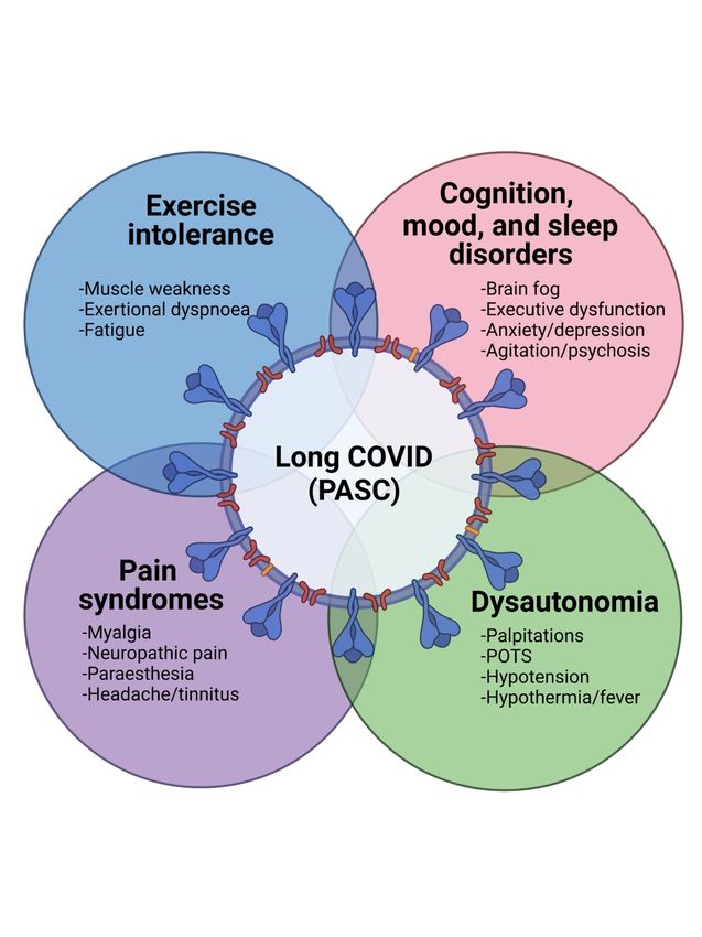
Indeed, it is probable that specific pathogenic processes underlie the individual neurological
presentations associated with COVID-19 in both the CNS (Figure 1A) and the PNS (Figure
1B). We review the different proposed mechanisms below.
Cerebrovascular disease/bioenergy failure. Microvascular injury characterized by thinning
of the basal lamina of endothelial cells, fibrinogen leakage, and microhemorrhages has been
described in the brainstem and olfactory bulb of deceased COVID-19 patients corresponding
Downloaded from https://academic.oup.com/brain/advance-article/doi/10.1093/brain/awab302/6353020 by guest on 20 December 2021
to visible MRI changes.122 These observations are also complemented by other neuroimaging
studies in which cerebral infarction was the most common finding on conventional brain
MRI.29 The majority of post-mortem analyses have shown signs of thrombotic
microangiopathy and endothelial injury with minimal evidence of prototypic vasculitis.53
This pattern is suggestive of endotheliitis. Though there have been several case reports of
CNS vasculitis associated with COVID-19, none have confirmed the diagnosis
histologically.132 A cohort of patients with stroke and COVID-19 in Wuhan, China, showed
elevated serum levels of IL-6,133 IL-8, and TNF-, a finding that has been replicated in
several subsequent studies.54 Both IL-8 and TNF- promote the release of von Willebrand
factor (vWF), a marker of endothelial damage that is elevated in both ICU and non-ICU
patients with COVID-19,134 while IL-6 inhibits cleavage of vWF leading to accumulation of
multimers that promote platelet aggregation.53 These changes are bolstered by findings of
damaged cerebral blood vessels or endotheliitis that was associated with extravasation of
fibrinogen.122 These mechanisms of disease are highly plausible because of the frequency of
coagulation–related events during COVID-19. Indeed, neuroimaging studies point to
abnormal energy metabolism, evinced by reduced FDG detection in frontal lobes of patients
with acute COVID-19.135
Viral neuroinvasion. SARS-CoV-2 infects respiratory cells via engagement of the
angiotensin-converting enzyme 2 (ACE2) receptor,136-138 with a higher binding affinity than
other coronaviruses such as SARS-CoV-1. The ACE2 receptor is present on type II alveolar
and respiratory epithelial cells, cardiomyocytes, neurons139 and astrocytes.138, 140 This
receptor is also present in pericytes and smooth muscle cells of cerebral blood vessels and is
expressed in the thalamus, cerebellum, and brainstem nuclei of humans.141-143 After binding
to ACE2, cleavage of the spike (S) protein of SARS-CoV-2 by transmembrane serine
protease 2 (TMPRSS2) facilitates cell entry.144 Alternative docking receptors including
neuropillin-1 (NRP-1)145 and basigin (BSG)/CD147146 are found at higher levels in the CNS.
ScholarOne, 375 Greenbrier Drive, Charlottesville, VA, 22901 Support (434) 964 4100Page 13 of 30 Brain
Similarly, alternative proteases including furin and cathepsin might permit viral entry in cells
with low levels of TMPRSS2 expression (e.g., brain).147
Several anatomic routes of neuroinvasion by SARS-CoV-2 have been proposed. The integrity
of the BBB is compromised in multiple conditions associated with mortality in COVID-19,
including hypertension, diabetes, and smoking, and stroke.148 Areas of increased vascular
Downloaded from https://academic.oup.com/brain/advance-article/doi/10.1093/brain/awab302/6353020 by guest on 20 December 2021
permeability or lack of BBB, such as the pituitary and median eminence of the hypothalamus
are also rich in ACE2, neuropilin-1, and TMPRSS2, thus representing possible portals of
entry into the CNS.149 SARS-CoV-2 infects nasal epithelium and perhaps olfactory bulb cells,
presenting another entry portal to the CNS, as suggested for other coronaviruses.150, 151 A
recent post-mortem analysis of humans with COVID-19 detected SARS-CoV-2 by RT-PCR
in neuroepithelium, the olfactory bulb, trigeminal ganglion, and brainstem, albeit at low
levels.16 Interestingly, olfactory nerves terminate in the frontal cortex as well as the
hypothalamus and amygdala, structures that are implicated clinically, radiographically, and
electrographically in the neurologic sequelae of COVID-19.149 The importance of the choroid
plexus in the development of COVID-19 associated neurological disease in conjunction with
neuroinflammation has been highlighted recently in a large study predicated on RNA deep
sequencing of brain-derived single cell nuclei transcriptomes.152 The lack of evidence for
productive infection of trafficking immune cells by SARS-CoV-2 to date makes a Trojan
Horse mechanism of neuroinvasion less likely. Nonetheless, viral proteins and RNA have
been detected in CD68+ macrophages isolated from bronchoalveolar lavage of COVID-19
patients.153 SARS-CoV-2 RNA levels in brain tissue detected by RT-PCR are low and
seemingly independent of the presence or absence of apparent neurological dysfunction and
histopathological alterations.16, 128 Immunodetection of SARS-Cov-2 viral antigens in
neurons from autopsied patients with COVID-19 underscores the potential for direct viral
invasion as an important disease determinant.154
Remdesivir, a nucleoside analogue that inhibits RNA-dependent replication of SARS-
CoV-2, is the only direct antiviral agent approved for COVID-19 treatment despite
preliminary results showing no impact on mortality or progression to mechanical
ventilation.155 Molnupiravir is orally available nucleoside analogue that induces coronavirus
lethal mutagenesis and is in Phase 2 and 3 trials for treatment of COVID-19.156 A recent
randomized control trial of the TMPRSS2 inhibitor, camostat mesylate, in hospitalized
patients with COVID-19 did not have any impact on recovery, progression to ICU, or
ScholarOne, 375 Greenbrier Drive, Charlottesville, VA, 22901 Support (434) 964 4100Brain Page 14 of 30
mortality.157
Host neuroimmune responses. Post-infectious neuro-inflammation triggered by expression
of viral antigens into the CNS is another proposed mechanism of encephalitis in COVID-19.
While human data supporting this hypothesis is limited, a recently published study using a
murine model showed a subunit of the SARS-CoV-2 spike protein (S1) crosses the blood-
brain barrier via absorptive transcytosis when administered intravenously and intra-nasally.158
Downloaded from https://academic.oup.com/brain/advance-article/doi/10.1093/brain/awab302/6353020 by guest on 20 December 2021
Indeed, neuropathological studies demonstrate glia activation and occasional leucocyte
infiltrates in patients with COVID-19 although the associated molecular pathways (e.g.,
cytokine, protease, or free radical release) induced are unclear. CSF studies suggest activation
of innate immune responses with elevated levels of beta-2-microglobulin and neopterin and
the presence of dedifferentiated monocytes.108, 118 This is associated with increased levels of
neurofilament suggesting neuronal injury.108 Autoimmune mechanisms including both
antibody- as well as cell-mediated immune injury of neural tissue are also plausible, given the
recognition of autoimmune processes in the systemic COVID-19 pathogenesis. The injury
and loss of endothelial cells in arterioles, venules, and capillaries represents another
neuropathogenic avenue via disruption on the blood-brain barrier and through endothelia
production of immune molecules159 in the lung, kidney, and heart of patients with COVID-
19. These latter events can be initiated by systemic immune activation as well as a
coagulation diathesis. An important qualification to the above mechanisms is that concurrent
clinical events including systemic hypoxia-ischemia might affect immune processes within
the nervous system. Among patients with COVID-19 associated cerebrovascular disease,
autoimmune processes have been directly implicated. For example, the contribution of
antiphospholipid antibodies (aPL-Ab) to ischemic stroke in patients with COVID-19 is
controversial. Zhang et al. described three COVID-19 patients with coagulopathy and multi-
territory infarcts and anticardiolipin and anti-β2 microglobulin antibodies.160 Subsequent
studies have reported lupus anticoagulant positivity in more than half of COVID-19
patients.161 Most case reports of aPL-Ab in COVID-19 do not include repeat assays 12 weeks
apart, which is required for the diagnosis of antiphospholipid antibody syndrome. Transient
elevation of lupus anticoagulant during systemic inflammation is common, and several
infections are associated with false positive antiphospholipid assays, including HIV, hepatitis
C virus, and syphilis, making current reports of aPL-Ab in COVID-19 difficult to interpret.162
Similarly, autoimmunity is also incriminated in COVID-19 associated GBS; anti-ganglioside
antibodies implicated in autoimmune polyradiculoneuropathies such as anti-Gq1b, -GM1163
ScholarOne, 375 Greenbrier Drive, Charlottesville, VA, 22901 Support (434) 964 4100Page 15 of 30 Brain
and -GD1b antibodies have been reported in patients with COVID-19 presenting with cranial
neuropathies, weakness, areflexia, and sensory ataxia.52 Anti-ganglioside antibodies are most
strongly associated with more aggressive axonal motor neuropathies and poorer functional
outcomes compared to AIDP.164 The rare presence of these antibodies raises concern about
potential molecular mimicry mediated by SARS-CoV-2 that could trigger autoimmune
responses with important implications for vaccine safety. The spike (S) protein of SARS-
Downloaded from https://academic.oup.com/brain/advance-article/doi/10.1093/brain/awab302/6353020 by guest on 20 December 2021
CoV-2 is highly glycosylated; thus, the development of anti-glycan antibodies may be
essential for an effective host immune response in COVID-19. In a microarray study of 800
human carbohydrate antigens, levels of anti-glycolipid antibodies associated with GBS,
including GM1a, GD1a, and GD1b significantly higher in COVID-19 patients compared to
healthy controls. In this latter study, there was no direct correlation with antibody titre and
clinical features of GBS. Anti-glycan antibodies are also observed in other viral and bacterial
infections (HIV, EBV, Neisseria meningitidis164, 165) as well as autoimmune diseases such as
Crohn’s disease166, and thus may merely be a marker of systemic inflammation. Of relevance,
there were no reported cases of GBS in the three major COVID-19 vaccine trials.167-169
While randomized control trials demonstrate dexamethasone and tocilizumab improve
respiratory outcomes in hospitalized patients, their effects on neurologic disease in COVID-
19 is presently supported only by case reports.50, 170-172 A subset of COVID-19 associated
encephalopathies are responsive to steroids and IVIg, and there is a single report of a young
patient with encephalitis and SARS-CoV-2 (on the basis of CSF lymphocytosis and
T2/FLAIR hyperintensities on MRI), which resolved after treatment with IVIg and
tocilizumab.173 In most cases with a positive response to immunosuppressive or modulatory
therapy, SARS-CoV-2 was not detected in CSF, further supporting a para-infectious/immune-
mediated basis for disease.
Future perspectives
Given the mounting impact of SARS-CoV-2 infection globally together with the increasing
recognition of associated neurological disorders, it is imperative to define the types of
COVID-19 related neurological syndromes, including those caused directly by viral infection
versus those arising from systemic illness, the impact of different viral variants on
neurological disease, as well as identifying informative diagnostic tools and effective
therapies. GWAS studies have identified susceptibility genes for severe respiratory illness
with COVID.174, 175 Similar studies to identify host factors associated with neurological
complications would also be useful. The long-term neurological sequelae of COVID-19
remain unclear and await delineation in longitudinal studies. The neurodevelopmental
ScholarOne, 375 Greenbrier Drive, Charlottesville, VA, 22901 Support (434) 964 4100Brain Page 16 of 30
impacts of COVID-19 are also unknown in utero as well as in infants or adolescents; this
issue could have substantial lasting effects that require further investigation. Finally, a more
comprehensive understanding of the pathogenic mechanisms underpinning the neurological
syndromes associated with COVID-19 will advance therapeutic options for affected patients.
Acknowledgements: The authors thank Brittney Hlavay for creation of figures, Nathalie
Downloaded from https://academic.oup.com/brain/advance-article/doi/10.1093/brain/awab302/6353020 by guest on 20 December 2021
Arbour for helpful discussions, Rebecca Folkerth, New York University for providing the
post-mortem tissue images, and Govind Nair for providing the MR images of the brain.
Funding: No specific funding was received towards this work.
Competing interests: The authors report no competing interests.
Literature search strategy and selection criteria: Studies were selected from the peer-reviewed
literature using NCBI and Google Scholar. We searched the databases using the following key words:
central and peripheral nervous systems, COVID-19, SARS-CoV-2, coronavirus, stroke,
encephalopathy, neurocognitive impairment, hypercoagulability, encephalitis, neurologic infection,
seizure, and neuroinflammation. We also reviewed bibliographies of relevant articles. Non-peer
reviewed studies and single case reports were not included as references unless they were highly
informative.
References
1. Al-Aly Z, Xie Y, Bowe B. High-dimensional characterization of post-acute sequelae of COVID-
19. Nature. Jun 2021;594(7862):259-264. doi:10.1038/s41586-021-03553-9
2. Huang C, Wang Y, Li X, et al. Clinical features of patients infected with 2019 novel
coronavirus in Wuhan, China. Lancet. 02 2020;395(10223):497-506. doi:10.1016/S0140-
6736(20)30183-5
3. Mao L, Jin H, Wang M, et al. Neurologic Manifestations of Hospitalized Patients With
Coronavirus Disease 2019 in Wuhan, China. JAMA Neurol. 06 2020;77(6):683-690.
doi:10.1001/jamaneurol.2020.1127
4. Varatharaj A, Thomas N, Ellul MA, et al. Neurological and neuropsychiatric complications of
COVID-19 in 153 patients: a UK-wide surveillance study. Lancet Psychiatry. 10 2020;7(10):875-882.
doi:10.1016/S2215-0366(20)30287-X
5. van den Pol AN. Viral infection leading to brain dysfunction: more prevalent than
appreciated? Neuron. Oct 2009;64(1):17-20. doi:10.1016/j.neuron.2009.09.023
6. Bradshaw MJ, Venkatesan A. Herpes Simplex Virus-1 Encephalitis in Adults: Pathophysiology,
Diagnosis, and Management. Neurotherapeutics. 07 2016;13(3):493-508. doi:10.1007/s13311-016-
0433-7
ScholarOne, 375 Greenbrier Drive, Charlottesville, VA, 22901 Support (434) 964 4100Page 17 of 30 Brain
7. Balcom EF, Roda WC, Cohen EA, Li MY, Power C. HIV-1 persistence in the central nervous
system: viral and host determinants during antiretroviral therapy. Curr Opin Virol. 10 2019;38:54-62.
doi:10.1016/j.coviro.2019.06.004
8. Bohmwald K, Gálvez NMS, Ríos M, Kalergis AM. Neurologic Alterations Due to Respiratory
Virus Infections. Front Cell Neurosci. 2018;12:386. doi:10.3389/fncel.2018.00386
9. Abdel-Mannan O, Eyre M, Löbel U, et al. Neurologic and Radiographic Findings Associated
With COVID-19 Infection in Children. JAMA Neurol. Jul 2020;doi:10.1001/jamaneurol.2020.2687
10. Cheung EW, Zachariah P, Gorelik M, et al. Multisystem Inflammatory Syndrome Related to
COVID-19 in Previously Healthy Children and Adolescents in New York City. JAMA. Jul
Downloaded from https://academic.oup.com/brain/advance-article/doi/10.1093/brain/awab302/6353020 by guest on 20 December 2021
2020;324(3):294-296. doi:10.1001/jama.2020.10374
11. Dufort EM, Koumans EH, Chow EJ, et al. Multisystem Inflammatory Syndrome in Children in
New York State. N Engl J Med. 07 2020;383(4):347-358. doi:10.1056/NEJMoa2021756
12. Feldstein LR, Rose EB, Horwitz SM, et al. Multisystem Inflammatory Syndrome in U.S.
Children and Adolescents. N Engl J Med. 07 2020;383(4):334-346. doi:10.1056/NEJMoa2021680
13. Lin JE, Asfour A, Sewell TB, et al. Neurological issues in children with COVID-19. Neurosci
Lett. 01 2021;743:135567. doi:10.1016/j.neulet.2020.135567
14. Pouga L. Encephalitic syndrome and anosmia in COVID-19: Do these clinical presentations
really reflect SARS-CoV-2 neurotropism? A theory based on the review of 25 COVID-19 cases. J Med
Virol. Jul 2020;doi:10.1002/jmv.26309
15. Yan CH, Faraji F, Prajapati DP, Ostrander BT, DeConde AS. Self-reported olfactory loss
associates with outpatient clinical course in COVID-19. Int Forum Allergy Rhinol. 07 2020;10(7):821-
831. doi:10.1002/alr.22592
16. Meinhardt J, Radke J, Dittmayer C, et al. Olfactory transmucosal SARS-CoV-2 invasion as a
port of central nervous system entry in individuals with COVID-19. Nat Neurosci. Nov
2020;doi:10.1038/s41593-020-00758-5
17. Antony AR, Haneef Z. Systematic review of EEG findings in 617 patients diagnosed with
COVID-19. Seizure. Oct 2020;doi:10.1016/j.seizure.2020.10.014
18. Anand P, Al-Faraj A, Sader E, et al. Seizure as the presenting symptom of COVID-19: A
retrospective case series. Epilepsy Behav. 11 2020;112:107335. doi:10.1016/j.yebeh.2020.107335
19. Lin L, Al-Faraj A, Ayub N, et al. Electroencephalographic Abnormalities are Common in
COVID-19 and are Associated with Outcomes. Ann Neurol. 05 2021;89(5):872-883.
doi:10.1002/ana.26060
20. Toniolo S, Di Lorenzo F, Scarioni M, Frederiksen KS, Nobili F. Is the Frontal Lobe the Primary
Target of SARS-CoV-2? J Alzheimers Dis. 2021;81(1):75-81. doi:10.3233/JAD-210008
21. Pensato U, Muccioli L, Pasini E, et al. Encephalopathy in COVID-19 Presenting With Acute
Aphasia Mimicking Stroke. Front Neurol. 2020;11:587226. doi:10.3389/fneur.2020.587226
22. Liotta EM, Batra A, Clark JR, et al. Frequent neurologic manifestations and encephalopathy-
associated morbidity in Covid-19 patients. Ann Clin Transl Neurol. 11 2020;7(11):2221-2230.
doi:10.1002/acn3.51210
23. Blazhenets G, Schröter N, Bormann T, et al. Slow but evident recovery from neocortical
dysfunction and cognitive impairment in a series of chronic COVID-19 patients. J Nucl Med. Mar
2021;doi:10.2967/jnumed.121.262128
24. Hosp JA, Dressing A, Blazhenets G, et al. Cognitive impairment and altered cerebral glucose
metabolism in the subacute stage of COVID-19. Brain. May 7 2021;144(4):1263-1276.
doi:10.1093/brain/awab009
25. Solomon IH, Normandin E, Bhattacharyya S, et al. Neuropathological Features of Covid-19. N
Engl J Med. 09 2020;383(10):989-992. doi:10.1056/NEJMc2019373
26. Farhadian S, Glick LR, Vogels CBF, et al. Acute encephalopathy with elevated CSF
inflammatory markers as the initial presentation of COVID-19. BMC Neurol. Jun 2020;20(1):248.
doi:10.1186/s12883-020-01812-2
ScholarOne, 375 Greenbrier Drive, Charlottesville, VA, 22901 Support (434) 964 4100Brain Page 18 of 30
27. Heming N, Mazeraud A, Verdonk F, Bozza FA, Chrétien F, Sharshar T. Neuroanatomy of
sepsis-associated encephalopathy. Crit Care. Mar 2017;21(1):65. doi:10.1186/s13054-017-1643-z
28. Bryce C, Grimes Z, Pujadas E, et al. Pathophysiology of SARS-CoV-2: targeting of endothelial
cells renders a complex disease with thrombotic microangiopathy and aberrant immune response.
The Mount Sinai COVID-19 autopsy experience. medRxiv. 2020:2020.05.18.20099960.
doi:10.1101/2020.05.18.20099960
29. Gulko E, Oleksk ML, Gomes W, et al. MRI Brain Findings in 126 Patients with COVID-19: Initial
Observations from a Descriptive Literature Review. AJNR Am J Neuroradiol. 12 2020;41(12):2199-
2203. doi:10.3174/ajnr.A6805
Downloaded from https://academic.oup.com/brain/advance-article/doi/10.1093/brain/awab302/6353020 by guest on 20 December 2021
30. Kantonen J, Mahzabin S, Mäyränpää MI, et al. Neuropathologic features of four autopsied
COVID-19 patients. Brain Pathol. Aug 2020;doi:10.1111/bpa.12889
31. Kremer S, Lersy F, Anheim M, et al. Neurologic and neuroimaging findings in patients with
COVID-19: A retrospective multicenter study. Neurology. 09 2020;95(13):e1868-e1882.
doi:10.1212/WNL.0000000000010112
32. Helms J, Kremer S, Merdji H, et al. Neurologic Features in Severe SARS-CoV-2 Infection. N
Engl J Med. 06 2020;382(23):2268-2270. doi:10.1056/NEJMc2008597
33. Helms J, Kremer S, Meziani F. More on Neurologic Features in Severe SARS-CoV-2 Infection.
Reply. N Engl J Med. 06 2020;382(26):e110. doi:10.1056/NEJMc2015132
34. Pilotto A, Masciocchi S, Volonghi I, et al. Clinical Presentation and Outcomes of Severe Acute
Respiratory Syndrome Coronavirus 2-Related Encephalitis: The ENCOVID Multicenter Study. J Infect
Dis. 01 2021;223(1):28-37. doi:10.1093/infdis/jiaa609
35. Pilotto A, Masciocchi S, Volonghi I, et al. SARS-CoV-2 encephalitis is a cytokine release
syndrome: evidences from cerebrospinal fluid analyses. Clin Infect Dis. Jan
2021;doi:10.1093/cid/ciaa1933
36. Alexopoulos H, Magira E, Bitzogli K, et al. Anti-SARS-CoV-2 antibodies in the CSF, blood-brain
barrier dysfunction, and neurological outcome: Studies in 8 stuporous and comatose patients.
Neurol Neuroimmunol Neuroinflamm. 11 2020;7(6)doi:10.1212/NXI.0000000000000893
37. Bodro M, Compta Y, Llansó L, et al. Increased CSF levels of IL-1β, IL-6, and ACE in SARS-CoV-
2-associated encephalitis. Neurol Neuroimmunol Neuroinflamm. 09
2020;7(5)doi:10.1212/NXI.0000000000000821
38. Espíndola OM, Gomes YCP, Brandão CO, et al. Inflammatory Cytokine Patterns Associated
with Neurological Diseases in Coronavirus Disease 2019. Ann Neurol. 05 2021;89(5):1041-1045.
doi:10.1002/ana.26041
39. Moriguchi T, Harii N, Goto J, et al. A first case of meningitis/encephalitis associated with
SARS-Coronavirus-2. Int J Infect Dis. May 2020;94:55-58. doi:10.1016/j.ijid.2020.03.062
40. Zuhorn F, Omaimen H, Ruprecht B, et al. Parainfectious encephalitis in COVID-19: "The
Claustrum Sign". J Neurol. Sep 2020;doi:10.1007/s00415-020-10185-y
41. Casez O, Willaume G, Grand S, et al. SARS-CoV-2 Related Encephalitis: MRI Pattern of the
Olfactory Tract Involvement. Neurology. Nov 2020;doi:10.1212/WNL.0000000000011150
42. Reichard RR, Kashani KB, Boire NA, Constantopoulos E, Guo Y, Lucchinetti CF.
Neuropathology of COVID-19: a spectrum of vascular and acute disseminated encephalomyelitis
(ADEM)-like pathology. Acta Neuropathol. 07 2020;140(1):1-6. doi:10.1007/s00401-020-02166-2
43. Poyiadji N, Shahin G, Noujaim D, Stone M, Patel S, Griffith B. COVID-19-associated Acute
Hemorrhagic Necrotizing Encephalopathy: Imaging Features. Radiology. 08 2020;296(2):E119-E120.
doi:10.1148/radiol.2020201187
44. Silva MTT, Lima MA, Torezani G, et al. Isolated intracranial hypertension associated with
COVID-19. Cephalalgia. Nov 2020;40(13):1452-1458. doi:10.1177/0333102420965963
45. Verkuil LD, Liu GT, Brahma VL, Avery RA. Pseudotumor cerebri syndrome associated with
MIS-C: a case report. Lancet. Aug 22 2020;396(10250):532. doi:10.1016/S0140-6736(20)31725-6
ScholarOne, 375 Greenbrier Drive, Charlottesville, VA, 22901 Support (434) 964 4100Page 19 of 30 Brain
46. Emamikhah M, Babadi M, Mehrabani M, et al. Opsoclonus-myoclonus syndrome, a post-
infectious neurologic complication of COVID-19: case series and review of literature. J Neurovirol. 02
2021;27(1):26-34. doi:10.1007/s13365-020-00941-1
47. Werner J, Reichen I, Huber M, Abela IA, Weller M, Jelcic I. Subacute cerebellar ataxia
following respiratory symptoms of COVID-19: a case report. BMC Infect Dis. Mar 2021;21(1):298.
doi:10.1186/s12879-021-05987-y
48. Dijkstra F, Van den Bossche T, Willekens B, Cras P, Crosiers D. Myoclonus and cerebellar
ataxia following Coronavirus Disease 2019 (COVID-19). Mov Disord Clin Pract. Aug
2020;doi:10.1002/mdc3.13049
Downloaded from https://academic.oup.com/brain/advance-article/doi/10.1093/brain/awab302/6353020 by guest on 20 December 2021
49. Cao A, Rohaut B, Le Guennec L, et al. Severe COVID-19-related encephalitis can respond to
immunotherapy. Brain. 10 2020;doi:10.1093/brain/awaa337
50. Cani I, Barone V, D'Angelo R, et al. Frontal encephalopathy related to hyperinflammation in
COVID-19. J Neurol. Jan 2021;268(1):16-19. doi:10.1007/s00415-020-10057-5
51. Matschke J, Lütgehetmann M, Hagel C, et al. Neuropathology of patients with COVID-19 in
Germany: a post-mortem case series. Lancet Neurol. 11 2020;19(11):919-929. doi:10.1016/S1474-
4422(20)30308-2
52. Guilmot A, Maldonado Slootjes S, Sellimi A, et al. Immune-mediated neurological syndromes
in SARS-CoV-2-infected patients. J Neurol. Jul 2020;doi:10.1007/s00415-020-10108-x
53. Hernández-Fernández F, Sandoval Valencia H, Barbella-Aponte RA, et al. Cerebrovascular
disease in patients with COVID-19: neuroimaging, histological and clinical description. Brain. 10
2020;143(10):3089-3103. doi:10.1093/brain/awaa239
54. Yaghi S, Ishida K, Torres J, et al. SARS-CoV-2 and Stroke in a New York Healthcare System.
Stroke. 07 2020;51(7):2002-2011. doi:10.1161/STROKEAHA.120.030335
55. Malas MB, Naazie IN, Elsayed N, Mathlouthi A, Marmor R, Clary B. Thromboembolism risk of
COVID-19 is high and associated with a higher risk of mortality: A systematic review and meta-
analysis. EClinicalMedicine. Dec 2020;29:100639. doi:10.1016/j.eclinm.2020.100639
56. Baldini T, Asioli GM, Romoli M, et al. Cerebral venous thrombosis and SARS-CoV-2 infection:
a systematic review and meta-analysis. Eur J Neurol. Jan 2021;doi:10.1111/ene.14727
57. Koralnik IJ, Tyler KL. COVID-19: A Global Threat to the Nervous System. Ann Neurol. 07
2020;88(1):1-11. doi:10.1002/ana.25807
58. Devasagayam S, Wyatt B, Leyden J, Kleinig T. Cerebral Venous Sinus Thrombosis Incidence Is
Higher Than Previously Thought: A Retrospective Population-Based Study. Stroke. 09
2016;47(9):2180-2. doi:10.1161/STROKEAHA.116.013617
59. Investigators I, Sadeghipour P, Talasaz AH, et al. Effect of Intermediate-Dose vs Standard-
Dose Prophylactic Anticoagulation on Thrombotic Events, Extracorporeal Membrane Oxygenation
Treatment, or Mortality Among Patients With COVID-19 Admitted to the Intensive Care Unit: The
INSPIRATION Randomized Clinical Trial. JAMA. Apr 27 2021;325(16):1620-1630.
doi:10.1001/jama.2021.4152
60. Tacquard C, Mansour A, Godon A, et al. Impact of high dose prophylactic anticoagulation in
critically ill patients with COVID-19 pneumonia. Chest. Jan 2021;doi:10.1016/j.chest.2021.01.017
61. ATTACC. ACTIV-4a & REMAP-CAP Multiplatform RCT: Results of Interim Analysis. 2021.
Accessed June 19th 2021. https://nhlbi-
connects.org/documents/mpRCT%20Interim%20Presentation.pdf
62. Kollias A, Kyriakoulis KG, Syrigos NK, Stergiou GS. Anticoagulation therapy in COVID-19: Is
there a dose-dependent benefit? Thromb Res. 03 2021;199:19-20.
doi:10.1016/j.thromres.2020.12.013
63. Moll M, Connors JM. When to use anticoagulation in COVID-19. Thromb Res. 2021;in press.
doi:https://doi.org/10.1016/j.thromres.2021.06.005
64. Dogra S, Jain R, Cao M, et al. Hemorrhagic stroke and anticoagulation in COVID-19. J Stroke
Cerebrovasc Dis. Aug 2020;29(8):104984. doi:10.1016/j.jstrokecerebrovasdis.2020.104984
ScholarOne, 375 Greenbrier Drive, Charlottesville, VA, 22901 Support (434) 964 4100You can also read