Effect of alcohol on the central nervous system to develop neurological disorder: pathophysiological and lifestyle modulation can be potential ...
←
→
Page content transcription
If your browser does not render page correctly, please read the page content below
AIMS Neuroscience, 8(3): 390–413.
                                                                  DOI: 10.3934/Neuroscience.2021021
                                                                  Received: 20 January 2021
                                                                  Accepted: 01 March 2021
                                                                  Published: 09 April 2021

http://www.aimspress.com/journal/neuroscience

Review

Effect of alcohol on the central nervous system to develop neurological
disorder: pathophysiological and lifestyle modulation can be potential
therapeutic options for alcohol-induced neurotoxication

Zinia Pervin1,*, Julia M Stephen2

1
    Department of Biomedical Engineering, University of New Mexico, Albuquerque, NM 87131, USA
2
    The Mind Research Network and Lovelace Biomedical and Environmental Research
    Institute, Albuquerque, NM 87106, USA

* Correspondence: Email: zinia@unm.edu; Tel: 5052725028.

Abstract: The central nervous system (CNS) is the major target for adverse effects of alcohol and
extensively promotes the development of a significant number of neurological diseases such as
stroke, brain tumor, multiple sclerosis (MS), Alzheimer’s disease (AD), and amyotrophic lateral
sclerosis (ALS). Excessive alcohol consumption causes severe neuro-immunological changes in the
internal organs including irreversible brain injury and it also reacts with the defense mechanism of
the blood-brain barrier (BBB) which in turn leads to changes in the configuration of the tight
junction of endothelial cells and white matter thickness of the brain. Neuronal injury associated with
malnutrition and oxidative stress-related BBB dysfunction may cause neuronal degeneration and
demyelination in patients with alcohol use disorder (AUD); however, the underlying mechanism still
remains unknown. To address this question, studies need to be performed on the contributing
mechanisms of alcohol on pathological relationships of neurodegeneration that cause permanent
neuronal damage. Moreover, alcohol-induced molecular changes of white matter with conduction
disturbance in neurotransmission are a likely cause of myelin defect or axonal loss which correlates
with cognitive dysfunctions in AUD. To extend our current knowledge in developing a
neuroprotective environment, we need to explore the pathophysiology of ethanol (EtOH) metabolism
and its effect on the CNS. Recent epidemiological studies and experimental animal research have
revealed the association between excessive alcohol consumption and neurodegeneration. This review
supports an interdisciplinary treatment protocol to protect the nervous system and to improve the
391
cognitive outcomes of patients who suffer from alcohol-related neurodegeneration as well as clarify
the pathological involvement of alcohol in causing other major neurological disorders.
Keywords: alcohol use disorder; central nervous system; oxidative stress response; neuropathology,
blood-brain barrier dysfunction; neuroimaging, antioxidant
1. Introduction
Alcohol is the most commonly used recreational beverage and drug of abuse among the adult
population, alcohol-related death is the third leading preventable cause of death in the United States
which accounts for more than 3.3 million global deaths annually [1,2]. According to the 2018-
National Survey on Drug Use and Health (NSDUH), 14.4 million people suffered from alcohol use
disorder (AUD) in the US, and over 100,000 deaths were attributable to alcohol [3]. The World
Health Organization reported that more than 200 health conditions including cancer, liver cirrhosis,
and neurocognitive impairment were also attributed to alcohol consumption [2]. These chronic health
conditions are progressive, cause a heavy economic burden to society, and decrease the quality of
life for both patients and caregivers [4].
According to the National Institute of Alcohol Abuse and Alcoholism (NIAAA), AUD is
defined as a chronic relapsing brain disease with an altered emotional state involving chronic alcohol
abuse. This disorder may contribute to a considerable proportion of dementia, neurocognitive deficits,
neuronal injury resulting from synaptic degeneration, nerve fiber demyelination, or blood-brain
barrier dysfunction [5,6]. In the presentation of the catastrophic or global loss of brain tissue,
significant cortical-subcortical volume loss including white matter shrinkage occurs in patients with
AUD, which is caused either by nutritional deficiency associated with alcoholic excitotoxicity or
oxidative stress which results in alteration of various types of normal brain function [7,8].
Furthermore, there is interest in the alcohol-induced metabolic disorder, Wernicke-Korsakoff
syndrome (WKS), which is associated with thiamine deficiency and may contribute to severe
neurological damage in the thalamus and hypothalamus [9,10]. A combined effect of nutritional
deficiency and ethanol toxicity may cause severe long-term effects and worsen the clinical
manifestation of neurological impairment [11]. In general, persistent alcohol consumption may lead
to gradual deterioration of psychological status with varying degrees of cognitive impairment
including severe dementia [10]. Alcohol is the second leading cause of dementia (10%) among the
adult population in the US after Alzheimer’s disease (40–60%) [12]. The severity of neurological
outcomes is associated in part with lifestyle factors including nutrition, amount, and term of alcohol
consumption. Chronic alcoholic patients may develop severe malnutrition because they usually
consume 50% of the calories from alcohol [13]. Alcohol consumption may have kindling effects and
may increase epileptic episodes, cerebral infections, cerebrovascular lesions, and alter
neurotransmitter systemic balance[14,15]. Cognitive impairment may affect high order executive
performance which may persist throughout the rest of life with secondary disabilities.
Neuron and myelin regeneration is a delicate process that requires different types of growth
factors (nerve growth factor and brain-derived neurotrophic factor) to regulate and maintain neuronal
homeostasis [16]. In AUD, essential growth factors of CNS homeostasis are downregulated by
AIMS Neuroscience Volume 8, Issue 3, 390–413.392
highly elevated alcohol metabolites acetaldehyde (AA) and reactive oxygen species (ROS), causing
neuronal injury that leads to neurodegeneration [17]. Neurodegeneration, the opposite of
regeneration, is when cells of the central nervous system stop working or die and usually perform
actions more poorly with time in the presence of toxic or pathological conditions [18,19]. Alcohol
triggers abnormal protein accumulation, lysosomal dysfunction, and DNA damage which promotes
neurodegeneration as well as accelerating the aging process of the brain [12,4]. In contrast to AD and
aging, alcohol’s effect on the brain may be possible to slow, halt, or even reversible with alcohol
abstinence because alcoholic brain shows shrinkage of brain tissue without significant loss of
neurons, however, disrupted neuronal function or connection can be reestablished by modifying
pathophysiology and lifestyle which can promote to maintain physiological homeostasis and
cognitive function [20,21].
Interestingly, previous research established the evidence of recovery and regeneration of
cortical volume including white matter thickness in short-term abstinence as well as improvement in
neurocognitive deficits particularly visuospatial abilities, working memory, and motor skills [22,23].
The mechanism of alcohol-induced degeneration and alcohol abstinence regeneration is a complex
phenomenon that is determined by a person’s genetic characteristics, dominant brain activity,
coexisting risk factors, and genetic process related to aging [24]. Sometimes, an immune-competent
status with a pharmacological trigger or lifestyle modification can be a way to prevent the alcohol-
induced neuronal insult and might play a significant role in brain recovery. This review will cover
possible mechanisms of neurotoxicity in AUD to support an effort to establish a multidisciplinary
therapeutic approach to prevent or reverse neurological damage.
2. Pathophysiology of alcohol metabolism and its consequence on BBB dysfunction
Despite thousands of published studies on alcohol-mediated neurological disturbance, the true
mechanism of alcohol-induced cell death remains ambiguous. Many chronic AUD patients
demonstrate neurocognitive and neurovascular injury associated with BBB dysfunction due to
ethanol metabolites [25]. The BBB is a highly selective semipermeable membrane formed by brain
microvascular endothelial cells (BMVEC). Pericytes and astrocytes connect the BMVEC assuring
BBB structural tightness by binding with a tight junction which not only acts as a natural protector
but also plays an important role to maintain normal brain homeostasis [26,27]. Ethanol metabolites
or neurotoxic substances may interact with the cytoskeletal structure of the brain to increase BBB
permeability to start neuroinflammation [28,29].
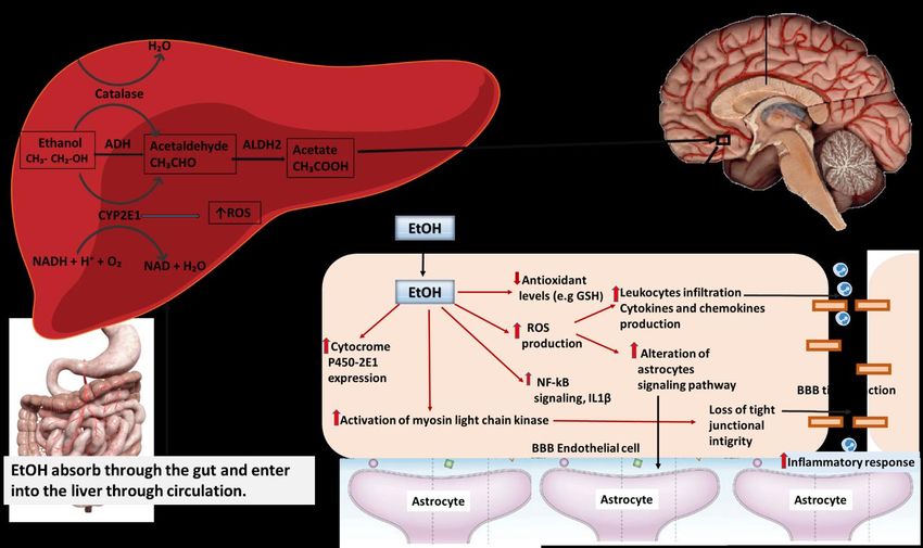
Before discussing the effect of alcohol on BBB damage, we have to look through alcohol
absorption and metabolism. The liver is the predominant organ for ethanol metabolism which
usually occurs via two oxidative pathways mediated by alcohol dehydrogenase (ADH) and
cytochrome P450 2E1 (CYP2E1) [30] (Figure 1). In brief, after drinking alcohol, absorption
Occurs in the gastrointestinal tract then the liver converts the alcohol to acetaldehyde through the
first-pass metabolism in the liver, this oxidation reaction is catalyzed by the alcohol dehydrogenase
enzyme [31,32]. After the first-pass metabolism, alcohol metabolites are distributed throughout the
body, it goes to the brain through the blood vessels then it enters into the endothelial cells from the
blood and alters the expression of signaling molecules which adhere to the BMVEC [33]. The
metabolism of EtOH in the brain is controversial than the metabolism of acetaldehyde due to
undetectable evidence of homogenous ADH activity in the whole brain. Animal experimental
AIMS Neuroscience Volume 8, Issue 3, 390–413.393
studies demonstrate the presence of cytochrome p4502E1 in the smooth endoplasmic reticulum of
brain cells that are capable of Ethanol metabolism in brain by catalyzing the H2O2 with catalase
enzyme [34]. However, the oxidation of acetaldehyde in brain cell is established because of ALDH
(aldehyde dehydrogenase) have been well known to be found in mitochondria of brain cells [35].
ALDH converts acetaldehyde to acetate, acetate has further effects on brain including increase
lipid peroxidation and free radicals production. EtOH exposure induces the catalytic expression of
oxidative metabolizing enzymes which is parallel to enhancing the production of ROS (Figure 1).
It is known that during oxidative stress conditions the levels of oxidants are higher than the
levels of antioxidants. So, ethanol indirectly decreases the antioxidant activity by increasing
oxidative stress response. Alcohol-induced ROS production is believed to be specific to EtOH
metabolism by cytochrome P450–2E1 (CYP2E1), which produces H2O2, superoxide, and free
radicals. These free radicals, in turn, activate Rho kinase(ROCK/JNK) signaling to induce the release
of vascular endothelial growth factor (VEGF) and inflammatory cytokines in brain endothelial cells
(e.g. upregulation of ICAM-1and E-selectin, the release of IL-6) [36,37].
Figure 1. Schematic of ethanol metabolism through the liver and hypothetical
involvement of ethanol metabolites for BBB dysfunction. In the presence of alcohol
dehydrogenase (ADH) and cytochrome P450 enzymes, alcohol undergoes 1st and 2nd pass
metabolism in the liver. Increased ROS and ethanol metabolites in the blood alter the
signaling pathways of BBB endothelial cells and down-regulate the tight junction, which
ultimately enhances leukocyte leakage and neuroinflammation [41,42,25].
Experimental studies on human brain tissue provide evidence of increased expression of
CYP2E1 after chronic ethanol exposure and as a result of CYP2E1 mediated metabolism induces
production of ROS and NO synthesis in the human brain [37,38]. However, actions of EtOH
metabolites depend on their concentration, ROS acts as active molecules at low concentration but at
high concentration, oxidants convert as a transducer of the oxidative stress response and
AIMS Neuroscience Volume 8, Issue 3, 390–413.394
neurodegenerative agents [39]. As a consequence of exaggerated actions of ROS, transcription
modulated lipid peroxidation is activated in neurons and increases the 4-HNE (lipid peroxidation
products) level as well as decreases the neuronal cytoskeletal proteins [38,40]. Disruption of neuron-
specific neurofilaments or neuronal death initiates the primary process of alcohol-related
neurodegeneration [37].
In the course of this phenomenon, further activation of astrocytes amplifies mitochondrial
phosphorylation with downregulation of the tight junction which enhances the permeability of the BBB
system. Thus, ethanol exposure results in BBB disruption by a complex immune-regulatory loop
between BMECs and astrocytes. Evidence from animal models and cell culture reports further
strengthens the idea that chronic excessive alcohol exposure downregulates the tight junction proteins
(claudin, occludin, zonula occludens) which are responsible for maintaining BBB integrity [43]. Both
acute and chronic alcohol exposure can increase the production of ROS and enhance peroxidation of
lipids, protein, and phosphorylation of mitochondria resulting in decreased ATP production by
disrupting phospholipid-containing cell membrane structure [44]. Astrocytes maintain the BBB
integrity by forming paracrine interactions to coordinates the CNS blood flow and neural function
between pericytes and CNS vasculature [45]. Alcohol-induced tight junction disassembly is usually
mediated via activation of expression protein kinase C (PKC) which subsequently allows toxic
substances to enter the brain which in turn affects CNS homeostasis. Incidental ablation occurs in
astrocytes, pericytes vascular basement membrane results in unyielding leakage of leukocytes and
immune complex molecules in and out of the brain, including secondary changes such as edema,
inflammation, and hyper excitability may have appeared in white matter and cortical regions [32,46]
(Figure 1). Loss of astrocytes function to maintain the neurovascular coupling is not recovered by the
proliferation of adjacent astrocytes resulting in long-term effect in neurovascular damage.
Empirical studies further show that ethanol-induced brain damage is mainly related to oxidative
stress response from proinflammatory cytokines activated during alcohol intoxication.
Proinflammatory cytokines NF-kB (transcription factor) mediate oxidative stress plays a role in the
induction of anti-inflammatory and immune response signals, which appear to underlie neuronal
degeneration and tissue atrophy [46,47]. Cytokines are large families of secreted proteins that are
transported from blood serum to neuronal tissue in response to oxidative stress-related alcohol
neuroinflammation [47]. Increased cytokines particularly tumor necrotic factor (TNFα), interleukin
IL-1β, and macrophage chemotactic protein 1 (CCL 2) expression cause neuroinflammation and
insult of nerve axons in nociceptive synaptic terminals which leads to intracortical network
miscommunication and neuropathy [48,49].
As a result of BBB dysfunction, abnormal expression of water channel aquaporin (AQP)
occurs which in turn causes cerebral edema by extravasating the water inside the brain tissue.
The swelling of the brain plays a critical role in the pathogenesis of an extensive variety of CNS
disorders including stroke, infection, and demyelination. Rodent brain studies provide evidence
of the association of abnormal expression of water channel AQP4 in ROS-induced BBB
dysfunction [50,51]. The distribution and expression of AQP4 are important to maintaining CNS
homeostasis. AQP4 is mainly arranged and organized in astrocytes and ependymal cells
alongside myelinated fiber tracts [29,52]. AQP4 may help astrocytes to maintain ion
concentration by taking excess K + inside the cell to activate the specific brain regions in
exchange for rapid transfer of water out of the cell [52]. Alcohol-induced oxidative response
interferes with the AQP4 activity and causes activity-related swelling of the extracellular space
AIMS Neuroscience Volume 8, Issue 3, 390–413.395
in white matter tracts (corpus callosum, optic chiasma, hippocampus, hypothalamic nuclei)
where perinodal astrocytes fail to regulate the intracellular junctions at the nodes of Ranvier [52].
Inconsistent water movement in between CSF and brain parenchyma causes edema which
appears to play a key role in the neurodegenerative process by facilitating a neuropathological
environment. In the case of thiamine deficiency in chronic alcoholic abusers causes Wernicke
korsakoff syndrome (WKS) due to its impaired metabolism of the mitochondrial oxidation to
produce the brain energy and causes increase oxidative stress response and neuronal intoxicat ion.
Glucose serves as a primary fuel to mitigate the high demand for energy production in the central
nervous system. The brain is highly vulnerable in a state of thiamine deficiency due to thiamine-
dependent enzymes are required to glucose metabolism as well as mitochondrial ATP production
for maintaining the CNS homeostasis, actions potentials, myelination and neuronal activity [53].
Impaired glucose metabolism decreases mitochondrial ATP production, thereby slow down the
firing of the neuronal action potential, in addition, trigger lipid peroxidation, oxidative damage
to CNS. Thus, Alcohol and its metabolites induce BBB disruption and neuroinflammation as
well as alter the CNS homeostasis.
3. Impairment of glucose transport system leads to neurodegeneration
Previous research suggests a strong correlation between the impairment of glucose metabolism
with subsequent neuronal loss at the interface of alcohol-induced BBB dysfunction which causes
neurodegeneration in CNS [54,55]. Therefore, disruption of BBB integrity may cause altered
expression of the glucose transport channel protein (GLUT 1 and GLUT 3) and reduce uptake of
glucose inside brain tissue [1]. About 90% of brain tissue depends on constant glucose supply as an
energy source to maintain a dynamic function. GLUT 1 glucose transporter facilitates glucose
transport from capillary endothelium to astrocytes then astrocytes metabolize some glucose
molecules and transport these to neurons as fuel for anti-oxidation and tissue plasticity regeneration
via the GLUT 3 transporter [56,54]. In the preclinical period of neurodegeneration, glucose
consumption is gradually diminished in neurons and glial cells of the hippocampus, corpus callosum,
cerebral cortex, which induce lactate production, aerobic glycolysis, and structural plasticity in
animal model [57,55]. Eventually, clinical symptoms emerge and are associated with ataxia,
spasticity, dementia, and mild to severe cognitive deficits. So, normal glucose homeostasis is
important to maintain brain function; if any alteration or disruption occurs then it leads to neuronal
toxicity with neuronal death results in neurodegenerative effect on cognitive function.
4. NMDA receptor-mediated neurotoxicity
N-methyl-D-aspartate (NMDA) is a primary excitatory brain neurotransmitter that binds to the
glutamate receptor usually found in nerve cells. Depolarization and activation of the nerve action
potential are maintained by the influx of different types of ions (Na+ and Ca2+) into the cell through
the NMDA receptors [58]. Under normal circumstances, NMDA receptors play an important role in
synaptic plasticity and signal transmission in the course of the cellular mechanism of learning,
visuospatial memory, and buildup of working memory by neuronal synchronization through the
intra-cortical communication of the central nervous system [59]. It is believed that alcohol acts as an
antagonist for the NMDA receptor, so in the case of AUD, it causes hypofunction of the NMDA
AIMS Neuroscience Volume 8, Issue 3, 390–413.396
receptor which may result in neuronal network impairment with loss of synaptic plasticity [60]. To
maintain normal neuronal function and homeostasis, the physiological actions of the NMDA receptor
are required. Several controversial studies implicated that NMDA receptors are strongly involved
with excitotoxicity which contributes to cell death and hamper the longevity of the cells [42,58].
Recent evidence supports the hypothesis that excitotoxic events of NMDA receptors play a role in
the formation of neurodegenerative diseases like Alzheimer’s and Huntington’s disease and affect
normal brain function [11]. However, there is no established theory that delineates the use of alcohol
as an NMDA receptor antagonist or medication for a neuroprotective role because successful
implementation of NMDA antagonists would require blocking the excessive activation without
interfering with the normal physiological function [61,42]. In contrast, prior studies had shown that
ethanol-induced blockage of the NMDA receptor could increase neurotoxicity by decreasing the
expression of brain-derived neurotrophic factor (BDNF) during chronic alcohol administration [62].
Therefore, more studies are needed to establish the role of the NMDA receptor in the mechanism of
neurodegeneration or neuro-regeneration in patients with AUD.
5. Astrocyte and oligodendrocyte associated neuronal dysfunction in AUD
Studies on the rodent and human brain delineated that excessive ethanol intake induces
neuronal injury during various developmental stages including neurodegeneration and this type of
ethanol-induced neurodegeneration seems to be connected with glial activation and
neuroinflammation [23,63,64]. Astrocytes and oligodendrocytes play a crucial role in the
molecular mechanism of signal conduction and neurotransmission in both gray and white matter.
Besides, astrocytes, oligodendrocytes, and myelin protein take part in the maintenance of plasticity
of gray and white matter [65]. In alcohol-related brain damage, ethanol and its metabolites have the
potential to disrupt glial physiology and neurobiology in gray and white matter. Ethanol triggers the
TLR4 receptor-dependent or -independent pathways of microglial activation which stimulates the
NF-kB, interleukins IL1, IL6, CCL2, and in turn, evokes the expression of proinflammatory
cytokines surrounding the astrocytes and oligodendrocytes [49,64]. If this leads to an agglomeration
of pro-inflammatory and neurotoxic mediators for a prolonged period in the glial environment, then
it leads to neuroinflammation and neurodegeneration [63,66]. In AUD, ethanol metabolites alter the
expression of astrocytes and oligodendrocytes which leads to impaired cell to cell communication.
Signal transmission and cell interaction are accomplished by the formation and maintenance of the
myelin sheath which is usually disrupted by alcohol metabolites. Alcohol interferes with the neuronal
homeostasis process including the ability to form colonies, integrate, differentiate, and mainly
proliferate [11].
CNS inflammatory sequelae are believed to play a vital role in neuronal death as the pathway of
neurodegeneration and inflammatory feedback is mainly mediated by microglial activation. In AUD,
brain immune defense cells, microglia, are activate and express many proinflammatory genes
including tumor necrotic factor α (TNF α), cyclo-oxygenase, NADPH enzymes which change the
brain immune system and nerve cell functions [67,68]. In the case of normal infection, this
immunomodulation is limited and controlled by further immune signals but in AUD, chronic
activation of microglia and sustained increases in microglial specific cellular markers activate
inflammatory gene expression which may, in turn, cause neuronal death and disrupt the cellular
integrity, ultimately leading to neurodegeneration. Therefore, a number of researchers believe that
AIMS Neuroscience Volume 8, Issue 3, 390–413.397
suppression of microglial activation could be a potential therapeutic to treat inflammation-mediated
neurodegenerative disease [46].
6. Neuroimaging evidence of alcohol-induced neuroinflammation and neurodegeneration
Neuroimaging technology can observe the dynamic brain in a living body and allows
researchers to conduct meticulous studies to gain insights into the effect of AUD on the human brain
throughout the periods of chronic drinking, relapse, and abstinence. Brain images can be used to
predict the severity of AUD by measuring the connectivity of neuronal features corresponding to the
executive control network associated with different brain regions [69]. To explore treatment options
for AUD, it is required to identify the status of neuronal injury and distinguish the reversible and
irreversible neuronal loss with connectivity network in the course of alcohol intake from abstinence
to heavy alcohol use. Structural MRI helps to visualize different cortical regions of the brain (gray,
white matter, cortex, and midbrain) to examine the region-specific effects of chronic alcohol
consumption [70]. Structural MRI findings in AUD provide evidence of mammillary body damage
with hippocampal volume deficits that are also associated with the decreased axonal diameter in
white matter, increased glial loss, or incorporation of newly formed astrocytes [71]. Structural MRI
studies have also revealed the shrinkage of the frontal cortex, pons, and cerebellar hemispheres along
with thinning of the corpus callosum as well as alcohol-related cortical abnormalities [72,70]. These
structural abnormalities give rise to the clinical symptoms of psychological impairment, dementia,
amnesia, and motor dysfunction in patients with AUD [73]. Basal ganglia play an important role in
regulating emotional and behavioral control but structural MRI images exhibit volume decreases of
the hippocampus and basal ganglia in patients with AUD which may cause mental impairment with
uncontrollable emotional aggression [74,75].
In particular, MRI studies of individuals with AUD demonstrate widespread diffuse loss of both
cortical white and gray matter thickness where disproportionate deficits of gray and white matter are
more visible in older age compared to young patients [86]. The mechanism of neuronal damage and
volume deficits in chronic drinking patterns that have been suggested is neuronal death with the
destruction of glial structure which may be caused by the induction of pro-inflammatory cytokines
and oxidative enzymes [87]. As a consequence of this damage, Wallerian degeneration and shrinkage
of white matter occur in AUD which further leads to irreversible brain damage. Previous research
provides evidence of neurogenesis in the adult brain as a process of pathological recovery, they have
reported that the delicate process of neuro-generation occurs in the dentate gyrus of the hippocampus
and persists into old age, after 65 years of age where the aging process usually halt the recovery
process in the brain [88–90]. However, this physiological process can be interrupted by ethanol
consumption before or after 65 years of age where ethanol metabolites hinder the growth of the
progenitor’s dendritic arbor to regulate the complexity of synaptic connections and thus may
contribute to neurodegeneration [91,92].
AIMS Neuroscience Volume 8, Issue 3, 390–413.398
Table 1. Evidence-based study about the relationship between alcohol and neurodegeneration.
Neurodegenerative Study type The number of subjects Brief Description of neurodegenerative References
disorder with alcohol exposure risk.
history. Cases/control
Alzheimer’s disease Population-based 111/3,202 The increased risk, an excessive [76], [77]
longitudinal study amount of alcohol enhances tau
phosphorylation and β- amyloid
accumulation in CNS.
Parkinson’s NIH-AARP diet and 1,113/ 306,895 Moderate risk, AUD activates [78], [79]
disease health cohort study cytochrome P450 2E1 and causes
dopamine toxicity with the aggregation
of α-synuclein in neuronal tissue.
Amyotrophic Population-based 1557/2922 No influence, inconsistent risk. [80], [81]
lateral sclerosis case-control study
(ALS)
Generalized dementia Ginkgo evaluation 512/3021 Considerable evidence, evidence of [82], [83]
of memory study marked white matter disturbances, and
alteration of glucose metabolism with
decreasing neuronal density and
volume decreases may be responsible
factors for dementia in AUD
Huntington’s disease Small study (42 *** Alcohol abuse has a strong effect on [84], [85]
subjects at johns- onset of motor symptoms in
Hopkins hospitals) Huntington’s disease, concurrent with
depression syndromes.
Multiple sclerosis Population based About 450/500000 Considerable evidence of elevated risk [81], [84]
cohort study on concurrent alcohol abuse with
cigarette smoking, heavy alcohol
consumption may cause inflammatory
demyelination and axonal degeneration.
Note: *** no data available.
Multimodal imaging may be useful in predicting the cognitive outcomes and therapeutic success
of substance use induced neurological disorder. The impacts of long term and short term alcohol use
on cognitive functioning and neurodegeneration can be studied extensively by resting-state fMRI
(functional magnetic resonance imaging) and task-based fMRI [69,93]. Resting-state fMRI
demonstrates the atypical dynamics in severe AUD [94] and task-based fMRI suggests altered
neuronal network activity in executive control regions (Basal ganglia, SN) during task performance
such as risk-taking, impulse control, and emotional oriented tasks [95,96].
In general, structural MRI detects the proton of the hydrogen atom contained in fat and water of
human body and reveals the tissue composition while diffusion tensor imaging (DTI) measures the
diffusion of water protons in the brain tissue which demonstrates the integrity of white matter fiber
AIMS Neuroscience Volume 8, Issue 3, 390–413.399
tracts [97,98]. Thus, the disruption of white matter microstructure has been extensively studied by
DTI which reveals the axonal density, cytoskeletal structure, and myelin sheath characteristics along
the long axis of projection fibers, which might be affected in AUD [99]. Ultimately DTI may be
useful to examine BBB stability by detecting the anisotropic changes to quantify the permeability of
water through the blood-brain barrier membrane [71]. DTI images bring evidence of osmotic
demyelination which may be caused by the rapid osmotic fluid shift in the malnourished brain of
individuals with AUD [75]. Central pontine and extra pontine myelinolysis is commonly seen in
brain images of individuals with AUD with as assessed with DTI and occasionally represent different
clinical manifestations [87]. Typical clinical symptoms of demyelination associated with AUD is the
deterioration of mental status with seizures, dysarthria, paresis, sometimes linked to movement
disorder such as catatonia, dystonia, and parkinsonism [100,70]. Outcomes of brain damage are
variable in patients with AUD and likely depend on the inflammatory or non-inflammatory loss of
the myelin sheath. Eliminating alcohol from the diet may reverse the axonal damage with the
regeneration of the myelin sheath in susceptible areas, but most cases of AUD brain damage cause
inflammation-mediated demyelination which is contributed to irreversible damage like a diffuse
axonal injury in AUD patients [101,102]. In conclusion, alcohol-related irreversible brain damage in
response to cerebral fluid shift mediated BBB dysfunction can be observed as a long term effect in
WKS with diffusion tensor imaging [103].
Magnetic resonance spectroscopy (MRS) provides additional information about the molecular
concentration and ethanol metabolites in the brain [104]. Proton-MRS can explore region-specific
neurobiological status in combination with genetic mediated neurocognitive decline which has
potential efficacy for future clinical management of AUD [105]. The largest MRS signals arise from
N-acetyl aspartate (NAA), glutamate, glutamine, and choline-containing compounds (Cho) which are
considered to measure neuronal integrity and normal brain function [106,70]. MRS studies of the
human brain have revealed a reduced level of NAA in several brain regions of patients with AUD
which indicates neuronal injury. Similarly, studies in AUD patients have shown an elevated level of
choline-containing compounds that usually provide evidence of demyelination but it is not consistent
with alcohol withdrawal syndrome [71,11]. According to earlier studies, alcohol withdrawal seizures
commonly occur due to an imbalance between glutamatergic and GABAergic neurotransmission
which can be detected by MRS of the human brain [107]. Proton-MRS can explore region-specific
neurobiological status in combination with genetic mediated neurocognitive decline which has
potential efficacy for future clinical management of AUD [105].
The main goal of neuroimaging techniques is to diagnose cognitive and functional abnormalities
of the brain. To further capture these problems magnetoencephalography (MEG) with a prosaccade
task can detect pathological alteration of neuronal activity in alcoholic patients compared to the
normally developing healthy controls [108]. Schulte et al. demonstrated cognitive processing
disturbance with neuronal desynchronization in adults with AUD in MEG study [100]. MEG data
exhibits altered oscillatory neuronal activity and delayed evoked response in the alcoholic group that
indicates the restricted ability to process somatosensory and multisensory response of high order
cognitive performance during day-to-day interactions which may persist throughout life and
ultimately leads to permanent cognitive impairment in chronic alcoholism [109,110]. Accordingly,
neuroimaging tools are required to observe the pathological changes and disease progression to
figure out an applicable treatment agreement for AUD.
AIMS Neuroscience Volume 8, Issue 3, 390–413.400
7. The potential therapeutic approach to prevent neurodegeneration
Despite the negative consequence of drinking alcohol, there is still hope for the recovery of
alcohol-induced neurodegeneration. Neuro-regeneration (neuronal stem cell proliferation and
formation of new neurons) generally depends on alcohol dosage, drinking duration, nutritional
deficiency, stage of neuronal damage, and cellular components that correspond with cognitive
functioning impairment. In AUD, alcohol alters the physiological status of the nervous system, may
cause interruption of neuroprotective functions, and interfere with the absorption of certain nutrients
which are necessary to maintain CNS homeostasis and brain cell development [111]. These factors
may then result in loss of structure and function of multiple brain regions which induce alcoholic
neurodegeneration [6]. Surprisingly alcohol abstinence could help individuals recover from the
pathological state as well as improve cognitive function with sustained abstinence [67]. During
abstinence, neural stem cells proliferate, differentiate, migrate and integrate into existing brain
circuits to regenerate new neurons and re-establish the dendritic-axonal connection that contributes
to learning [112,67]. The longer the period of abstinence, the greater the chance of sustaining a
healthy recovery of hippocampal dentate gyrus neurons, mammillary bodies, and return of executive
functions including learning, memory, and other forms of cognition [75,113]. Thus, abstinence
regeneration is likely involved in blocking the pro-inflammatory gene expression and enhancing the
high signaling cascades which contribute to the genesis of progenitor cells of neural stem cells,
astrocytes, microglia, and oligodendrocytes in the course of trophic brain growth.
7.1. Reducing ROS by antioxidant and anti-inflammatory therapy
The generation of ethanol metabolites and ROS related oxidative damage is believed to be
induced by the pathogenesis of several neurodegenerative diseases such as Alzheimer’s, Parkinson’s,
Huntington’s diseases [25]. This oxidative damage is mainly mediated by O2‧−, H2O2, and the highly
reactive hydroxyl radical (HO‧) which are byproducts of ethanol metabolism [114] (Figure 2).
Therefore, maintaining the ROS level in the brain is required to regulate normal brain activity.
Antioxidant activity is considered as enzymatic or non-enzymatic based on the mechanism of action
involved to destroy the free radicals from the body [115]. The activity of antioxidant enzymes is
significantly altered in the CNS of people who are chronically intoxicated with ethanol. There are
some antioxidant enzymes mediated by protective signaling pathways such as superoxide dismutase
(SOD) and heme oxygenase (HO−1) which can protect the neuronal tissue from oxidative damage by
regulating transcription factor expression [116] (Figure 2).
Several transcription factors such as nuclear factor-erythroid 2 (NF-E2) related factor 2 (Nrf2),
and peroxisome proliferator-activated receptor-coactivator (PGC-1) are responsible for
upregulating the endogenous antioxidant enzyme systems to save the brain from alcohol-induced
severe neuronal injury [113,116]. Antioxidant enzyme heme oxygenase is thought to be highly
associated with AD pathology and upregulated by the Nrf2 transcription factor. In the AD brain,
GFAP positive astrocytes expressed heme oxygenase enzymes in the response to pharmacological
activation of Nrf2 [117]. Overexpression of heme oxygenase can significantly decrease the
intracellular cholesterol concentrations as well as a decrease in the exacerbations of amyloid-beta
(Aβ) deposition in the neurodegenerative process of AD [118]. Also, heme oxygenase has another
potential ability to cleave the tau protein by ubiquitin protease system which helps to inhibit the
AIMS Neuroscience Volume 8, Issue 3, 390–413.401
neurodegenerative process [119,120]. Antioxidant SOD is regulated by PGC-1 factors and
associated with human amyloid precursor protein (hAPP)-/Aβ-induced impairments in the AD
brain [77]. Overexpression of SOD reduces the neurotoxicity by amyloid precursor protein and
prevents memory deficits by reducing the activity of hippocampal superoxide. The most important
exogenous antioxidants in the CNS are vitamin E, C, Omega 3 fatty acids, and selenium but both
vitamin E and C levels in the CNS fall after alcohol exposure by ROS activity [121,122].
We can trigger the antioxidant system by exogenous supplementation that can protect BBB
from alcohol-induced toxicity. Some gases such as nitric oxide, CO, hydrogen sulfide known as
medical gases can also be used as triggering factors of endogenous antioxidant enzymes (SOD) to
scavenge the ROS from brain tissues [116]. Nitric oxide is a signaling molecule that is responsible to
maintain physiological, immunological, and endothelial function also plays important role in
inflammation as a precursor of ROS [123]. Under physiological limit, nitric oxide acts as a vasodilator
and improves the oxygenation to the cells when tissue can defend themselves against oxidative damage
through the antioxidant system. However, an imbalance presence of endogenous free radicals (NO) and
antioxidants results in a pathological response of cells that contribute to the oxidative response. The
anti-oxidative capacity of the CNS also depends on endogenous enzymatic antioxidant activity and
exogenous antioxidants obtained by the organism through dietary intake. The endogenous enzymatic
activity can be evoked by anaerobic exercise, depending on the antioxidant enzyme, changes of activity
differ in time after the end of the anaerobic exercise [124]. Previous study findings revealed that an
antioxidant named Butylated hydroxytoluene (BHT) precisely blocks elevation of alcohol-induced
DNA binding of NF-κB, proinflammatory gene induction, and degradation of alcohol-induced DNA
binding of cAMP-responsive element-binding protein (CREB) [125]. AUD may lead to thiamine
deficiency or malnutrition because alcohol blocks the person’s ability to absorb vitamins and
nutrition’s in the body [126]. Wernicke encephalopathy is a reversible condition in heavy alcohol use,
usually caused by inadequate absorption or deficiency of thiamine that can be recovered by thiamine
supplementation. However, Chronic thiamine deficiency in AUD leads to irreversible clinical
condition known as Korsakoff syndrome that causes irreversible damage and are not improved with
thiamine supplementation. Accordingly, rehabilitation treatment with vitamin supplementation (D,
B6, A, C, thiamine, and B12) may help to improve the quality of life and halt the degenerative
process. Stabilization of oxidative response and maintenance of physiological magnitude of
endogenous antioxidant system play a key role to control the AUD associated neurotoxicity.
7.2. Pharmacological and lifestyle modifications
Currently, only five FDA-approved drugs are available to diminish the progression of
neurodegenerative conditions. Four of them donepezil, rivastigmine, galantamine, tacrine, are based on
acetylcholinesterase inhibition, and one of them, memantine, is an NMDA receptor antagonist [119].
Cognitive-behavioral therapy in conjunction with pharmacological options is developing interest as a
treatment regime to enhance alcohol abstinence along with relapse prevention. The therapeutic agent,
disulfiram was discovered for the treatment of alcohol dependence that blocks the conversion of
acetaldehyde to acetic acid irreversibly results in accumulates the intermediate toxic product to
develop an aversion to alcohol rather than proceed neurochemical actions of alcohol [127]. The
adverse effect of disulfiram is outrageous over the clinical success towards preventing alcohol
relapse. Anti-craving agents acamprosate and naltrexone are emerging concepts to control drinking.
AIMS Neuroscience Volume 8, Issue 3, 390–413.402
Naltrexone is an opioid receptor antagonist, found to be more effective to prevent relapse and
maintain abstinence that reduces the rewarding effect of alcohol by generating fewer withdrawal
effects [127,128]. Acamprosate enhance the tolerance of alcohol withdrawal symptom by stabilizing
the activity of N-methyl-D-aspartate (NMDA)-mediated glutamatergic excitation during early
abstinence. However, their full clinical success has not been established and it depends on the
administration, target, and severity of the disease.
Figure 2. Alcohol-induced oxidative response which enhances the formation of certain
free radicals (H2O2, OḢ, and HOCl), causes cell damage and neuronal degeneration.
However, increased expression of the antioxidant system can inhibit the process of
cellular dysfunction and trigger the tissue repair system.
Besides, immune therapy, N terminus-based antibodies immunization has a significant role in
clearing the misfolded protein (Aβ and tau protein) but it is only effective at the earliest stage of
disease [77]. Active Aβ immunotherapy (AN1792 vaccine trial) provided evidence of progression
of mild to moderate dementia prior to death with a clearance of brain plaque in post-mortem
follow-up [129,130]. Tau Immunotherapy ACI-35 blocks the oligomerization of tau protein and
improves motor activity, neurological deficiency with extended life expectancy in the tau
transgenic rodent model [131,132]. Therefore, novel therapeutic options are needed to treat the
single or multi-target molecules of misfolded protein formation, oxidative stress damage, cognitive
impairments, and synaptic integrity as well as the pro-inflammatory response in alcohol-induced
neurodegeneration. Anti-inflammatory and neuroprotective agents can be one of the novel
therapeutic options to treat or diminish the progression of neurodegenerative disease. Neuro-
inflammation is activated by glial cells and produces inflammatory cytokines and toxic factors that
AIMS Neuroscience Volume 8, Issue 3, 390–413.403
cause neuronal death so if any anti-inflammatory drug controls the micro-glial activation and
cytokine production then it may be another potential way to treat neurodegenerative disease [117,17].
A case-control study on AUD suggests that baclofen has played an important role as a neuronal
predictor in AUD; this study provides evidence of increased activation of the right anterior cingulate
cortex and dorsolateral prefrontal cortex (which are involved in cognitive control) following baclofen
treatment in comparison with the control group [133]. There is evidence of the success of
neuroprotective agents over neurodegeneration because neuroprotective agents have the ability to
activate neuronal restoration and survival pathways by correcting the oxidative damage or
mitochondrial dysfunction, reducing microglial activation, and synaptogenesis or axonal genesis [134].
Among these factors, glial cell line-derived neurotrophic factor (GDNF) and mesencephalic astrocyte-
derived neurotrophic factor (MANF) play a key role as neuroprotective agents in neuro restoration and
neurogenesis to protect the neuron from oxidative damage [119,112].
Interestingly, treatment options are not confined to pharmacology, conventional and clinical
neurocognitive event-related potentials may have a significant role to improve the decision making,
judgment, and social interaction ability which are impaired by addiction in heavy alcoholic
individuals [135]. Delayed evoked response latency and decrease amplitude in ERP are commonly
seen in AUD due to inhibitory mechanism and functional impairment associated with excessive
alcohol consumption. Brain imaging (fMRI, MEG) cognitive ERP could be used to evoke the
electrophysiological stimulus in the neuronal network to process the sensory, motor response to
executing the high order event-related cognitive task [136]. Accordingly, cognitive ERP can help to
dopamine release in cortical-striatal circuit results in hyper activation of the sensory-motor cortex
over the alcoholic-cue inhibitory phenomenon. It is expected that this would have a constructive
impact on the alcoholic individual’s self-esteem, inducement, and compliance with the anticipated
outcome. Transcranial magnetic stimulation (TMS) may be a relevant option to regenerate neurons
and dendritic axon fibers by producing electric fields that spur action potential throughout the central
and peripheral neurons [137]. TMS along with fMRI can be used to study neuronal connectivity and
excitability, recently this treatment option was used to reduce dementia and induce remission for
individuals with the major depressive disorder [138,139]. Sometimes it is also experimentally used
as a stimulant for myelination and to restore neuronal function in neurodegenerative diseases like
multiple sclerosis and Alzheimer’s disease [22,140]. It is believed by several researchers that each
glial cell has the ability to respond to the currents induced by TMS and play a role in enhancing the
proliferation of adult progenitor and neuronal stem cells but there is less evidence on cell survival
and cell differentiation process [66,108]. Further research is needed to observe the direct effect of
TMS on the blood-brain barrier and neuronal activity synchronization, which may introduce TMS as
a complementary treatment option for AUD.
Lifestyle modification is also one of the most promising initiatives to reduce alcohol or age-
related neurodegeneration as well as possible intervention strategies to control chronic disease or
prevent the onset of dementia. Several lifestyle factors like aerobic and anaerobic exercise, an
antioxidant-rich diet, limited alcohol consumption, neuropsychological therapy, and cognitive
training have been demonstrated to improve cognitive function or postpone disease progression in
AUD [141,142]. The association between lifestyle modification and neurodegeneration in AUD is
outlined in Table 2.
AIMS Neuroscience Volume 8, Issue 3, 390–413.404
Table 2. The association between lifestyle modification and neurodegeneration in AUD.
Lifestyle and Risk assessment in AUD for developing Protective strategy References
etiological factors neurodegeneration.
Age The brain is highly susceptible to induced Alcohol abstinence with [4], [143], [82]
neurodegeneration in old age (>65) with a history of antioxidants supplements can
chronic alcoholism. reduce the aging or degenerative
process.
Genetic ApoE 4 genotype is a strong risk factor for developing Lower risk of developing dementia [77], [144]
susceptibility AD. Moderate and heavy alcohol consumption during among ApoE 2 allele carriers.
old age causes dementia with a major decline in
learning ability among ApoE4 allele carriers.
Smoking Concurrent heavy smoking with alcohol drinking Control drinking and smoking risk [145], [146]
increases the incidence of dementia, AD. with vitamin A, C supplementation
to decrease the risk of dementia
Substance misuse Cocaine use associated with AUD to facilitates Stop drug use and add nutritional [147], [148]
neurodegeneration. supplements
Comorbid Cardiovascular disease, liver cirrhosis, stroke, Alcohol abstinence with treatment [149], [150]
conditions traumatic brain injury can exaggerate the alcohol and control of the comorbid
effects on the CNS. condition.
Hypertension and High blood pressure and high lipidemia have a Reduce cholesterol and BP by [149], [151]
hypercholesteremia relation with AUD to develop neurodegeneration in controlling alcohol consumption
the elderly.
Nutritional Alcohol interferes with vitamin absorption in the Choline, folate, Vitamin A, C, B1, [77], [152]
hypothesis body and causes nutritional (thiamine, folate) B6 supplementation can postpone
deficiency which induces CNS degeneration the alcohol-related degeneration.
Physical exercise Less physical activity enhances the chance to develop Aerobic and anaerobic exercise [124], [153]
dementia in AUD triggers the body’s enzymatic
antioxidants production and
prevents neurodegeneration.
Psychosocial status Less education, depression, work complexity Increased mental activity and social [154], [149]
enhances neurotoxicity in AUD. networking, cognitive training, and
education can help to prevent
dementia.
8. Conclusions
This review provides insight into alcohol mediated brain damage and establishes evidence that
changes in the pathophysiology and lifestyle modifications can be an option for recovery and cell
restoration in alcohol-induced neurodegeneration. Chronic alcohol abuse initially induces oxidative
reduction response which leads to inflammatory activation with cytoskeletal destabilization of BBB
integrity which further activates astrocytes to amplify the VEGF generation and increase the AQP4
expression, and thus finally causing BBB disruption and neuronal death. With the application of
antioxidant therapy to control the oxidative response mediated inflammation, we expect to improve the
outcome of neurocognitive function and structural stability of BBB with re-myelination and regrowth
of neuronal processes to diminish neurodegeneration in patients with AUD. This review enhances the
AIMS Neuroscience Volume 8, Issue 3, 390–413.405
current state of knowledge regarding preventive approaches to alcohol-induced neurodegeneration by
outlining the current understanding of alcohol-induced neurotoxicity while establishing possible
therapeutic interventions to reduce further neurological impairment of patients with AUD.
Conflict of interest
The authors declare no conflict of interest.
References
1. Muneer PMA, Alikunju S, Szlachetka AM, et al. (2011) Inhibitory effects of alcohol on glucose
transport across the blood-brain barrier leads to neurodegeneration: Preventive role of acetyl-L-
carnitine. Psychopharmacology (Berl) 214: 707–718.
2. Alcohol Facts and Statistics, National Institute on Alcohol Abuse and Alcoholism (NIAAA),
2016, Natl Inst Alcohol Abus Alcohol 1–7.
3. Ferrari AJ, Norman RE, Freedman G, et al. (2014) The burden attributable to mental and
substance use disorders as risk factors for suicide: Findings from the Global Burden of Disease
Study 2010. PLoS One 9: e91936.
4. Wyss-Coray T (2016) Ageing, neurodegeneration and brain rejuvenation. Nature 539: 180–186.
5. Lieber CS, Victor M (1992) The Effects of Alcohol on the Nervous System. Med Nutr
Complicat Alcohol 413–457.
6. Sutherland GT, Sheedy D, Kril JJ (2014) Neuropathology of alcoholism. Handbook of Clinical
Neurology, Elsevier BV, 603–615.
7. Peng B, Yang Q, Joshi RB, et al. (2020) Role of alcohol drinking in Alzheimer’s disease,
Parkinson’s disease, and amyotrophic lateral sclerosis. Int J Mol Sci 21: 2316.
8. Harper C (2009) The Neuropathology of Alcohol-Related Brain Damage. Alcohol Alcohol 44:
136–140.
9. Alcaide ML, Jayaweera D, Espinoza L, et al. (2003) Wernicke’s encephalopathy in AIDS: A
preventable cause of fatal neurological deficit. Int J STD AIDS 14: 712–713.
10. Kwok CL (2016) Central Nervous System Neurotoxicity of Chronic Alcohol Abuse. Asia Pac J
Med Toxicol 2: 70–71.
11. Crews FT, Nixon K (2009) Mechanisms of Neurodegeneration and Regeneration in Alcoholism.
Alcohol Alcohol 44: 115–127.
12. Crews FT (1999) Alcohol and neurodegeneration. CNS Drug Rev 5: 379–394.
13. Diamond I, Francisco S (1993) Neurologic Effects of Alcoholism. West J Med 161: 279–287.
14. Dam AM, Fuglsang‐Frederiksen A, Svarre‐Olsen U, et al. (1985) Late‐Onset Epilepsy:
Etiologies, Types of Seizure, and Value of Clinical Investigation, EEG, and Computerized
Tomography Scan. Epilepsia 26: 227–231.
15. Freedland ES, McMicken DB (1993) Alcohol-related seizures, part I: Pathophysiology,
differential diagnosis, and evaluation. J Emerg Med 11: 463–473.
16. Przedborski S, Vila M, Jackson-Lewis V (2003) Series Introduction: Neurodegeneration: What
is it and where are we? J Clin Invest 111: 3–10.
17. Tabrizi S (2006) Neurodegenerative diseases neurobiology pathogenesis and therapeutics. J
Neurol Neurosurg Psychiatry 77: 284.
AIMS Neuroscience Volume 8, Issue 3, 390–413.406
18. Collins MA, Corso TD, Neafsey EJ (1996) Neuronal degeneration in rat cerebrocortical and
olfactory regions during subchronic ―binge‖ intoxication with ethanol: Possible explanation for
olfactory deficits in alcoholics. Alcohol Clin Exp Res 20: 284–292.
19. Przedborski S (2008) Neurodegeneration, Neuroimmune Pharmacology. Springer US, 229–237.
20. Tyas SL (2002) Alcohol Use and the Risk of Developing Alzheimer’s Disease. Alcohol Res
Health 25: 299–306.
21. Joseph J, Cole G, Head E, et al. (2009) Nutrition, brain aging, and neurodegeneration. J
Neurosci 29: 12795–12801.
22. Brust JCM (2010) Ethanol and cognition: Indirect effects, neurotoxicity and neuroprotection: A
review. Int J Environ Res Public Health 7: 1540–1557.
23. Abrahao KP, Salinas AG, Lovinger DM (2017) Alcohol and the Brain: Neuronal Molecular
Targets, Synapses, and Circuits. Neuron 96: 1223–1238.
24. Kranzler HR, Zhou H, Kember RL, et al. (2019) Genome-wide association study of alcohol
consumption and use disorder in 274,424 individuals from multiple populations. Nat Commun
10: 1499.
25. Haorah J, Knipe B, Leibhart J, et al. (2005) Alcohol-induced oxidative stress in brain
endothelial cells causes blood-brain barrier dysfunction. J Leukoc Biol 78: 1223–1232.
26. Walker I, Coleman MD (1995) The blood-brain barrier: In vitro methods and toxicological
applications. Toxicol Vitr 9: 191–204.
27. Daneman R, Prat A (2015) The Blood-Brain Barrier. Cold Spring Harb Perspect Biol 7: a020412.
28. Haorah J, Heilman D, Knipe B, et al. (2005) Ethanol-induced activation of myosin light chain
kinase leads to dysfunction of tight junctions and blood-brain barrier compromise. Alcohol Clin
Exp Res 29: 999–1009.
29. Liu X, Sui B, Sun J (2017) Blood-brain barrier dysfunction induced by silica NPs in vitro and in
vivo: Involvement of oxidative stress and Rho-kinase/JNK signaling pathways. Biomaterials
121: 64–82.
30. Zakhari S (2012) Alcohol metabolism and epigenetics changes. Alcohol Res Curr Rev 35: 6–16.
31. Edenberg HJ, McClintick JN (2018) Alcohol Dehydrogenases, Aldehyde Dehydrogenases, and
Alcohol Use Disorders: A Critical Review. Alcohol Clin Exp Res 42: 2281–2297.
32. Wu DF, Cederbaum AI (2003) Alcohol, oxidative stress, and free radical damage. Alcohol Res
Heal 27: 277–284.
33. Hernández JA, López-Sánchez RC, Rendón-Ramírez A (2016) Lipids and Oxidative Stress
Associated with Ethanol-Induced Neurological Damage. Oxid Med Cell Longev 2016: 1–15.
34. Warner M, Gustafsson JÅ (1994) Effect of ethanol on cytochrome P450 in the rat brain. Proc
Natl Acad Sci U S A 91: 1019–1023.
35. NIAAA Publications. Available from: https://pubs.niaaa.nih.gov/publications/arh294/266-273.htm.
36. Zehendner CM, Librizzi L, Hedrich J, et al. (2013) Moderate Hypoxia Followed by
Reoxygenation Results in Blood-Brain Barrier Breakdown via Oxidative Stress-Dependent
Tight-Junction Protein Disruption. PLoS One 8: e82823.
37. Haorah J, Ramirez SH, Floreani N, et al. (2008) Mechanism of alcohol-induced oxidative stress
and neuronal injury. Free Radic Biol Med 45: 1542–1550.
38. Maffi SK, Rathinam ML, Cherian PP, et al. (2008) Glutathione content as a potential mediator
of the vulnerability of cultured fetal cortical neurons to ethanol-induced apoptosis. J Neurosci
Res 86: 1064–1076.
AIMS Neuroscience Volume 8, Issue 3, 390–413.You can also read