Gallocatechin gallate from green tea rescues cognitive impairment through restoring hippocampal silent synapses in post menopausal depression ...
←
→
Page content transcription
If your browser does not render page correctly, please read the page content below
www.nature.com/scientificreports
OPEN (−)‑Gallocatechin gallate
from green tea rescues cognitive
impairment through restoring
hippocampal silent synapses
in post‑menopausal depression
Sukjin Ko1,4, Won Seuk Jang2,4, Ji‑Hyun Jeong1, Ji Woong Ahn1, Young‑Hwan Kim3,
Sohyun Kim1, Hyeon Kyeong Chae3 & Seungsoo Chung1*
Post-menopausal depression (PMD) is a common psychological disorder accompanied by a cognitive
deficit, which is caused by a series of uncontrolled emotional disruptions by strong environmental
stressors during menopause. To overcome PMD-induced cognitive deficit, Green tea has been
suggested as a dietary supplement because of its ameliorating effect on cognitive dysfunction
induced by normal aging or neurodegenerative syndromes; however, its clinical use to improve
PMD-accompanied cognitive deficit is still limited due to the controversy for the active ingredients
and ambiguous mechanism of its action. Here, we developed modified high-temperature-processed
green tea extract (HTP-GTE), which showed lower neuronal toxicity than the conventional green
tea extract (GTE). We also demonstrated that HTP-GTE administration prevented the development
of learned helplessness (LH) in a rat post-menopausal model. Additionally, HTP-GTE improved
LH-induced cognitive impairments simultaneously with rescued the long-term synaptic plasticity. This
occurred via the restoration of silent synapse formation by increasing the hippocampal BDNF-tyrosine
receptor kinase B pathway in the helpless ovariectomized (OVX) rats. Likewise, we also identified that
(−)-gallocatechin gallate was the main contributor of the HTP-GTE effect. Our findings suggested that
HTP-GTE has a potential as a preventive nutritional supplement to ameliorate cognitive dysfunctions
associated with PMD.
Depressive disorder is a common but serious condition with severe symptoms such as uncontrolled emotional
disruptions and cognitive dysfunction. Depression occurs in women twice as often as in men1 especially during
menopause with loss of ovarian hormonal f unctions2–4. Depression is often caused after a series of uncontrolled
emotional disruptions triggered by strong environmental stressors when the individuals are in clinically vul-
nerable conditions such as unbalanced hormonal state. In this case, we refer this specific depression as post-
menopausal depression (PMD).
PMD plays a critical role in the alteration of synaptic efficacy of neuronal circuits in various brain regions
including the cortex, hippocampus, and a mygdala5–7. Of note, the disruption of hippocampal synaptic function,
which under healthy conditions contributes to explicit memory and l earning8,9, may result in cognitive impair-
ments. Hippocampal volume and neurogenesis have been reported to be reduced in patients with major depres-
sive disorder10–13. In addition, stress induces a decrease in spine density, and attenuates synaptic strength by both
suppressing long-term potentiation (LTP) and by augmenting long-term depression (LTD) in the hippocampal
circuit. This results in impairments of learning and memory, which has been demonstrated to increase the risk
of depressive and apathetic behavior in r odents14–17. Taken together, this strongly suggests that the restoration of
synaptic function in impaired hippocampal circuits is a potential therapeutic strategy for improving depression-
induced learning and memory deficits.
1
Brain Korea 21 Plus Project for Medical Science, Department of Physiology, Yonsei University College of
Medicine, Seoul 03722, Republic of Korea. 2Department of Medical Engineering, Yonsei University College of
Medicine, Seoul 03722, Republic of Korea. 3BnH Research Co., LTD., Goyang‑si, Gyeonggi‑do 10594, Republic of
Korea. 4These authors contributed equally: Sukjin Ko and Won Seuk Jang. *email: sschung@yuhs.ac
Scientific Reports | (2021) 11:910 | https://doi.org/10.1038/s41598-020-79287-x 1
Vol.:(0123456789)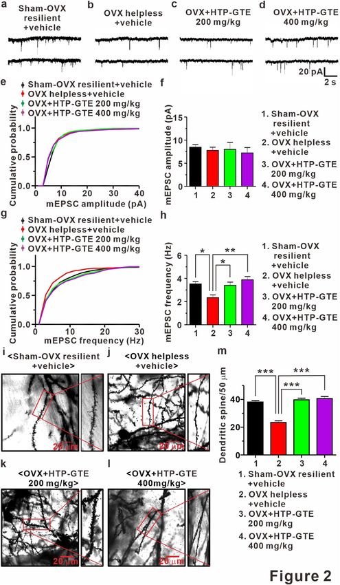
www.nature.com/scientificreports/
Green tea (Camellia sinensis) is a widely consumed beverage, especially in Asia. It has been reported to have
therapeutic effects such as amelioration of cognitive dysfunction induced by normal aging or neurodegenerative
syndromes18,19. Akin to these data, green tea consumption could be a therapeutic solution for cognitive declines
in psychopathological conditions, including post-menopausal depressive disorders. The main constituents are
reported to be catechins and theanine20, responsible for the bioactive functions of green tea. Among them,
(−)-epigallocathechin-3-gallate (EGCG) is a major catechin21,22 while theanine is a free amino acid present in
high levels in green t ea23. The studies on the effects of green tea on cognitive function are not always consistent
because the concentrations of green tea and its constituents vary in each experiment depending on the manufac-
turing process. In fact, catechin content in canned green tea is relatively low because of the conversion of green
tea catechins to their epimers (e.g. from EGCG to (−)-gallocatechin gallate (GCG)) during the heating processes
of brewing and sterilization24,25. GCG has been reported to be more stable26 and bioactive than EGCG27–29 despite
its small portion in the composition of unprocessed green tea. In addition, when green tea is administered as
an extract with high doses of its bioactive ingredients, EGCG has been reported to be associated with other side
effects, including hepatotoxicity and abnormal thyroid enlargement5,30–33. However, other isomers of green tea
catechins, such as (−)-catechin (C), (−)-gallocatechin (GC), and GCG have yet been explored to test their func-
tions in improving the impaired cognitive functions.
Nonetheless, the clinical use of green tea, especially as a dietary supplement are still limited due to the
unknown mechanisms directly correlated to the cognitive functions. Thus, there is a need to develop a safe
green tea derivative, which confers potent cognitive improvement without causing adverse side effects such as
hepatotoxicity, even at high concentrations. Furthermore, detailed studies should be performed on potential
derivatives to elucidate the signaling mechanisms of active ingredients responsible for improving cognitive
impairments in depression.
We developed (−)-gallocatechin gallate (GCG)-enriched green tea extract, a modified green tea derivative in
which GCG, an epimer of EGCG, was enriched by epimerization via heating and pH a djustment24. In addition,
we used the learned helplessness (LH) model of depression to test the effects of HTP-GTE on improving the
cognitive function in ovariectomized (OVX) rats. The LH paradigm has been proven to be a very useful technique
to assess the cognitive dysfunction for understanding human depression34. To elucidate the mechanisms under-
lying the effects of HTP-GTE on PMD-induced cognitive dysfunction, we investigated the effect of HTP-GTE
on the synaptic impairment at Schaffer collateral (SC)-CA1 synapses of the hippocampus from helpless OVX
rats using electrophysiological and immuno-histochemical spine count techniques. We also examined whether
GCG component itself can affect cognitive dysfunctions in helpless OVX rats by measuring spatial learning and
memory using the Morris water maze t est35. In this study, we demonstrated that HTP-GTE can prevent PMD-
related cognitive impairments more effectively than conventional green tea (GTE). Furthermore, HTP-GTE was
found to be functionally effective by restoring long-term synaptic plasticity. It induces the activation of silent
synapses, which was mediated by the brain-derived neurotrophic factor (BDNF)/ tyrosine receptor kinase B
(TrkB) signaling pathway. GCG was identified as a major bioactive constituent responsible for the main effect of
HTP-GTE. Our findings suggested that GCG-enriched green tea derivatives are potential therapeutic candidates
to prevent the development of cognitive deficits induced by PMD.
Results
HTP‑GTE is safer and less neurotoxic than GTE. We developed HTP-GTE for the present study
through a process involving epimerization that involved heating and pH adjustment24. The detailed process of
manufacturing this modified green tea extract is described in the method section. To determine the bioavail-
ability of HTP-GTE as a potential therapeutic for depression, we performed a toxicological investigation of
HTP-GTE compared to the conventional green tea extract (GTE) under in vivo conditions. Either HTP-GTE
or GTE was administered orally every day at a dose of 5 g/kg to a group of five male and five female rats that
were fasted overnight. Mortality, clinical signs, body weights, and gross necropsy findings were evaluated. The
results showed that there was no unscheduled death in HTP-GTE-treated rats. Three female rats and one of five
male rats fed with GTE died during the study (Fig. S1a,b). Regarding the clinical signs and body weights, no
treatment-related changes were observed in HTP-GTE-fed rats (Fig. S1c). At the end of the observation period
all animals were euthanized by CO2 gas overdose. Any gross abnormalities in organs including kidney, liver, and
spleen were observed with details of the location, color, shape, and size. No remarkable abnormalities were found
at necropsy on the 15th day in HTP-GTE-fed rats (data not shown).
We also investigated the toxicity assay under in vitro conditions. By using 3-(4,5-dimethylthiazol-2-yl)-
2,5-diphenyltetrazolium bromide (MTT) assay, we evaluated the neuronal cell viability in SH-SY5Y cells (human
neuroblastoma cell line) and cultured rat hippocampal neurons following HTP-GTE or GTE treatment at various
doses (3—100 μg/mL) for 2 h. In SH-SY5Y cells treated with GTE, cell viability decreased in a dose-dependent
manner, but no significant difference in cell viability was observed between control and HTP-GTE-treated
cells, except at high doses (100 μg/mL) (Fig. S1d). Similarly, GTE treatment significantly reduced hippocampal
neuronal viability in a dose-dependent manner relative to control cells, but HTP-GTE administration had little
effect on the neuronal viability, except at high doses (100 μg/mL) (Fig. S1e). These results strongly imply that
HTP-GTE is a safer and less toxic green tea-based therapeutic than GTE.
HTP‑GTE administration improves resilience in OVX rats. To compare the effect of either HTP-GTE
or GTE on the development of depression during menopause, we orally administered the green tea extracts in
rats for 30 days before the LH induction protocol (Fig. S2c). Vehicle-fed OVX rats were firstly exposed to 50
randomly applied inescapable/unpredictable shocks for 3 days so that we could confirm the effectiveness of our
shock protocol for LH induction in post-menopausal models. With these conditions, the same shock parameters
Scientific Reports | (2021) 11:910 | https://doi.org/10.1038/s41598-020-79287-x 2
Vol:.(1234567890)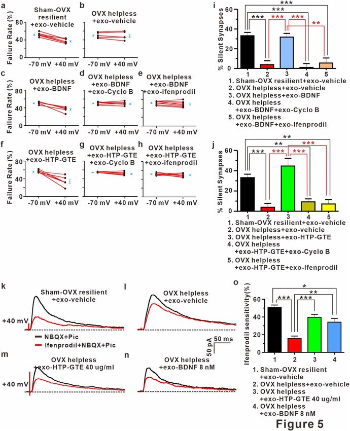
www.nature.com/scientificreports/
were used for the rats fed with either HTP-GTE or GTE. On the 4th day of the LH protocol, the rats were tested
for escape behavior to evaluate the induction of helplessness. Rats showing more than 20 escape failures during
the course of 30 trials were referred to be "helpless"36,37. As shown in Fig. S2a–c, the shock protocol effectively
induced LH with an incidence rate of 84.2% in vehicle-fed OVX rats (Fig. S2f–g). The administration of HTP-
GTE almost completely suppressed the acquisition of LH in a dose-dependent manner in OVX rats (Fig. S2d–f).
GTE (200 mg/kg) potently augmented resilience against the induction of LH in OVX rats to a similar degree of
resilience seen with the same dose of HTP-GTE. However, GTE at a high dose (400 mg/kg) was not as effective
as HTP-GTE in preventing the induction of LH in OVX rats (Fig. S2g–h), implying the superiority of HTP-GTE
over GTE in attenuating the symptoms of PMD. It was imperative to minimize the contribution of HTP-GTE-
independent resilience in the experiments involving HTP-GTE-treated OVX groups. Thus, we maximized the
number of resilient rat populations by orally administering HTP-GTE at doses of 200 and 400 mg/kg, which
almost completely prevented the induction of LH induction in OVX rats (Fig. S2h). These resilient rats were
further categorized as HTP-GTE-fed OVX (OVX + HTP-GTE) group. To investigate the comparative effects of
GTE and HTP-GTE on the synaptic alterations by LH in OVX rats, the same dose of GTE was administered,
and the resilient rats selected were further categorized as GTE-fed OVX (OVX + GTE) group. For comparison,
we used only “helpless” rats as vehicle-treated OVX (OVX) group. Sham-operated control (Sham resilient) is a
group female rats which were exposed to foot shock chamber under the same LH induction protocol.
Locomotor activities in OVX rats. To validate if there is any change in the motor function by surgical
ovariectomy, rotarod test was performed. As shown in Fig. S3a, we confirmed that there was no change in the
mobility of rats before and after ovariectomy. A complete ovariectomized state was also confirmed by enzyme-
linked immunosorbent assay (ELISA) of 17β-estradiol (E2 estrogen) levels in serum. We measured the amount
of E2 before and after ovariectomy and, as shown in Fig. S3b, OVX state is confirmed by lowered E2 levels in the
serum after the surgery.
HTP‑GTE rescues the synaptic impairments induced by LH‑induced stress via recovering LTP
at Schaffer collateral‑CA1 synapses in OVX rats. In an attempt to investigate the effect of HTP-GTE
feeding on the synaptic alterations in the hippocampi of OVX rats induced by LH procedure, we compared
the synaptic strengths and LTP induction at Schaffer collateral (SC)-CA1 circuits in the acute hippocampal
brain slices by recording field excitatory postsynaptic potential (fEPSP). First, to rule out whether OVX-induced
hormonal alteration may be directly involved in the effect of HTP-GTE on hippocampal synaptic function in
helpless OVX rats, we prepared no shock control groups (Sham no shock, Sham no shock + HTP-GTE, OVX no
shock, OVX no shock + HTP-GTE). The synaptic strengths and LTP induction were measured and compared
between the groups. As shown in Fig. S4a–f, we confirmed that the synaptic strength at the SC-CA1 circuit
was not attenuated in all the test groups with no shock. Similarly, LTP induction at SC-CA1 circuit was not
suppressed in the OVX no shock group when compared with sham no shock group (Fig. S4g–k). These results
strongly suggested that cognitive dysfunction due to PMD is not due to the hormone loss. Furthermore, we
confirmed that the synaptic strength at the SC-CA1 circuit was significantly attenuated in helpless OVX rats
compared to sham resilient controls (Figs. 1a,b,e,f, and S5a–d), which was consistent with a previous r eport34.
This impaired synaptic strength was dramatically restored in HTP-GTE-fed OVX rats to the level exhibited by
the sham resilient controls (Fig. 1c–f). Similarly, LTP induction at SC-CA1 circuit was significantly suppressed
in the helpless OVX rats when compared with sham resilient controls (Figs. 1g,h,k and S5e–g), and this impair-
ment was almost completely ameliorated in HTP-GTE-treated OVX rats (Fig. 1i–k). For comparison, we tested
the LTP induction at SC-CA1 synapses in GTE-fed OVX rats. As shown in Fig. S6a–h, hippocampal LTP induc-
tion was also restored in GTE-fed OVX rats, but less potently than in the HTP-GTE-fed OVX group with the
equivalent dose. To rule out the involvement of presynaptic alterations in the probability of transmitter release
(Pr) in the HTP-GTE-induced synaptic improvements, the paired pulse ratio (PPR) was measured in the help-
less OVX rats at SC-CA1 pyramidal cell synapses in all groups. As shown in Figs. 1i–p, and S5h–j there was no
significant difference in PPR in all groups tested, indicating that the probability of presynaptic release was not
altered by HTP-GTE feeding. These results suggest that Post-menopausal state is a "vulnerable factor” which
makes the females be more susceptible to the stressors which may cause depression more easily as well as the
associated cognitive impairments. In addition, HTP-GTE may ameliorate the hippocampal synaptic dysfunction
due to the shock-triggered depression by the restoration of post-synaptic LTP in OVX rats.
HTP‑GTE augments the number of functional synapses without affecting synaptic strength
at the hippocampal circuit in OVX rats. New spines and associated synapses are formed following LTP
induction in the hippocampus38,39, and LTP-induced synaptogenesis occurs only in newly-emerging spines that
are in contact with axon terminals activated by LTP s timuli39. Therefore, it is possible that HTP-GTE may aug-
ment the synaptic strength, which is suppressed in helpless OVX rats, by improving the functional connectivity
resulting from the restoration of LTP-induced synaptogenesis. To test this, we measured AMPA receptor-medi-
ated miniature excitatory postsynaptic currents (mEPSCs) at CA1 pyramidal neurons in the acute hippocampal
brain slices from each group. As shown in Figs. 2a,b,g,h and S6a,b,e,f the mean frequency of mEPSCs was sig-
nificantly reduced in helpless OVX rats when compared with sham resilient controls. This attenuation of mEPSC
frequency was significantly ameliorated in HTP-GTE-fed OVX rats (Fig. 2c,d,g,h). However, the mean ampli-
tudes of mEPSCs were not altered in any of the experimental groups (Figs. 2a–f and S6a–d). Furthermore, CA1
dendritic spine density was significantly decreased in helpless OVX rats when compared with sham resilient
controls, consistent with the previous report (Figs. 2i,j,m and S6g–i)34. Reduced spine density was almost com-
pletely recovered to sham resilient control levels in HTP-GTE-fed OVX rats (Fig. 2i–m). These results strongly
Scientific Reports | (2021) 11:910 | https://doi.org/10.1038/s41598-020-79287-x 3
Vol.:(0123456789)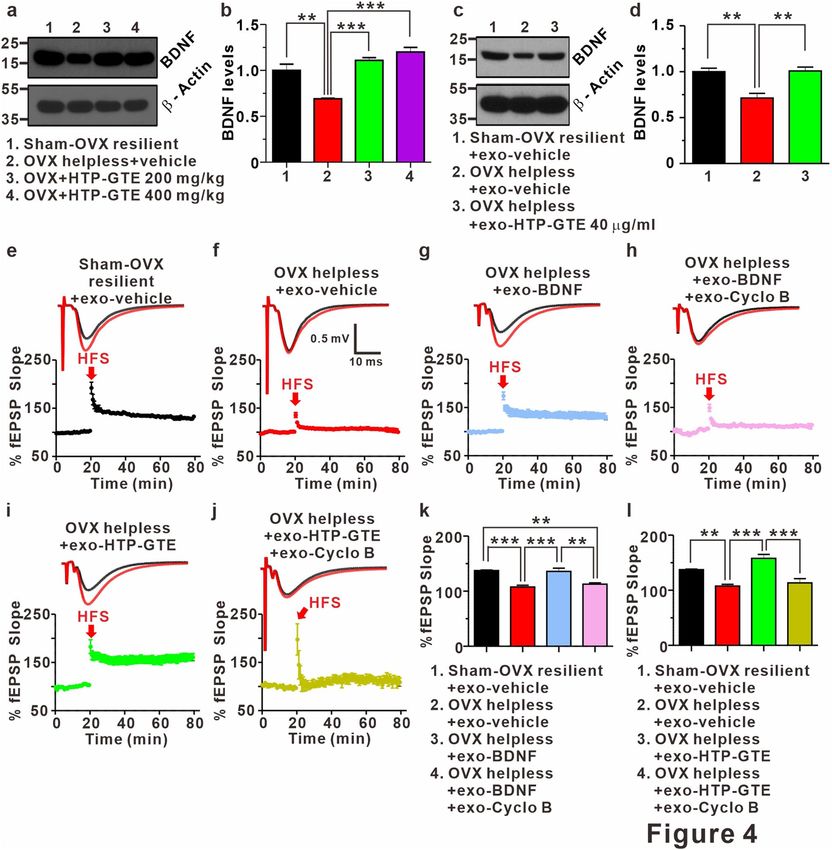
www.nature.com/scientificreports/
Figure 1. HTP-GTE rescues LH-induced synaptic impairments by restoring LTP at Schaffer collateral-CA1 ▸
synapses in OVX rats. (a–d) Representative traces of fEPSPs from hippocampal slices from representative
experiments at four increasing stimulus intensities. (e) The scatter plot of the Input and Output (I/O)
relationship corresponding to the recorded fEPSPs in A. (f) The average of slope I/O relationship for sham-
OVX resilient + vehicle, OVX helpless + vehicle, OVX + HTP-GTE 200 mg/kg, and OVX + HTP-GTE 400 mg/
kg group (Sham-OVX resilient + vehicle: 2.27 ± 0.07, n = 6 slices/3 rats; OVX helpless + vehicle: 1.62 ± 0.07, n = 6
slices/3 rats; OVX + HTP-GTE 200 mg/kg: 2.17 ± 0.06, n = 5 slices/3 rats; OVX + HTP-GTE 400 mg/kg: 2.22 ±
0.11, n = 5 slices/3 rats). (g–j) Top: representative traces showing field EPSPs before (average of 20 traces, black
line) and after (average of 180 traces, red line) high-frequency stimulus. Bottom: average time courses for field
EPSP amplitude during LTP induction in all groups. Data are shown as mean ± SEM. (k) Quantified graph was
shown (Sham-OVX resilient + vehicle: 153.3 ± 3.39, n = 5 slices/3 rats; OVX helpless + vehicle: 109.2 ± 3.65, n = 6
slices/3 rats; OVX + HTP-GTE 200 mg/kg: 166.6 ± 6.66, n = 6 slices/3 rats; OVX + HTP-GTE 400 mg/kg: 143.3 ±
10.59, n = 6 slices/3 rats). (l–o) Representative traces of paired pulse-stimulation evoked EPSCs (50 Hz; average
of 10 trials) for sham-OVX resilient + vehicle, OVX helpless + vehicle, OVX + HTP-GTE 200 mg/kg, OVX + GTE
200 mg/kg, OVX + HTP-GTE 400 mg/kg, and OVX + GTE 400 mg/kg. (p) Quantified graph was shown (Sham-
OVX resilient + vehicle: 1.35 ± 0.07, n = 6 cells/3 rats; OVX helpless + vehicle: 1.29 ± 0.05, n = 6 cells/3 rats;
OVX + HTP-GTE 200 mg/kg: 1.31 ± 0.06, n = 7 cells/3 rats; OVX + HTP-GTE 400 mg/kg: 1.43 ± 0.10, n = 8
cells/4 rats). Data are represented as mean ± SEM (One-way ANOVA/Tukey’s post hoc test, *p < 0.05, **p < 0.01,
***p < 0.001).
suggest that HTP-GTE ameliorates the synaptic impairments by increasing a new functional connectivity, but
not by potentiating the synaptic strength in already existing functional synapses.
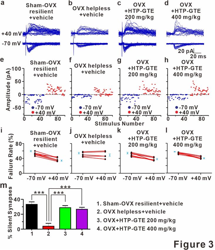
Silent synapses re‑emerge following HTP‑GTE administration in OVX rats. A silent synapse is
defined as one possessing post-synaptic NMDA receptors, but no AMPA receptors, which makes it unable to
mediate synaptic transmission under physiological c onditions40–42. Silent synapses occur in early developmental
stage (critical period) and advanced aging, serving as a reservoir for experience-dependent synaptic plasticity,
including LTP, in hippocampus43. LTP converts silent synapses into functional ones by inserting AMPA recep-
tor into the postsynaptic membrane40–42. In addition, LTP-induced functionalization of silent synapses usually
results in an increase in the number of functional synapses (i.e. increase in mEPSC frequency) without altering
presynaptic release p robability44,45, which is consistent with the effect of HTP-GTE on the quantal contents in
SC-CA1 pyramidal cell synapses shown in Figs. 2 and S7. Therefore, it is possible that the reemergence of silent
synapses may contribute to HTP-GTE-induced restoration of LTP induction, which is suppressed in helpless
OVX rats. To test this hypothesis, the proportion of silent synapses was measured using a minimal stimulation
protocol by comparing failure rates at holding potentials of − 70 mV and + 40 mV42. As shown in Figs. 3a, b,e,f,i,j
and S8 the silent synapses were present at SC-CA1 synapses in the brain slices from sham resilient control but
were significantly reduced in those from helpless OVX rats. Reduction in silent synapse formations in the help-
less OVX rats was significantly reversed by HTP-GTE administration (Fig. 3c,d,g,h,k,l,m). We confirmed this
result by measuring silent synapses through a minimal stimulation intensity at which no AMPA EPSCs were
detected (at − 70 mV) and a subsequent depolarization of the target neuron to + 40 mV, so that we could detect
NMDA-only EPSCs40,46,47. NMDA-only EPSCs were detected in all SC-CA1 synapses recorded in sham resilient
controls, but not in helpless OVX rats (Fig. S9a,b,e,f,i). The suppression of NMDA-only EPSCs was significantly
abrogated by HTP-GTE application in the OVX models (Fig. S9c,d,g,h,i), implying a possible role of the acti-
vated silent synapses by HTP-GTE-induced recovery of the long-term plasticity impairments in OVX rats.
Regulation of BDNF‑TrkB pathway underlies the HTP‑GTE‑dependent amelioration of hip‑
pocampal synaptic impairments in helpless OVX rats. BDNF has been known as an important regu-
lator of long-term synaptic plasticity processes in the hippocampus underlying learning and memory during
adulthood48–50. Down-regulation of BDNF levels in the hippocampus plays an important role in depression-
related symptoms, including cognitive impairment51–53. Therefore, it is possible that the amelioration of hip-
pocampal synaptic impairment induced by HTP-GTE may be mediated by increased BDNF levels, resulting in
reversing LTP impairments in helpless OVX rats. To test this hypothesis, we compared the hippocampal BDNF
expression in the helpless OVX and HTP-GTE-fed OVX rats using an immunoblot. In addition, we tested the
effect of exogenous HTP-GTE treatment on BDNF expressions in acute hippocampal slices from the helpless
OVX rats. As shown in Fig. 4a,b, and Fig. S10a–d the hippocampal BDNF expression was significantly sup-
pressed in helpless OVX rats when compared with sham resilient controls. Suppressed BDNF expression was
recovered to sham resilient control levels in HTP-GTE-treated OVX rats. Moreover, surprisingly, the exogenous
treatment of HTP-GTE (40 μg/mL) for 2 h significantly augmented BDNF expression in the acute hippocampal
slices from the helpless OVX rats when compared with sham resilient controls (Fig. 4c,d). We further tested the
effect of exogenous BDNF and HTP-GTE on LTP induction at SC-CA1 synapses in acute hippocampal slices
from the helpless OVX rats. As shown in Fig. 4e–h,k, exogenous BDNF application effectively ameliorated the
impairment of LTP induction at SC-CA1 synapses in helpless OVX rats. The ameliorating effect of exogenous
BDNF on hippocampal LTP was blocked by co-treatment with 1 μM cyclotraxin B (cyclo B), a potent inhibitor
of TrkB54. Similar to BDNF effect, the exogenous application of HTP-GTE (40 μg/mL) rescued the impairments
of LTP at SC-CA1 synapses in the acute hippocampal slices from the helpless OVX rats, however, this recovery
by HTP-GTE was nearly completely prevented by 1 μM cyclo B (Fig. 4i,j,l). These results suggest that HTP-GTE
Scientific Reports | (2021) 11:910 | https://doi.org/10.1038/s41598-020-79287-x 4
Vol:.(1234567890)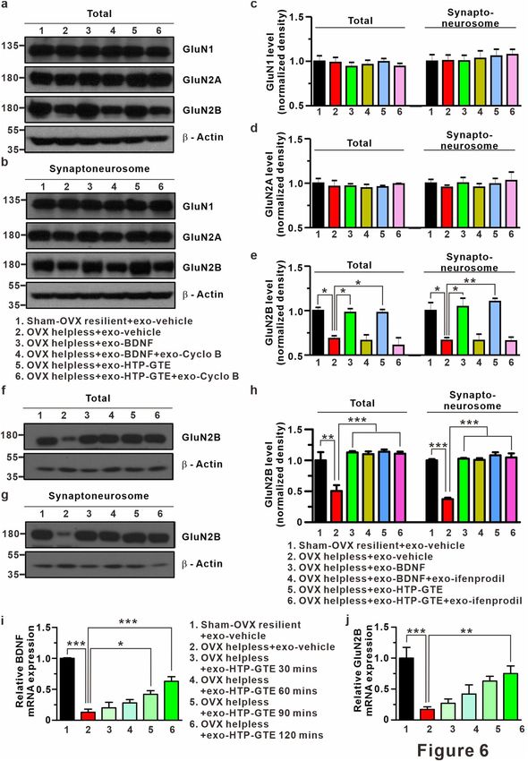
www.nature.com/scientificreports/

Scientific Reports |   (2021) 11:910 |   https://doi.org/10.1038/s41598-020-79287-x        5

                                                                                      Vol.:(0123456789)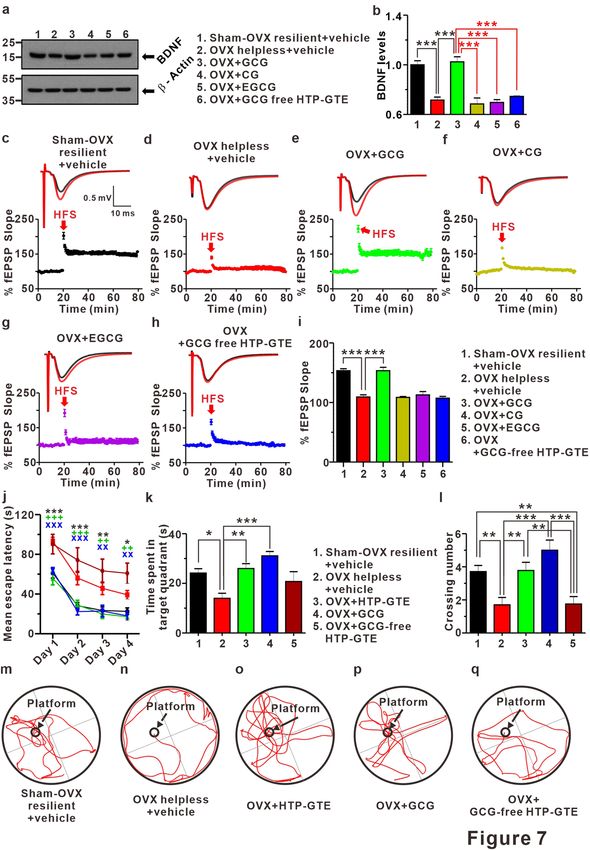
www.nature.com/scientificreports/
Figure 2. HTP-GTE augments the number of functional synapses without affecting synaptic strength at ▸
hippocampal circuit in OVX rats. (a–d) Sample traces showed miniature EPSCs for both groups. (e) Cumulative
probability plots of mEPSC amplitude. mEPSC amplitude nearby sham resilient (Sham-OVX resilient + vehicle
vs OVX helpless + vehicle: p = 0.9829; Sham-OVX resilient + vehicle vs OVX + HTP-GTE 200 mg/kg: p = 0.3581;
Sham-OVX resilient + vehicle vs HTP-GTE 400 mg/kg: p = 0.9829, Kolmogorov–Smirnov two-sample test).
(f) The mean amplitude of mEPSC for sham-OVX resilient + vehicle, OVX helpless + vehicle, OVX + HTP-
GTE 200 mg/kg, and OVX + HTP-GTE 400 mg/kg group (Sham-OVX resilient + vehicle: 8.47 ± 0.57, n = 6
slices/3 rats; OVX helpless + vehicle: 7.78 ± 0.69, n = 7 slices/3 rats; OVX + HTP-GTE 200 mg/kg: 7.99 ± 1.52,
n = 7 slices/3 rats; OVX + HTP-GTE 400 mg/kg: 7.24 ± 1.16, n = 6 slices/3 rats). (g) Cumulative probability
plots of mEPSC frequency. mEPSC frequency nearby sham resilient (Sham-OVX resilient + vehicle vs OVX
helpless + vehicle: p < 0.01; Sham-OVX resilient + vehicle vs OVX + HTP-GTE 200 mg/kg: p = 0.1863; Sham-
OVX resilient + vehicle vs HTP-GTE 400 mg/kg: p = 0.557, Kolmogorov–Smirnov two-sample test). (h) The
mean frequency of mEPSC for all groups (Sham-OVX resilient + vehicle: 3.52 ± 0.19, n = 6 slices/3 rats; OVX
helpless + vehicle: 2.33 ± 0.24, n = 7 slices/3 rats; OVX + HTP-GTE 200 mg/kg: 3.39 ± 0.28, n = 7 slices/3 rats;
OVX + HTP-GTE 400 mg/kg: 3.88 ± 0.28, n = 6 slices/3 rats). (i–l) Representative golgi-stained dendritic
segments of CA1 pyramidal neuron from animals on sham-OVX resilient + vehicle, OVX helpless + vehicle,
OVX + HTP-GTE 200 mg/kg, and OVX + HTP-GTE 400 mg/kg groups. (m) Quantitative analysis of spine
density for all groups (Sham-OVX resilient + vehicle: 38.00 ± 1.47; OVX helpless + vehicle: 23.63 ± 1.89;
OVX + HTP-GTE 200 mg/kg: 39.14 ± 2.25; OVX + HTP-GTE 400 mg/kg: 40.29 ± 2.99, 3–4 brains/group). Data
are represented as mean ± SEM (One-way ANOVA/Tukey’s post hoc test *** p < 0.001).
may improve depression-related cognitive dysfunction by restoring LTP via the activation of the BDNF-TrkB
signaling pathway.
BDNF‑TRkB‑mediated signaling contributes to the reemergence of silent synapses in
HTP‑GTE‑treated OVX rats. Functionalization of silent synapses is a principal mechanism for NMDAR
dependent LTP, and BDNF/TrkB-mediated signaling is required for functionalization of silent synapses55–57.
In addition, GluN2B-containing NMDAR plays a critical role in the formation and maintenance of silent
synapses58,59. Therefore, it is possible that BDNF-TrkB signaling may be involved in the HTP-GTE-mediated
reemergence of silent synapses in the hippocampus in the helpless OVX rats. This was checked by determining
the effect of cyclo B (1 μM) and ifenprodil (5 μM: a potent GluN2B antagonist) on HTP-GTE-induced reemer-
gence of silent synapses at SC-CA1 circuits using the same protocol as in Fig. 3 in the acute hippocampal slices
taken from helpless OVX rats. Co-treatment with cyclo B or ifenprodil effectively prevented HTP-GTE-induced
reemergence of silent synapses in helpless OVX rats (Fig. 5a,b,f–h,j). Similar to the results of HTP-GTE treat-
ment, the exogenous BDNF significantly increased the number of silent synapses at SC-CA1 circuits as seen in
the studies with the acute hippocampal slices from the helpless OVX rats (Fig. 5a–e,i). The effect of BDNF on
the proportion of silent synapses was nearly completely inhibited by co-treatment with cyclo B or ifenprodil
(Fig. 5d,e,i). In term to the effect of ifenprodil, it is also possible that it could be that new silent synapses prefer-
entially have GluN2B and so adding ifenprodil mean it can no longer detect them even if they still exist in newly
formed silent synapses. To rule out this possibility, we evaluated ifenprodil sensitivity with the percentage of
GluN2B-containing NMDA receptor by calculating the changes in the responses before and after the adminis-
tration of drug in order to functionally confirm the HTP-GTE- or BDNF-induced increase of GluN2B expres-
sion at SC-CA1 circuits in acute hippocampal slices from the helpless OVX rats. Furthermore, we investigated
whether direct ex vivo application of HTP-GTE or BDNF could cause an increase in the functional numbers of
dendritic spines by measuring the mEPSC frequency in the acute hippocampal slices from the helpless OVX rats.
In the helpless OVX rat, NMDA EPSCs at SC-CA1 circuits confirmed a decrease in ifenprodil sensitivity when
compared with sham-OVX resilient controls. This decrease of ifenprodil sensitivity was significantly restored
to the levels of sham-OVX resilient controls after the exogenous HTP-GTE- or BDNF- treatment (Fig. 5k–o).
In addition, the suppression of mEPSC frequencies by LH was recovered to the levels of sham-OVX resilient
control in the acute hippocampal slices from helpless OVX rats treated with exogenous HTP-GTE (40 μg/mL)
or BDNF (8 nM) (Fig. S11a–c,j–l,h–i,q–r). However, the mean amplitudes of mEPSCs were not altered by HTP-
GTE or BDNF administration, and this result was demonstrated in the acute hippocampal slices from helpless
OVX rats (Fig. S11a–c,j–l,f–g,o–p). Furthermore, the ameliorating effect of exogenous BDNF or HTP-GTE on
the suppressed mEPSC frequencies was blocked by co-treatment with cyclo B or ifenprodil (Fig. S11d–i,m–o,r),
which implied the involvement of GluN2B-containing NMDA receptors in HTP-GTE-induced increase of func-
tional synaptic connection in helpless OVX rats.
Expression levels of BDNF and GluN2B at hippocampus in HTP‑GTE treated‑OVX rats. With
the electrophysiological evidences supporting the involvement of GluN2B in BDNF-TrkB signaling, we also
found that the exogenous BDNF or HTP-GTE increased the expression of GluN2B proteins without altering
GluN1 or GluN2A expressions in the hippocampal synaptosomes from the helpless OVX rats. HTP-GTE- or
BDNF-mediated increase in GluN2B expression was significantly suppressed by cyclo B co-treatment (Fig. 6a–
e). However, HTP-GTE- or BDNF-mediated increase GluN2B expression was not suppressed by ifenprodil co-
treatment (Fig. 6f–h). We also assessed the role of HTP-GTE in the genomic levels of BDNF and GluN2B by
real-time quantitative polymerase chain reaction (qPCR), which was conducted to quantify the time-dependent
mRNA expression levels at every 30 min. The effect of exogenous HTP-GTE treatment on the acute brain slice
was observed through the genomic expression levels of target genes, and the expression of BDNF was detected at
Scientific Reports | (2021) 11:910 | https://doi.org/10.1038/s41598-020-79287-x 6
Vol:.(1234567890)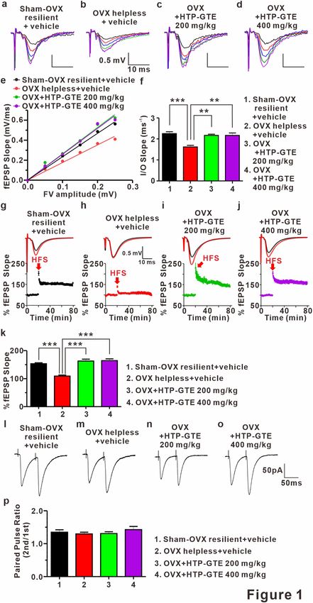
www.nature.com/scientificreports/

Scientific Reports |   (2021) 11:910 |   https://doi.org/10.1038/s41598-020-79287-x        7

                                                                                      Vol.:(0123456789)
www.nature.com/scientificreports/
Figure 3. Silent synapses are re-emerged following HTP-GTE administration in OVX rats. (a-d) Representative
traces for EPSCs evoked by minimal stimulation for 50 trials at holding potentials of -70 mV or + 40 mV
in the slices from sham-OVX resilient + vehicle, OVX helpless + vehicle, OVX + HTP-GTE 200 mg/kg, and
OVX + HTP-GTE 400 mg/kg groups. (e–h) Time course of EPSC amplitudes in representative cells shown
in collected -70 mV (blue symbols) and + 40 mV (red symbols). (i–l) Failure rates for EPSCs at − 70 mV
or + 40 mV in slices for all groups. (m) Percentage of silent synapse proportions for all groups (Sham-OVX
resilient + vehicle: 33.19 ± 3.38, n = 7; OVX helpless + vehicle: 3.925 ± 3.89, n = 6; OVX + HTP-GTE 200 mg/kg:
28.78 ± 2.82, n = 7; OVX + HTP-GTE 400 mg/kg: 26.63 ± 2.67, n = 6).
Scientific Reports | (2021) 11:910 | https://doi.org/10.1038/s41598-020-79287-x 8
Vol:.(1234567890)www.nature.com/scientificreports/
Figure 4. Regulation of BDNF-TrkB pathway underlies HTP-GTE-dependent amelioration of hippocampal
synaptic impairments in helpless OVX rats. (a, b) Western blot of BDNF level in hippocampus on sham-OVX
resilient + vehicle, OVX helpless + vehicle, OVX + HTP-GTE 200 mg/kg, and OVX + HTP-GTE 400 mg/kg
groups in oral administration of HTP-GTE or vehicle solution daily for 4 weeks (Sham-OVX resilient + vehicle:
1.00 ± 0.07; OVX helpless + vehicle: 0.69 ± 0.01; OVX + HTP-GTE 200 mg/kg: 1.11 ± 0.03; OVX + HTP-GTE
400 mg/kg: 1.20 ± 0.05, n = 3 brains/group). Full-length blot is presented in Supplementary Fig. S13. (c,d)
Western blot of BDNF level in hippocampus of exogenous application of HTP-GTE for 2 h (Sham-OVX
resilient + exo-vehicle: 1.0 ± 0.04; OVX helpless + exo-vehicle: 0.71 ± 0.05; OVX helpless + exo-HTP-GTE
40 μg/ml: 1.01 ± 0.04, n = 3 brains/group). Full-length blot is presented in Supplementary Fig. S13. (e–j) Top:
representative traces showing EPSPs before (average of 20 traces, black line) and after (average of 180 traces,
red line) high-frequency stimulus. Bottom: average time courses for EPSP amplitudes during LTP induction
in all groups. (k,l) Quantified graph was shown (Sham-OVX resilient + exo-vehicle: 137.4 ± 1.02, n = 5 slices/3
rats; OVX helpless + exo-vehicle: 107.6 ± 3.18, n = 8 slices/3 rats; OVX helpless + exo-BDNF (8 nM): 136.0 ±
5.74, n = 6 slices/3 rats; OVX helpless + exo-BDNF (8 nM) + exo-cyclo B: 112.8 ± 2.27, n = 6 slices/3 rats; OVX
helpless + exo-HTP-GTE 40 μg/ml: 157.9 ± 7.21, n = 7; OVX helpless + exo-HTP-GTE 40 μg/ml + cyclo B:
113.4 ± 7.64, n = 6). Data are represented as mean ± SEM (One-way ANOVA Tukey’s post hoc test, **p < 0.01,
***p < 0.001).
Scientific Reports | (2021) 11:910 | https://doi.org/10.1038/s41598-020-79287-x 9
Vol.:(0123456789)www.nature.com/scientificreports/
Scientific Reports | (2021) 11:910 | https://doi.org/10.1038/s41598-020-79287-x 10
Vol:.(1234567890)www.nature.com/scientificreports/
◂ Figure 5. BDNF-mediated augmentation of GluN2B expressions contributes to the activation of silent synapses
in HTP-GTE-treated OVX rats. (a–h) Failure rates for EPSCs at -70 mV or + 40 mV in slices from sham-OVX
resilient + exo-vehicle, OVX helpless + exo-vehicle, OVX helpless + exo-BDNF, OVX helpless + exo-BDNF + exo-
cyclo B, OVX helpless + exo-BDNF + ifenprodil, OVX helpless + exo-HTP-GTE, OVX helpless + exo-HTP-
GTE + exo-cyclo B, and OVX helpless + exo-HTP-GTE + exo-ifenprodil. (i,j) Summary of the percentage of silent
synapse for all groups (Sham-OVX resilient + exo-vehicle: 33.19 ± 3.38, n = 7; OVX helpless + exo-vehicle: 3.93 ±
3.89, n = 6; OVX helpless + exo-BDNF: 31.79 ± 3.80, n = 6; OVX helpless + exo-BDNF + exo-cyclo B: 1.08 ± 3.88,
n = 7; OVX helpless + exo-BDNF + exo-ifenprodil: 5.52 ± 5.12, n = 7; OVX helpless + exo-HTP-GTE: 44.61 ±
7.64, n = 6; OVX helpless + exo-HTP-GTE + exo-cyclo B: 9.30 ± 2.93, n = 7; OVX helpless + exo-HTP-GTE + exo-
ifenprodil: 7.18 ± 4.28, n = 7). (k–n) Representative traces for the effect of infeprodil on the NMDA EPSCs in
hippocampal slices from sham-OVX resilient + exo-vehicle, OVX helpless + exo-vehicle, OVX helpless + exo-
HTP-GTE, and OVX helpless + exo-BDNF groups. o. Percentage of ifenprodil-sensitivity current for all groups
(Sham-OVX resilient + exo-vehicle: 50.62 ± 2.91, n = 6; OVX helpless + exo-vehicle: 15.68 ± 2.77, n = 5; OVX
helpless + exo-HTP-GTE 40 μg/ml: 39.61 ± 3.35, n = 6; OVX helpless + exo-BDNF 8 nM: 34.11 ± 4.26, n = 5).
90 min after the drug administration while GluN2B was detected at 120 min (Fig. 6i,j). These results suggested
that BDNF-TrkB signaling contributes to HTP-GTE-activated silent synapses by regulating GluN2B-containing
NMDARs.
GCG, not EGCG, is a major contributor to the HTP‑GTE‑induced improvement of synaptic
and cognitive impairments in the helpless OVX rats. We developed HTP-GTE for the present study
through a process involving epimerization that involved heating and pH a djustment24. To compare the composi-
tions of catechins in HTP-GTE and GTE in detail, we analyzed the constituents of both green tea derivatives by
Ultra-performance liquid chromatography photometric diode array (UPLC-PDA). As shown in Table S1, EGCG
and EGC were the major catechins present in both conventional green tea (GTE) and modified green tea (HTP-
GTE). However, the levels of GCG and CG (i.e. epimers of EGCG and EGC, respectively) were much higher in
HTP-GTE than in GTE. Firstly, each rat was individually fed with GCG, CG, EGCG, or HTP-GTE containing
no GCG (i.e. GCG-free HTP-GTE), and exposed to the protocol shown in Fig. S2, so that we can independently
assess the effect of each HTP-GTE catechin component on the LH models of OVX rats. Each animal group
was subsequently tested for the escape behaviors to evaluate the induction of LH. As shown in Fig. S12a,b, the
administration of GCG and EGCG, and not CG or GCG-free HTP-GTE, significantly increased the rodent
resilience against LH in OVX rats. This is an important finding to primarily indicate that HTP-GTE-induced
resilience against LH in OVX rats is due to either GCG or EGCG. Therefore, in the current study, we designed
our experimental protocols to use the resilient rats only in the GCG- or EGCG-fed groups. Meanwhile, the
helpless OVX rats were enrolled into two groups to study the CG-only HTP-GTE or GCG-free HTP-GTE-fed
effects. To identify the major active components of the HTP-GTE effect, we investigated the effects of equivalent
doses of GCG (5.58 mg/kg), CG (0.64 mg/kg), and EGCG (25 mg/kg) present in HTP-GTE on the hippocampal
BDNF expressions and LTP induction in OVX rats, which were fed with the individual components for 30 days.
Consistent with the results shown in Figs. 4a,b and 7a,b showed that the hippocampal BDNF expression was sig-
nificantly suppressed in the helpless OVX rats when compared with sham-OVX resilient controls. Notably, only
GCG-fed OVX rats exhibited a significant recovery of the suppressed hippocampal BDNF expressions while nei-
ther CG-fed nor EGCG-fed OVX rats showed any changes in BDNF expressions. In addition, GCG-free HTP-
GTE-fed OVX rats showed no statistically significant alteration in the hippocampal BDNF expressions when
compared with the helpless OVX rats (Fig. 7a,b). Administration of GCG nearly completely rescued impairment
of LTP induction at SC-CA1 synapses in OVX rats, but CG, EGCG, and GCG-free HTP-GTE treatment showed
little effect (Fig. 7c–i). Similarly, the exogenous administration of GCG restored LTP induction to sham-OVX
resilient control levels, as observed in the acute hippocampal slices from the helpless OVX rats. These results
suggested that both GCG and EGCG are effective in promoting and maintaining resilience against the induction
of depression by LH protocol, but GCG on its own was by far more potent in recovering the depression-related
synaptic impairments than EGCG. In an attempt to further evaluate the effects of HTP-GTE and its major active
component, GCG, on memory and learning (which are usually malfunctioning during depression), all animal
groups were trained to find the hidden platform in a water pool for 4 days, and the Morris water maze test was
performed on the 5th day. As shown in Fig. 7j–l,m–q, the helpless OVX rats spent more time to find the hid-
den platform and performed fewer platform crossings than sham-OVX resilient controls. On the other hand,
the oral administration of HTP-GTE or GCG in the OVX rats significantly restored their ability to perform the
tasks to the level of sham-OVX resilient controls. Akin to this finding, GCG-free HTP-GTE had no effect on
the improvement of performance, and this further emphasizes the fundamental role of GCG as the major active
substance in HTP-GTE underpinning the HTP-GTE-induced rescue of synaptic and cognitive impairments in
postmenopausal depression.
Discussion
LH is mediated by the failure to learn behavioral responses induced by inescapable aversive events. Hippocam-
pus, the primary locus for learning and memory, has been known to play a major role in the development of
LH where it is associated with its own functional and anatomical changes38,60,61. LH can occur with inescapable
stress, which disrupts hippocampal LTP induction in vivo. However, cumulative evidences have raised ques-
tions regarding the role of the hippocampus in the development of depression. Passive response to inescapable
shock is mediated by the serotonergic activity of the dorsal raphe nucleus (DRN), which results in escape failure.
Scientific Reports | (2021) 11:910 | https://doi.org/10.1038/s41598-020-79287-x 11
Vol.:(0123456789)www.nature.com/scientificreports/
Scientific Reports | (2021) 11:910 | https://doi.org/10.1038/s41598-020-79287-x 12
Vol:.(1234567890)www.nature.com/scientificreports/
◂Figure 6. BDNF-mediated augmentation of GluN2B expression contributes to activation of silent synapses
in HTP-GTE-treated OVX rats. (a,b) Western blot of GluN1, GluN2A, GluN2B levels in brain lysate for
total and synaptoneurosomal fraction from hippocampus. Full-length blots are presented in Supplementary
Fig. S13. (c–e) Quantification of GluN1, GluN2A, GluN2B levels (GluN2B, total: Sham-OVX resilient + exo-
vehicle: 1.00 ± 0.04; OVX helpless + exo-vehicle: 0.69 ± 0.03; OVX helpless + exo-BDNF: 0.98 ± 0.04; OVX
helpless + exo-BDNF + exo-cyclo B: 0.66 ± 0.07; OVX helpless + exo-HTP-GTE: 0.98 ± 0.04; OVX helpless + exo-
HTP-GTE + exo-cyclo B: 0.61 ± 0.09; Synaptoneurosome: Sham-OVX resilient + exo-vehicle: 1.00 ± 0.09; OVX
helpless + exo-vehicle: 0.66 ± 0.03; OVX helpless + exo-BDNF: 1.04 ± 0.09; OVX helpless + exo-BDNF + exo-
cyclo B: 0.66 ± 0.07; OVX helpless + exo-HTP-GTE: 1.10 ± 0.04; OVX helpless + exo-HTP-GTE + cyclo B: 0.66
± 0.04, n = 3 brains/group). (f,g) Western blot of GluN2B levels in brain lysate for total and synaptoneurosomal
fraction from hippocampus. Full-length blots are presented in Supplementary Fig. S13. (h) Quantification of
GluN2B levels (GluN2B, total: Sham-OVX resilient + exo-vehicle: 1.00 ± 0.13; OVX helpless + exo-vehicle:
0.51 ± 0.09; OVX helpless + exo-BDNF: 1.13 ± 0.02; OVX helpless + exo-BDNF + exo-ifenprodil: 1.11 ±
0.04; OVX helpless + exo-HTP-GTE: 1.14 ± 0.04; OVX helpless + exo-HTP-GTE + ifenprodil: 1.11 ± 0.03;
Synaptoneurosome: Sham-OVX resilient + exo-vehicle: 1.00 ± 0.02; OVX: 0.37 ± 0.02; OVX helpless + exo-
BDNF: 1.01 ± 0.01; OVX helpless + exo-BDNF + exo-ifenprodil: 1.01 ± 0.03; OVX helpless + exo-HTP-GTE:
1.08 ± 0.05; OVX helpless + exo-HTP-GTE + exo-ifenprodil: 1.05 ± 0.07, n = 3 brains/group). (i,j) Quantitative
real-time PCR. Data represent the BDNF and GluN2B gene expressions normalized to GAPDH gene. Values
are mean ± SEM of three independent experiments. Data are represented as mean ± SEM (One-way ANOVA
Tukey’s post hoc test, *p < 0.05, **p < 0.01, ***p < 0.001).
This passivity is overcome by learning control via the ventromedial prefrontal cortex (vmPFC), which inhibits
DRN activity, making the rats possible to learn the fact that an escape from aversive stress is possible. Thus, the
alterations in the vmPFC-DRN pathway serve as the main influence on the development of LH62. Consistent
with these findings, we showed that both EGCG and GCG can effectively increase the resilience to escape the
stress against foot shock-induced LH; however, only GCG can ameliorate hippocampal synaptic dysfunction
and cognitive deficits induced by LH, and this difference between EGCG and GCG was clearly observed in
Morris water maze tests in our results (Figs. 7 and S12). These results demonstrated that the failure of escaping
behavior due to LH is mediated by a neural circuit, which is different from the hippocampal circuit responsible
for the cognitive impairments by LH. Likewise, the administration of GCG is more effective in ameliorating
depression-induced behavior than EGCG.
Although many studies have stressed the role of hippocampal synaptic plasticity as a possible mechanism
underlying the cognitive impairment commonly accompanying depression14–17, a detailed synaptic and molecular
mechanism remains to be elucidated. In the present study, we provided a possible mechanism to account for
depression-induced cognitive deficits in terms of synaptic plasticity for the first time. The proportion of silent
synapses containing NMDA receptor, but not AMPA receptor, in the hippocampus is significantly reduced in
a PMD rodent model (Fig. 3), strongly implying a reduction in the actual formation of silent synapses in the
cases of a depression-induced cognitive dysfunctions. In addition, the reemergence of silent synapses serves as
the fundamentally mandatory mechanism for HTP-GTE-induced restoration of LTP in the hippocampus of a
depressive animal model (Figs. 1 and 7).
BDNF plays a pivotal role in regulating a long-term synaptic plasticity in the h ippocampus48–50, and the
down-regulation of BDNF levels contributes to the depression-related cognitive i mpairments5,51–53. Concurrently,
GluN2B-containing NMDA receptor plays a critical role in the formation and maintenance of silent synapses58,59.
In fact, GluN2B signaling limits AMPA receptor incorporation in the developing synapses, resulting in the
maintenance of a low AMPA/NMDA ratio at the immature glutamatergic s ynapses59. However, GluN2B deletion
increases the number of functional synapses, which in turn prevents premature synapse maturation until cor-
related activity allows the induction of functional s ynapses58. Despite this circumstantial evidence indicating the
involvement of BDNF in GluN2B-mediated regulation of silent synapses, the evidences to elucidate the relation-
ship between BDNF and silent synapse formation has not yet been provided. In this study, we demonstrated that
hippocampal BDNF expression is notably reduced in the helpless OVX rats, and that the suppressed BDNF-TrkB
signaling pathway interferes with the hippocampal GluN2B expression, resulting in the reduced formation of
silent synapses in the helpless OVX rats. Taken together, HTP-GTE rescues the dysfunctional long-term plasticity
by reversing this process by increasing the hippocampal GluN2B expressions through the activation of BDNF-
TrkB signaling pathway in the helpless OVX rats.
In conclusion, we developed a modified green tea extract, HTP-GTE, which is safer and more bioactive
than conventional green tea extract. We demonstrated its therapeutic efficacy in overcoming postmenopausal
depression as well as improving the cognitive dysfunctions associated with it. We also elucidated the potential
mechanism underlying the effect of HTP-GTE by identifying GCG as a major bioactive component responsible
for the effect. Our findings suggest that GCG-based green tea derivatives may serve as a potential therapeutic
agent to prevent or ameliorate the cognitive deficits induced by postmenopausal depression.
Materials and methods
Animals. Female Sprague–Dawley rats (6-week-old, 140–160 g) were randomized by weight and housed in
cages. They were housed three per cage and maintained with food and water on a 12-h light/dark cycle (lights on
at 7 a.m. and lights off at 7 p.m.) in a temperature-controlled environment (23 ± 2 °C). The animals were initially
distributed into two groups: placebo surgery (Sham) and ovariectomized (OVX) groups. On post-operative day
Scientific Reports | (2021) 11:910 | https://doi.org/10.1038/s41598-020-79287-x 13
Vol.:(0123456789)www.nature.com/scientificreports/
Scientific Reports | (2021) 11:910 | https://doi.org/10.1038/s41598-020-79287-x 14
Vol:.(1234567890)www.nature.com/scientificreports/
◂ Figure 7. GCG, not EGCG, plays a role as a major contributor to HTP-GTE-induced improvement of
synaptic and cognitive impairments in helpless OVX rats. (a,b) Western blot of BDNF level in the hippocampi
of sham-OVX resilient + vehicle, OVX helpless + vehicle, OVX + GCG, OVX + CG, OVX + EGCG, and
OVX + GCG-free HTP-GTE via oral administration of each green tea component daily for 4 weeks (Sham-OVX
resilient + vehicle: 1.00 ± 0.03; OVX helpless + vehicle: 0.72 ± 0.02; OVX + GCG: 1.02 ± 0.04; OVX + CG: 0.69
± 0.05; OVX + EGCG: 0.70 ± 0.02; OVX + GCG free HTP-GTE: 0.75 ± 0.02, n = 3 brains/group). Full-length
blot is presented in Supplementary Fig. S13. (c–h) Top: representative traces showing EPSPs before (average
of 20 traces, black line) and after (average of 180 traces, red line) high-frequency stimulus. Bottom: average
time courses for EPSP amplitude during LTP induction on all groups. (i) Quantified graph was shown (Sham-
OVX resilient + vehicle: 153.3 ± 3.39, n = 5 slices/3 rats; OVX helpless + vehicle: 109.2 ± 3.65, n = 6 slices/3 rats;
OVX + GCG: 153.4 ± 5.72, n = 6 slices/3 rats; OVX + CG: 108.5 ± 1.51, n = 6 slices/3 rats; OVX + EGCG: 112.8
± 5.58, n = 6 slices/3 rats; OVX + GCG free HTP-GTE: 107.3 ± 2.96, n = 6 slices/3 rats). (j) Morris water maze
learning curves of 4 consecutive days. Data points represent mean value of escape latency of each day (Two-way
repeated measured (RM) ANOVA Tukey’s post hoc test, *p < 0.05, **p < 0.01, ***p < 0.001). (k) Quantification of
time spent in target quadrant (Sham-OVX resilient + vehicle: 24.16 ± 1.75, n = 14; OVX helpless + vehicle: 14.00
± 2.01, n = 10; OVX + HTP-GTE: 25.99 ± 1.97, n = 9; OVX + GCG: 31.07 ± 1.78, n = 7; OVX + GCG free HTP-
GTE: 20.74 ± 3.98, n = 8). (l) Quantification of crossing number (Sham-OVX resilient + vehicle: 3.71 ± 0.37,
n = 14; OVX helpless + vehicle: 1.7 ± 0.45, n = 10; OVX + HTP-GTE: 3.78 ± 0.49, n = 9; OVX + GCG: 5.00 ± 0.62,
n = 7; OVX + GCG free HTP-GTE: 1.75 ± 0.45, n = 8). Data are represented as mean ± SEM (One-way ANOVA
Tukey’s post hoc test, *p < 0.05, **p < 0.01, ***p < 0.001). (m–q) Representative Morris water maze movement
track from all groups.
3, the OVX group was subdivided into three more groups which received vehicle (0.9% saline) or poly-phenolic
compounds dissolved in vehicle (two different doses). Animal care was performed in accordance with the Yonsei
University College of Medicine Animal Care (Project license number: #00,062; 2017–0070) and all experimental
protocols were approved by Yonsei University College of Medicine and use Committee or the NIH Guide for the
Care and Use of Laboratory Animals.
Preparation of high temperature processed‑green tea extract (HTP‑GTE). Fresh green tea
(Camellia sinensis, CS) leaves were collected in spring from Osulloc Tea Garden in Jeju, Korea and were dried
at 150 °C for 10 min. The dried CS leaf was extracted two times with 50% aqueous ethanol at 60 °C for 3 h. The
50% aqueous ethanol extract was decaffeinated by filtration with activated carbon and the incubated at high
temperature for epimerization of catechins. The HTP-GTE was concentrated with a rotary evaporator (Buchi,
Flawil, Switzerland) in vacuo and stored in a refrigerator (− 20 °C) prior to Ultra Performance Liquid Chroma-
tography (UPLC) analysis.
Preparation of GCG‑free HTP‑GTE. GCG-free HTP-GTE was prepared by the AmorePacific CO R&D
center (Yongin, South Korea). GCG was isolated using a β-cyclodextrin-bonded silica column, and the amount
of (−)-epigallocathechin-3-gallate (EGCG) was equalized to the conventional tea level by adding pure EGCG.
UPLC‑Photometric Diode Assay (UPLC‑PDA) analysis. Bioactive components in HTP-GTE were
determined by UPLC with a PDA detector using a Zorbax Eclipse XDB C18 column (2.1 mm × 100 mm, 1.8 μm;
Agilent Technologies). The mobile phases were 0.05% Trifluoroacetic acid in water for solvent A and methanol/
acetonitrile (7:3 (v/v) for solvent B. The mobile phase flow-rate was 1.0 mL/min and the injection volume was
2 μL.
Preparation of human neuroblastoma SH‑SY5Y cell and primary hippocampal neuron cul‑
tures. Primary hippocampal neuronal cell culture was produced and maintained using a method described
previously3, with some modifications. All experiments were performed in accordance with the Yonsei Univer-
sity College of Medicine Animal Care and Use Committee or NIH Guide for the Care and Use of Labora-
tory Animals. Briefly, primary hippocampal neuron cultures were prepared from Sprague–Dawley rat embryos
(embryonic day 18) of either sex. The hippocampi were dissected and the cells were dissociated by trituration
using a fire-polished Pasteur pipette. The cells were then plated onto cover glasses coated with poly-L-lysine.
The hippocampal cells were grown in neurobasal medium (Thermo Fisher Scientific, Waltham, CA, USA) sup-
plemented with B-27 (2%) and L-glutamine (2 mM, Gibco, Waltham, MA, USA). The cultures were maintained
at 37 °C in 5% CO2/95% air. Human neuroblastoma SH-SY5Y cells purchased from American Type Culture
Collection (ATCC, Manassas, VA, USA) were maintained in Dulbecco’s Modified Eagle’s Medium (DMEM;
Gibco, Waltham, MA, USA) supplemented with 10% fetal bovine serum (HyClone, GE healthcare, Chicago, IL,
USA), 100 mg/mL streptomycin, and 100 U/mL penicillin (Gibco, Waltham, MA, USA) at 37 °C in a humidified
incubator with 5% CO2.
Surgical procedures of ovariectomy (OVX). Six-week-old female rats were anesthetized with isoflu-
rane (5% isoflurane, 95% O2) and bilateral ovariectomy was performed. The test group of female rats underwent
ovariectomy (ovaries removed by surgical method) and the other group underwent sham surgery (the skin was
cut and sewed back in place). The surgery consisted of a dorsolateral incision of the skin between the last rib
and pelvis and muscle dissection to expose periovarian fat. Forceps were used to find the ovaries surrounded
by variable amount of fat. The ovary was pulled out and the junction between the fallopian tube and uterine
Scientific Reports | (2021) 11:910 | https://doi.org/10.1038/s41598-020-79287-x 15
Vol.:(0123456789)www.nature.com/scientificreports/
horn was cut. Bleeding was usually light and stopped soon. The horn and periovarian fat were returned into the
abdominal cavity. The muscle wound was first sutured shut and the skin incision was closed. In sham surgery,
the rats underwent the same incision but no ovaries were removed. To reduce pain, ketoprofen (2.5 mg/kg IM)
was administered after surgery and the rats recovered within 3 days.
Preparation of learned helplessness (LH) rat model by exposure to inescapable electric foot
shock. The rats in the shock-exposed group received inescapable and unpredictable foot shock in an electric
foot shock chamber (20.5 × 20 × 24 cm; Panlab, Barcelona, Spain). The shock chamber was equipped with metal
rod (stainless steel) flooring connected to a shock generator and a shock control box (LE10026, LE900; Panlab,
Barcelona, Spain). Inescapable foot shocks were delivered to rats repeatedly 50 times at an amplitude of 0.8 mA,
a duration of 4 s, and randomized inter-shock intervals of 30–90 s over three consecutive days. Learned helpless-
ness (LH) was assessed 24 h after the third shock procedure by testing escape performance, which comprised
30 escape trials. Each trial adopted a single 0.8 mA foot shock administered for a maximum duration of 15 s.
For each escape trial, shock onset was accompanied by a sound/light cue that signaled door opening to permit
escape into the adjacent compartment. Each trial was terminated when the rat escaped to the non-shock side
of the shuttle box or the maximum duration (15 s) was reached. If the rat did not escape during shock, it was
counted as a ‘‘failed’’ trial. Thirty shocks lasting for 15 s each were applied with an inter-trial time of 20 s. The
rats with more than 20 escape failures in the 30 trials were regarded as being "helpless" and as having attained a
state of learned helplessness36,63.
Toxicity test. In vivo Toxicity test was carried out in compliance with the OECD Guidelines for the testing
of chemicals, Acute Oral Toxicity – Fixed Dose Procedure (No. 420), and the Testing Guidelines for Safety Evalu-
ation of Drugs (Notification No. 2009–19) issued by the Ministry of Food and Drug Safety. All rats were given a
single oral dose of test articles after a 14-h fasting period. Doses (g/kg) were adjusted according to body weight
recorded just before the examination. An initial dose volume of 10 mL/kg was used. A single dose of 5 g/kg was
administered to animals. All visible signs of reaction to treatment and mortality were recorded daily. On the first
day of treatment for each dose level, the animals were observed at the following approximate time points: at the
end of dosing and 0.5, 1, 2, 3, 4, 5, and 6 h after dosing. Thereafter, any changes in clinical signs and/or mortality
of test animals were observed daily during the test period. Then, body weights were recorded on the day of dos-
ing, Day 1,4, 7, 10, 13, 16, 19, 22, 25 and 28. At the end of the observation period all animals were euthanized by a
CO2 gas overdose. In terms of necropsy and gross pathology, any abnormalities were recorded, including details
of location, color, shape, and size, and then appropriately sampled and identified.
Cell viability test. In vitro toxicity test was done as follows. Cell viability was measured by the CCK-8 assay
(Dojindo laboratories, Shanghai, China). SH-SY5Y cells were seeded at a density of 5 × 10⁴ cells per well in a
96-well plate and cultured until 70% confluence was attained. At 24-h post-treatment with vehicle [dimethyl
sulfoxide (DMSO)], conventional green tea extract (GTE), or modified GCG-enriched green tea extract (HTP-
GTE), the cells were incubated with 10 μL CCK-8 reagent for 2 h at 37 °C. The optical density was measured at
a wavelength of 450 nm using a microplate reader (Molecular devices, San Jose, CA, USA). Cell viability was
calculated by dividing the optical density of the treated group by that of the control group. Primary hippocam-
pal neurons (2 × 10⁶ cells per well, 24-well plate) were cultured for 14 days to ensure complete maturation. At
2-h post-treatment with GTE or HTP-GTE, the medium was carefully replaced with fresh neurobasal medium
containing dilute MTT (Sigma Aldrich, St. Louis, MO, USA) (1:10, 10% MTT) and incubated for another 2 h
at 37 °C. After removing the incubation medium, the formazan crystals were dissolved in 200 μL DMSO. MTT
reduction was quantified by measuring the light absorbance at 570 nm using the microplate reader64. The cell
viability was calculated by dividing the optical density of the treated group by that of the control group.
Hippocampal slice preparation. Hippocampal slices (400 μm thick) were prepared from learned help-
lessness (LH)-tested 11-week-old female Sprague Dawley rats. The rats were anesthetized with isoflurane (5%
isoflurane, 95% O 2) and perfused with ice-cold sucrose artificial cerebrospinal fluid (aCSF) with the following
concentrations in mM: 195.5 sucrose, 2.5 KCl, 1 N aH2PO4, 32.5 N aHCO3, 11 glucose, 2 Na pyruvate, and 1 Na
ascorbate (all chemicals from Sigma-Aldrich, St. Louis, MO, USA) bubbled with 95% O 2/5% CO2 at a pH of
7.4. After perfusion, the brains were quickly removed from the skull and slices were cut on a vibratome (Leica
biosystems, Wetzlar, Germany). The slices were transferred to an incubation chamber containing incubation
solution with the following concentrations in mM: 119 NaCl, 2.5 KCl, 1 NaH2PO4, 26.2 N aHCO3, 11 glucose,
2 Na pyruvate, 1 Na ascorbate, 3 M gSO4, and 1.5 C aCl2 at 35 °C for 15 min. After incubation, the slices were
transferred to a container filled with aCSF solution at 23—24 °C for 1 h.
Electrophysiology. Field recordings were made with a concentric bipolar electrode positioned in the stra-
tum radiatum of the CA1 region using an extracellular glass pipette (3—5 MΩ) filled with aCSF. Stimulation was
delivered through a bipolar electrode (FHC, Bowdoin, ME, USA) placed in the Schaffer collateral-CA1 (SC).
The SC circuit was visualized using differential interference contrast (DIC) microscopy at 4 × magnification and
identified by the ability to evoke short and constant latency field excitatory postsynaptic potentials (fEPSPs) at
CA1 synapses by SC input stimulation. The test stimulation in all fEPSP experiments was measured prior to the
beginnging of all the experiments (30—300 μA) and a test-pulse stimulation strength that evoked 50% of the
maximum fEPSP was used. Baseline synaptic responses were recorded for 30 min, and then long-term poten-
tiation (LTP) was induced by high-frequency stimulation (HFS; 100 Hz, 4 trains, 1 s duration, 20 s inter-train
interval). Recordings were made every 10 s for 1 h using Axopatch 1D amplifier (Molecular Devices, San Jose,
Scientific Reports | (2021) 11:910 | https://doi.org/10.1038/s41598-020-79287-x 16
Vol:.(1234567890)You can also read