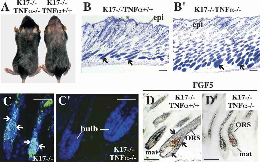
Keratin 17 modulates hair follicle cycling in a TNF -dependent fashion
←
→
Page content transcription
If your browser does not render page correctly, please read the page content below
Downloaded from genesdev.cshlp.org on March 17, 2015 - Published by Cold Spring Harbor Laboratory Press
Keratin 17 modulates hair follicle cycling
in a TNF␣-dependent fashion
Xuemei Tong1 and Pierre A. Coulombe1,2,3
1
Department of Biological Chemistry and 2Department of Dermatology, The Johns Hopkins University School of Medicine,
Baltimore, Maryland 21205, USA
Mammalian hair follicles cycle between stages of rapid growth (anagen) and metabolic quiescence (telogen)
throughout life. Transition from anagen to telogen involves an intermediate stage, catagen, consisting of a
swift, apoptosis-driven involution of the lower half of the follicle. How catagen is coordinated, and spares the
progenitor cells needed for anagen re-entry, is poorly understood. Keratin 17 (K17)-null mice develop alopecia
in the first week post-birth, correlating with hair shaft fragility and untimely apoptosis in the hair bulb. Here
we show that this abnormal apoptosis reflects premature entry into catagen. Of the proapoptotic challenges
tested, K17-null skin keratinocytes in primary culture are selectively more sensitive to TNF␣. K17 interacts
with TNF receptor 1 (TNFR1)-associated death domain protein (TRADD), a death adaptor essential for
TNFR1-dependent signal relay, suggesting a functional link between this keratin and TNF␣ signaling. The
activity of NF-B, a downstream target of TNF␣, is increased in K17-null skin. We also find that TNF␣ is
required for a timely anagen–catagen transition in mouse pelage follicles, and that its ablation partially
rescues the hair cycling defect of K17-null mice. These findings identify K17 and TNF␣ as two novel and
interdependent regulators of hair cycling.
[Keywords: Intermediate filament; hair cycle; apoptosis; anagen; catagen]
Supplemental material is available at http://www.genesdev.org.
Received October 21, 2005; revised version accepted March 13, 2006.
The hair-producing tissue, known as the hair follicle, is During anagen, matrix epithelial cells in the prominent
fascinating from developmental, histological, and physi- hair bulb proliferate rapidly, giving rise to progenitors for
ological standpoints. The various types of hair present in the layers comprising the hair shaft and IRS (Paus and
mammalian skin undergo a unique cycle throughout life, Cotsarelis 1999; Fuchs et al. 2001; Millar 2002). The hair
with phases of growth (anagen) interspersed with phases bulb and proximal region above it are destroyed via ap-
of involution (catagen) and rest (telogen) (Hardy 1992). optosis during catagen, producing a short, quiescent telo-
This cycle recapitulates several aspects of hair morpho- gen hair (Fig. 1A). The mesenchymal dermal papilla is
genesis, a process initiated in the embryo, and requires spared from apoptosis, and migrates upward during cata-
integration, in time and space, of multiple stimulatory gen to maintain physical proximity with the bulge, a
and inhibitory signals (Paus and Cotsarelis 1999; Fuchs local swelling of the ORS that houses a significant frac-
et al. 2001; Millar 2002). Premature hair loss due to vari- tion of skin epithelial stem cells (Fig. 1A; Paus and Cot-
ous factors, including disease, hormonal imbalance, and sarelis 1999; Fuchs et al. 2001; Millar 2002). Anagen re-
drug regimens, affects a large fraction of the population entry requires the action of a papilla-derived signal
(Cotsarelis and Millar 2001; Hadshiew et al. 2004). Most (Hardy 1992) that causes bulge epithelial stem cells to
nonmelanoma skin cancers, whose incidence is on the proliferate, migrate downward, and differentiate de novo.
rise, arise clonally from progenitor cells located in hair In recent years, much progress has been made in iden-
follicles (Lavker et al. 1993). Thus, there are compelling tifying the signals and mechanisms contributing to hair
reasons to understand the fundamentals of hair biology. follicle morphogenesis during development. These path-
A mature hair follicle comprises eight distinct epithe- ways, which include Wnts, sonic hedgehog (SHH), trans-
lial layers organized in concentric circles around the forming growth factor (TGF)- family members (includ-
main axis of the hair. These layers form three major ing bone morphogenetic proteins or BMPs), and others,
compartments: the innermost hair shaft, the inner root also operate at the time of anagen re-entry in adult skin
sheath (IRS), and the outer root sheath (ORS) (Fig. 1A). (Millar 2002; Jamora et al. 2003; Paus and Foitzik 2004).
Numerous studies helped define, with cellular and mo-
lecular resolution, how competing cues are integrated by
3
Corresponding author. progenitor cells so as to give rise to the intricate archi-
E-MAIL coulombe@jhmi.edu; FAX (410) 614-7567.
Article and publication are at http://www.genesdev.org/cgi/doi/10.1101/ tecture of a hair follicle. Considerably less is known,
gad.1387406. however, about the anagen–catagen transition. While
GENES & DEVELOPMENT 20:1353–1364 © 2006 by Cold Spring Harbor Laboratory Press ISSN 0890-9369/06; www.genesdev.org 1353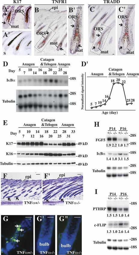
Downloaded from genesdev.cshlp.org on March 17, 2015 - Published by Cold Spring Harbor Laboratory Press Tong and Coulombe Figure 1. Hair cycling defect in K17−/− skin. (A) Schematic of anagen, catagen, and telogen phases of the hair cycle. Adapted from Beaudoin et al. (2005) (© 2005 Na- tional Academy of Sciences). (B,B⬘) Histo- logic appearance of K17+/+ (wild-type; B) and K17−/− (B⬘) mouse backskin at P14. Shown are hematoxylin/eosin-stained lon- gitudinal sections from backskin of age- and gender-matched mice. Arrows point to hair bulbs. Bars, 50 µm. (C–C⬙) TUNEL (green) and Hoechst (blue) stainings of wild- type (C) and K17−/− (C⬘,C⬙) mouse skin at P14. Arrows point to TUNEL-positive nu- clei. Bars, 50 µm. (D–D⬘) In situ hybridiza- tion analysis of FGF5 mRNA in K17+/+ and K17−/− hair follicles at P7. Four percent paraformaldehyde-fixed frozen sections were hybridized with antisense (D,D⬘) or sense (D⬙) probes. Asterisks in D–D⬙ denote melanin pigment. Arrows point to hybrid- ization signals. Bars, 50 µm. (E) Northern blot analysis of RNA (20 µg) prepared from backskin of K17+/+ and K17−/− mice at P5, P7, P10, P16, and P20, and probed for FGF5 and -tubulin mRNAs. Signal was quanti- tated in a PhosphorImager and normalized to -tubulin. Average values of two inde- pendent experiments are reported below each lane. (F–F⬙) In situ hybridization analysis of PTHRP mRNA in K17+/+ and K17−/− hair follicles at P12. Four percent paraformaldehyde-fixed frozen sections were hybridized with antisense (F,F⬘) or sense (F⬙) probes. Asterisks in F–F⬙ denote melanin pigment. Arrows point to hybrid- ization signals. Bars, 50 µm. (G–G⬙) Dual immunostaining for BrdU (green) and keratin 14 (red) in hair bulbs from K17+/+ (G) and K17−/− (G⬘) mice at P5. Quantitation of BrdU-positive nuclei per surface area of bulb tissue is shown in G⬙. (*) P < 0.01. Bars, 20 µm. (H,H⬘) Histologic appearance of K17+/+ (H) and K17−/− (H⬘) mouse backskin at P22. Shown are hematoxylin/eosin-stained longitudinal sections from backskin of age- and gender-matched mice. Arrows point to dermal papillae (H) or hair bulbs (H⬘). Bars, 50 µm. (Bulb) Hair bulb; (DP) dermal papilla; (epi) epidermis; (IRS) inner root sheath; (mat) matrix; (ORS) outer root sheath; (SG) sebaceous gland. several key signals including fibroblast growth factor genes are cotranscribed in a pairwise, tissue-type, and (FGF)-5 (Hebert et al. 1994), parathyroid hormone-related differentiation-related fashion, providing useful markers peptide (PTHRP) (Cho et al. 2003), TGF-1 (Foitzik et al. to study epithelia in health and disease (Fuchs 1995; 2000), and interferon (IFN)-␥ (Hirota et al. 2002, 2003) Omary et al. 2004). In K17-null mice, pelage hair loss have been implicated, much remains to be learned about correlates in part with hair shaft fragility, consistent the mechanisms regulating this carefully controlled tis- with the major function of structural support shared by sue regression. all keratins. In addition, abnormal apoptosis occurs in Unlike their wild-type littermates, mice null for the hair bulb tissue, possibly reflecting the novel “prosur- type I keratin 17 (K17) gene fail to develop a full pelage vival” role evidenced for select keratins (Oshima 2002; hair coat during the first week after birth (McGowan et Coulombe and Wong 2004). Of note, the pelage skin phe- al. 2002). This finding was unexpected, as more than half notype of K17-null mice is reversible and strain-depen- of the ∼50 known keratin genes are expressed in hair dent, correlating with a compensatory up-regulation in follicle tissue alone, often in overlapping patterns (Lang- K16 (McGowan et al. 2002). Here, we show that the un- bein et al. 2003; Tong and Coulombe 2004). Keratins are timely apoptosis in hair follicles of K17-null mice, be- intermediate filament-forming proteins, and represent ginning at postnatal day 5 (P5), corresponds to a prema- major cellular constituents in complex epithelia. Two ture anagen–catagen transition. K17-null keratinocytes types of keratin sequences, I and II, can be distinguished, in primary culture are selectively more sensitive to reflecting a biochemical requirement for both during the TNF␣-induced apoptosis. We further show that TNF␣ is multistep assembly reaction that produces a 10–12-nm- required for a timely anagen–catagen transition in pelage wide keratin filament (Fuchs 1995). Type I and II keratin hair follicles, that TNF␣ signaling is enhanced in K17- 1354 GENES & DEVELOPMENT
Downloaded from genesdev.cshlp.org on March 17, 2015 - Published by Cold Spring Harbor Laboratory Press

                                                                                         A role for keratins during hair follicle cycling

null skin, and finally, that loss of TNF␣ partially rescues        of K16 in hair matrix epithelial cells and normalization
the hair cycling defect of K17-null mice. The interaction          of the pelage phenotype (McGowan et al. 2002), the
between K17 and the death adaptor protein TRADD                    length of the second anagen is not significantly altered in
(TNFR1 [TNF receptor 1]-associated death domain pro-               K17-null hair follicles (data not shown). These data sug-
tein) provides further evidence for a functional link be-          gest that, in the absence of effective compensatory
tween this keratin and TNF␣ signaling. These findings              mechanisms, loss of K17 selectively affects the anagen–
identify K17, a structural protein, and TNF␣, a proin-             catagen transition of the hair cycle.
flammatory cytokine, as two novel and interdependent                 The finding that nullifying a single keratin gene per-
regulators of hair cycling.                                        turbs hair cycling was unexpected. To assess whether
                                                                   this defect specifically results from loss of K17, we rein-
                                                                   troduced K17 protein in K17-null mice. All aspects of the
Results                                                            skin phenotype, including hair fragility, hair cycling de-
                                                                   fect, and premature apoptosis in hair follicles, are nor-
Loss of K17 causes premature catagen entry in pelage
                                                                   malized in the K17 replacement mice (Supplementary
hair follicles
                                                                   Fig. 1). This confirms that the hair alterations associated
Four lines of evidence suggest that hair follicles prema-          with the K17-null mutation are specifically due to loss of
turely enter catagen in K17-null mouse skin. First, rou-           K17 protein.
tine histology conducted at P14 shows that wild-type
hair follicles are in anagen, as evidenced by their maxi-
                                                                   K17-null keratinocytes are selectively more sensitive
mal length and bell-shaped bulb region (Fig. 1B). In con-
                                                                   to TNF␣-mediated apoptosis
trast, K17-null hair follicles are in catagen, as shown by
their shortening and the narrowing and destruction of              To explore the molecular basis of untimely apoptosis in
bulb tissue (Fig. 1B⬘). Second, TUNEL assay shows the              K17-null hair follicles, we examined the response of ke-
presence of many apoptotic cells in the lowermost por-             ratinocytes in primary culture to a Fas-, TNF␣-, and
tion of involuting hair follicles in K17-null, but not wild-       TRAIL-induced apoptotic challenge in the presence of
type, skin (Fig. 1C–C⬙). Third, two key markers of the             cycloheximide (CHX) (Caulin et al. 2000). Each of these
anagen–catagen transition, FGF5 and PTHRP, are mis-                pathways is operational in skin keratinocytes (Wehrli et
regulated in K17-null backskin. The FGF5 mRNA is nor-              al. 2000). Additionally, defects in K8/K18 filaments in-
mally low during early and mid-anagen, begins to rise at           crease the susceptibility of trophectoderm and adult
approximately P10 in the hair matrix and ORS, and                  liver hepatocytes to specific proapoptotic signals includ-
peaks in late anagen (Hebert et al. 1994). By in situ hy-          ing TNF␣ and FasL, a finding that is reproduced in epi-
bridization, we find that FGF5 is up-regulated in a subset         thelial cell lines (Caulin et al. 2000; Gilbert et al. 2001;
of hair follicles at P7 in K17-null skin, while no signal is       Jaquemar et al. 2003; Ku et al. 2003). In the context of
detected in wild type (Fig. 1D–D⬙). By Northern analysis,          primary culture, newborn mouse skin keratinocytes ex-
FGF5 is slightly up-regulated at P5, and more so at P7             press K17 and K14 uniformly, and K16 in a patchy fash-
and P10 in K17-null skin, compared with wild type (Fig.            ion (Wawersik and Coulombe 2000). Expression of K17
1E). While FGF5 reaches peak levels at P16 in the con-             and K16 in this setting likely reflects the elaboration of
trol, it has already decreased in K17-null skin (Fig. 1E).         a wound repair program in serum-exposed keratinocytes
The PTHRP mRNA is normally expressed during anagen                 (Weiss et al. 1984).
and is down-regulated at catagen entry (Cho et al. 2003).             Treatment with dimethyl sulfoxide (DMSO) (CTL)
In situ hybridization shows that PTHRP is down-regu-               produced very little apoptosis over 24 h in wild-type and
lated at P12 in K17-null hair follicles, while it is still         K17-null keratinocytes (Fig. 2A,B,C,C⬘). Of all the treat-
expressed in the ORS of wild-type follicles (Fig. 1F–F⬙).          ment combinations tested, TNF␣ and TRAIL gave rise to
Finally, exit from anagen requires a decrease in cell pro-         a greater degree of apoptosis. K17-null cells (24.1 ± 3.5%)
liferation in the hair matrix. At P5, we find a significant        showed more apoptosis than wild type (15.8 ± 0.9%) in
reduction (∼30%) in the frequency of BrdU-positive cells           response to TNF␣ (Fig. 2A,B). This difference was obvi-
in this region of K17-null hair follicles, relative to wild        ous in TUNEL-stained preparations (Fig. 2C–D⬘). No dif-
type (Fig. 1G–G⬙). Altogether, these findings establish            ference was observed for any other treatment (Fig. 2A,B),
that hair follicles prematurely commit to the catagen              and following UVB exposure (data not shown). This dif-
phase of the hair cycle, starting at P5/P7, in K17-null            ferential TNF␣ responsiveness is likely underestimated
backskin.                                                          since (1) these cultures contain mostly epidermis-de-
   Histology was conducted on backskin tissue at later             rived, instead of hair follicle-derived, keratinocytes (Mar-
time points to determine whether additional aspects of             celo et al. 1978); and (2) K14 and K16 are expressed as
hair cycling are affected in K17-null mice. At P20, wild-          well, raising the prospect of phenotypic rescue in the
type and K17-null hair follicles are in telogen and early          setting of primary culture (see below).
anagen, respectively (data not shown). At P22, wild-type              To assess whether K17 acts cell-autonomously in this
follicles are still in telogen (Muller-Rover et al. 2001),         setting, K17-null keratinocytes were transiently trans-
whereas K17-null follicles have progressed further in              fected with CMV promoter-K17 cDNA, and subjected 36
anagen, as evidenced by the presence of a well-defined             h later to TNF␣/CHX treatment. A greater fraction of
bulb (Fig. 1H–H⬘). Coinciding with the ectopic induction           K17-null keratinocytes (31.5 ± 2.0%) showed apoptosis

                                                                                               GENES & DEVELOPMENT                 1355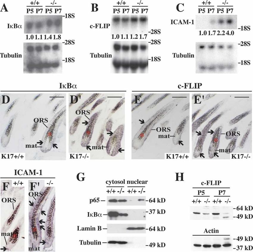
Downloaded from genesdev.cshlp.org on March 17, 2015 - Published by Cold Spring Harbor Laboratory Press
Tong and Coulombe
Figure 2. K17-null keratinocytes show increased apo-
ptosis following TNF␣ treatment. (A,B) Response of
wild-type and K17−/− keratinocytes in primary culture to
various proapoptotic treatment combinations. (CTL)
DMSO-treated controls; (CHX) cycloheximide. Other
treatments are self-explanatory. A shows the percentage
of apoptotic cells based on TUNEL and Hoechst stain-
ing, while B shows a caspase activity assay (RFU, rela-
tive fluorescence units). Both A and B show that K17−/−
keratinocytes are selectively more sensitive to
TNF␣ + CHX treatment than wild type. (*) p < 0.01. (C–
D⬘) Representative micrographs from wild-type and
K17−/− cultures treated with DMSO vehicle (CTL) or
TNF␣ + CHX. Dual TUNEL (green) and Hoechst (blue)
stainings are shown. Arrows denote TUNEL-positive
nuclei. Bars, 20 µm. (E) Comparing the sensitivity of
K17−/− keratinocytes (K17-null N-TF) to TNF␣ + CHX
treatment after transfection with wild-type K17 cDNA
(K17-null TF). Percentage of apoptotic cells is based on
TUNEL and Hoechst stainings (see A). Cells re-express-
ing K17 are significantly less sensitive to treatment than
K17−/− cells. P < 0.001.
under these conditions (Fig. 2E), likely owing to the are present in endogenous TRADD immunoprecipitates
stress of lipofectamine-mediated transfection. Neighbor- obtained from primary cultures of mouse skin keratino-
ing K17-re-expressing keratinocytes showed signifi- cytes (Fig. 3A,A⬘). In mouse keratinocytes transfected
cantly less apoptosis (18.2 ± 2.3%; p < 0.001), as expected with GFP fusion constructs, both full-length TRADD
(Fig. 2E). Together, these findings show that loss of K17 (residues 1–312) (Fig. 3B–B⬙) and its C-terminal moiety
cell-autonomously increases the susceptibility of cul- (residues 105–312) (Fig. 3C–C⬙) partially colocalize with
tured skin keratinocytes to TNF␣-mediated apoptosis, K17 in the cytoplasm. Similar results were obtained in
suggesting that such may be the case as well in vivo. preparations double-stained for K14 and TRADD (data
not shown). In contrast, the N terminus of TRADD (resi-
dues 1–105) shows a diffuse distribution in the cyto-
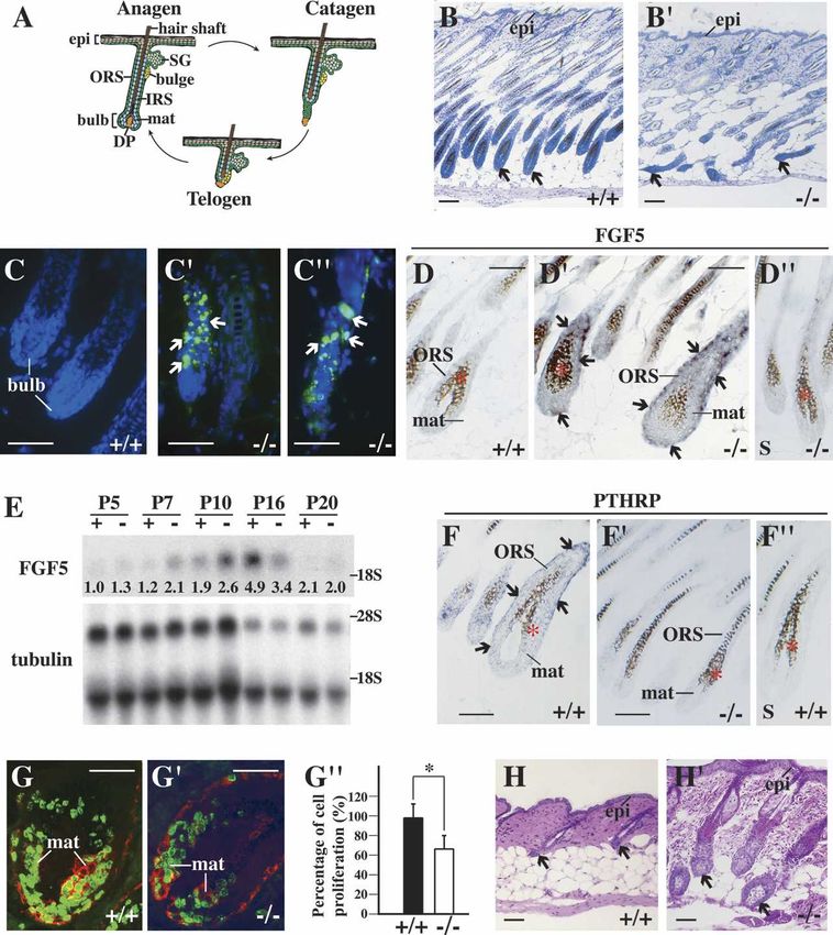
K17 interacts with TRADD, a death adaptor, in mouse plasm and nucleus (data not shown). Partial colocaliza-
skin keratinocytes tion between TRADD, a signaling adaptor present at low
That K17, a cytoskeletal protein, influences the respon- levels, and the abundant K17 does not preclude a physi-
siveness of keratinocytes to TNF␣ is a priori surprising. ologically relevant interaction. The finding that TRADD
The previously documented interaction between K18 potentially interacts with K14 and K16 in vivo is signifi-
and TRADD (Inada et al. 2001), a death adaptor protein cant, as K16 has been implicated in the reversibility and
essential for signal relay downstream from TNF receptor strain-dependence of the hair phenotype in K17-null
1 (TNFR1) (Micheau and Tschopp 2003), provides a pos- mice (McGowan et al. 2002). Additional studies are re-
sible mechanism. Inada et al. (2001) found that the K18– quired to ascertain whether altered regulation of
TRADD interaction affords a protection against TNF␣- TRADD is, indeed, involved in the increased suscepti-
induced apoptosis, possibly via the sequestration of bility of K17-null keratinocytes to TNF␣-induced apo-
TRADD away from TNFR1. TRADD also binds K14 in a ptosis.
human keratinocyte cell line (Yoneda et al. 2004). Map-
ping studies showed that TRADD binds the 1A subdo-
TNF␣ signaling contributes to hair cycle regulation
main in K18 (Inada et al. 2001) and K14 (Yoneda et al.
in vivo
2004), a region that is highly conserved in other type I
keratins. The physiological significance of these interac- There is as yet no definitive evidence implicating the
tions in vivo is unknown. TNF␣/TNFR1 signaling pathway in mature hair follicles
Coimmunoprecipitation assays using two distinct an- of postnatal mouse skin. Several conditions need to be
tibodies revealed that, as expected, K17, K16, and K14 met to support the possibility that TNF␣/TNFR1 play a
1356 GENES & DEVELOPMENT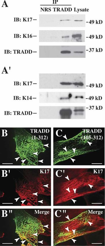
Downloaded from genesdev.cshlp.org on March 17, 2015 - Published by Cold Spring Harbor Laboratory Press
A role for keratins during hair follicle cycling
a cell-autonomous, K17-dependent modulation of
TNFR1/TRADD signaling in hair tissue. Using in situ
hybridization, we found that both TNFR1 (Fig. 4B,B⬘) and
TRADD (Fig. 4C,C⬘) mRNAs occur in the hair matrix
and ORS compartments of anagen-stage wild-type fol-
licles, in a pattern that overlaps significantly with that of
K17 (Fig. 4A,A⬘).
Next, we indirectly measured the activity of TNF␣/
TNFR1-dependent signaling during normal hair cycling
through monitoring IB␣ mRNA levels between P5 and
P28 in wild-type mouse skin. The IB␣ gene is a direct
transcriptional target of NF-B, which is activated fol-
lowing the engagement of TNFR1 (Hoffmann et al. 2002;
Micheau and Tschopp 2003) and related receptors
(Moynagh 2005). The IB␣ mRNA occurs at low levels
between P5 and P14, when follicles are in anagen (Fig.
4D,D⬘). Near the point of catagen entry (approximately
P16), its level rises and reaches a plateau that is main-
tained between P18 and P22. At P25, near the onset of
the next anagen phase, IB␣ mRNA levels have returned
to precatagen levels (Fig. 4D,D⬘). In light of our finding of
delayed catagen entry in mice lacking TNF␣ (see below),
the hair cycle dependency of IB␣ regulation, at the
mRNA level, likely reflects parallel fluctuations in
TNFR1 signaling in vivo. By comparison, K17 protein is
regulated in a reciprocal fashion during hair cycling: Its
level increases progressively during anagen in wild-type
backskin, reaching peak levels between P12 and P16, but
is markedly reduced during catagen and telogen (be-
tween P18 and P25) (Fig. 4E). K17 levels reincrease at
P28, coinciding with the next anagen (Fig. 4E). These
Figure 3. Association between K17 and TRADD in skin kera- findings significantly extend previous studies focusing
tinocytes. (A,A⬘) Immunoprecipitates from lysates prepared on K17 mRNA (Panteleyev et al. 1997; see also Lin et al.
from wild-type mouse skin keratinocytes in primary culture, 2004). Whereas its level also increases during anagen,
using normal rabbit serum (NRS) and two distinct anti-TRADD K16 is distinct from K17 in that it peaks during the P18–
polyclonal antibodies, obtained from Santa Cruz Biotechnology P25 interval (Fig. 4E); that is, during catagen/telogen
(A) and Dr. M. Inagaki (Aichi Cancer Research Institute, (Bernot et al. 2002). Other keratin proteins, including K5,
Nagoya, Japan) (Inada et al. 2001) (A⬘). Precipitates were sub- do not show such hair cycle-related fluctuations (data
jected to Western blotting with anti-K17, anti-K16, anti-K14, or not shown). These observations indicate that K17 is
anti-TRADD polyclonal antibody, as indicated at left. The mi-
poised to attenuate TNF␣/TNFR1-dependent signaling
gration of 37- and 49-kDa markers is shown at right. (B–C⬙)
during anagen in pelage hair follicles.
Indirect immunofluorescence of wild-type keratinocytes in pri-
mary culture transiently transfected with EGFP-TRADD fusion Next, we analyzed C57Bl/6 mice null for the TNF␣
contructs. (B–B⬙) Full-length TRADD (312 residues long). (C–C⬙) gene to assess whether the loss of this cytokine impacts
TRADD’s C terminus (residues 105–312). The signal detected hair follicle cycling. TNF␣-null mice are viable and do
(GFP, K17, or merge) is identified in the upper right corner. not exhibit an obvious phenotype unless immunologi-
Arrowheads point to instances of colocalization. Bars, 30 µm. cally challenged (Pasparakis et al. 1996). Histologically,
we find that TNF␣-null hair follicles show a delayed en-
role in hair cycling, and specifically, in the defect exhib- try into catagen. At P16, hair follicles are still in anagen
ited by K17-null mice: (1) Key pathway components such in TNF␣-null backskin, whereas they have already initi-
as TNFR1 and TRADD should be coexpressed with K17 ated catagen, as expected, in control TNF␣+/− littermates
in follicles (consistent with a cell-autonomous mecha- (Fig. 4F,F⬘). Accordingly, no apoptosis can be detected in
nism); (2) inactivation of this pathway should delay the TNF␣-null hair follicles at P16 (Fig. 4G⬘,G⬙), while con-
anagen–catagen transition in otherwise normal skin; (3) trols showed significant TUNEL staining and narrowing
TNF␣/TNFR1-dependent signaling should be enhanced in the bulb region (Fig. 4G). The morphological findings
in K17-null skin; and (4) abrogation of TNF␣ should ame- are substantiated by Northern analyses. Thus, higher
liorate if not rescue the hair cycling defect in K17-null levels of two markers of late anagen, FGF5 and PTHRP,
skin. occur at P16 in TNF␣-null skin, relative to control (Fig.
We first sought to determine whether TNFR1 and 4H,I). Conversely, the mRNAs for two direct NF-B tar-
TRADD are expressed in K17-expressing keratinocytes get genes, IB␣ and c-FLIP (Kreuz et al. 2001), are de-
within hair follicles (Fig. 4A,A⬘). This is a prerequisite for creased at P14, and especially at P16 (by twofold), in
GENES & DEVELOPMENT 1357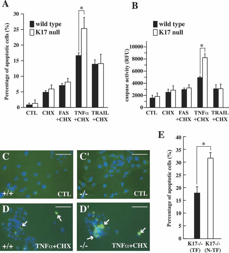
Downloaded from genesdev.cshlp.org on March 17, 2015 - Published by Cold Spring Harbor Laboratory Press
Tong and Coulombe
TNF␣-null skin (Fig. 4H,I), providing further evidence activity can be used as an indicator of TNF␣-dependent
that NF-B activity is a reliable indicator of TNF␣/ signaling in mouse postnatal skin. To assess NF-B ac-
TNFR1 activity in mouse postnatal skin. These findings tivity at the time of phenotype onset in K17-null skin,
establish that loss of TNF␣ causes a modest but consis- we examined the expression of several of its target genes.
tent delay in catagen entry in mouse backskin (by ∼1.5 d, By Northern analysis, the IB␣ mRNA level is increased
based on histology), thus revealing a previously un- by 40% at P5 and 80% at P7 in K17-null tissue relative to
known role for this cytokine in hair cycling. Since the controls (Fig. 5A). The c-FLIP mRNA level is increased
ablation of FGF5, so far defined as the most significant by 20% and 70% at P5 and P7, respectively, in K17-null
signal in this context, delays catagen entry by only ∼2–3 tissue (Fig. 5B). Greater increases, that is, 120% at P5 and
d (Hebert et al. 1994), the finding that loss of TNF␣ does 135% at P7, occur for yet another well-known NF-B
not abrogate this key step altogether could be antici- target gene, ICAM-1 (Fig. 5C; Kondo and Sauder 1997;
pated. Min et al. 2005). In situ hybridization studies, conducted
at P7, showed that IB␣ (Fig. 5D⬘), c-FLIP (Fig. 5E⬘), and
ICAM-1 mRNAs (Fig. 5F⬘) are markedly elevated in the
Alteration in NF-B activity in K17-null skin matrix and ORS in a subset of hair follicles in K17-null
skin, thereby enhancing the significance of the Northern
Although subject to a complex regulation in response to
findings. Only weak hybridization for these mRNAs was
engagement of receptors other than TNFR1 (Moynagh
detected in control skin tissue (Fig. 5D–F). Next, we per-
2005), the findings reported above suggest that NF-B
formed subcellular fractionation of backskin tissue
coupled with Western analysis to assess translocation of
p65 (relA) to its site of action, the nucleus (Scott et al.
1993). Coinciding with the increased expression of NF-
B target genes starting at P5, there is a significant in-
crease in nuclear relA (p65) along with a decrease in cy-
toplasmic levels of IB␣ protein in K17-null skin relative
to the control (Fig. 5G). RelA (p65) could not be localized
in mouse skin owing to a lack of antibodies suitable for
immunostaining of tissue sections (our unpublished
data).
Figure 4. Characterization of TNF␣ signaling in wild-type
mouse skin. (A–C⬘) In situ hybridization for K17 (A,A⬘), TNFR1
(B,B⬘), and TRADD (C,C⬘) mRNAs in hair follicles of P5–P7
wild-type mice. Four percent paraformaldehyde-fixed frozen
sections were hybridized with antisense (A,B,C) or sense probes
as controls (A⬘; data not shown) for the designated transcripts.
The boxed areas in B and C are shown at higher magnification
in B⬘ and C⬘, respectively. Asterisks denote melanin pigment.
Arrows point to hybridization signals. (Bulb) Hair bulb; (epi)
epidermis; (mat) matrix; (ORS) outer root sheath. Bars, 20 µm.
(D,D⬘) Northern blot analysis of total RNA (20 µg) prepared
from backskin of wild-type mice at different ages, and probed for
IB␣ and -tubulin (loading control) mRNAs. (D⬘) Normalized
signal intensity for IB␣ mRNA, based on average values of two
independent experiments. The P5 value, used as a reference, has
been normalized to 1. Peak levels of IB␣ mRNA (3.86 ± 0.02)
occur at P20. (E) Western blot analysis of K17 and K16 levels in
total protein extracts (2.5 µg) prepared from backskin of wild-
type mice at different ages. The tubulin blot serves as a loading
control. (F,F⬘) Histology of TNF␣+/− (F) and TNF␣−/− (F⬘) skin at
P16. Shown are hematoxylin/eosin-stained staining longitudi-
nal sections from backskin of age- and gender-matched mice.
Arrows point to hair bulbs. Bars, 50 µm. (G–G⬙) TUNEL (green)
and Hoechst (blue) stainings of TNF␣+/− (G) and TNF␣−/− (G⬘,G⬙)
skin tissue at P16. Bars, 50 µm. (H,I) Northern blot analysis of
total RNA (20 µg) prepared from backskin of TNF␣+/− or
TNF␣−/− at P14 and P16, and probed for FGF5 and IB␣ (in H),
PTHRP and c-FLIP (in I) mRNAs. Signal was quantitated by
PhosphorImager and normalized to -tubulin. Average values
from two independent experiments are reported below each
lane.
1358 GENES & DEVELOPMENT
Downloaded from genesdev.cshlp.org on March 17, 2015 - Published by Cold Spring Harbor Laboratory Press
A role for keratins during hair follicle cycling
Figure 5. Increased NF-B activity in K17-null skin.
(A–C) Northern blot analysis of total RNA (20 µg) pre-
pared from backskin of K17+/+ and K17−/− mice at P5 and
P7, and probed for IB␣ (A), c-FLIP (B), and ICAM-1 (C)
mRNAs. Signal was quantitated by PhosphorImager and
normalized to -tubulin. The average values of two or
three independent experiments are reported below each
lane. (D–F⬘) In situ hybridization analysis for IB␣
(D,D⬘), c-FLIP (E,E⬘), and ICAM-1 (F,F⬘) mRNAs in back-
skin tissue from K17+/+ and K17−/− mice at P7. Sections
were hybridized with antisense (D–F⬘) or sense probes (as
control; data not shown) for the designated transcripts.
Asterisks denote melanin pigment. Arrows point to hy-
bridization signals. Bars, 50 µm. (G) Western blot analy-
sis of cytosol and nuclear fraction prepared from back-
skin of K17+/+ and K17−/− mice at P5 using anti-p65,
anti-IB␣, anti-lamin B, or anti-tubulin antibody. Lamin
B and tubulin serve as controls for both loading and frac-
tionation. Note that there is more relA (p65) in the
nuclear fraction and less IB␣ protein in the cytosol frac-
tion of K17−/− than K17+/+ control. (H) Western blot
analysis of total proteins (30 µg) extracted from backskin
of K17+/+ and K17−/− mice at P5 and P7 using an anti-c-
FLIP antibody. Actin serves as a loading control.
As part of a negative feedback loop, c-FLIP protein 1). A significant fraction of K17−/− TNF␣−/− mice (31%)
counteracts the proapoptotic role of the TRADD-, RIP1-, showed no apoptosis (Fig. 6C–C⬘) in their pelage follicles,
and TRAF2-containing complex following TNFR1 acti- correlating with well-developed, bell-shaped hair bulbs
vation (Micheau and Tschopp 2003; Wachter et al. 2004). characteristic of anagen-stage follicles (Fig. 6B–B⬘). Simi-
In contrast to its mRNA, intact c-FLIP protein is mark- larly, ∼34% of the K17−/− TNF␣−/− mouse samples ana-
edly decreased in K17-null, compared with wild type, at lyzed by in situ hybridization showed low levels of FGF5
P5 and P7 (Fig. 5H; Gilbert et al. 2004). Other than apo- mRNA, as is the case in wild-type mice (Fig. 6D–D⬘;
ptosis and NF-B activation, TNF␣ also cause phos- Table 1). In both instances, the difference between
phorylation and activation of JNK (Zhang et al. 2004). K17−/− TNF␣−/− and K17−/− TNF␣+/+ mice reached a high
We find, however, that the levels of activated JNK do not degree of statistical significance (Table 1), establishing
differ between genotypes at P5 or P7 in total skin protein the occurrence of a genetic interaction. Despite rescue of
extracts (data not shown). the hair cycle defect, only two K17−/− TNF␣−/− mice
showed a normal hair coat (e.g., Fig. 6A; Table 1). Con-
sistent with the latter, all K17−/− TNF␣−/− mice had frag-
Loss of TNF␣ can partially rescue the apoptotic ile hair shafts (Table 1). Apoptosis and hair fragility thus
phenotype of K17-null hair follicles represent independent phenomena in K17-null skin.
The model suggested by our findings predicts that the
hair cycling defect in K17-null mice should be normal-
Discussion
ized, to some degree, by loss of TNF␣. To put this notion
to a stringent test, we generated double-null mice The studies reported here identify two novel regulators
(K17−/− TNF␣−/−) in the C57Bl/6 strain background. Sev- of hair cycling, the cytoskeletal protein K17 and the cy-
eral parameters were assessed, between P7 and P14, in tokine TNF␣. Expression of K17 protein, or a functional
the 29 K17−/− TNF␣−/− mice produced for analysis (Table equivalent (e.g., K16) (McGowan et al. 2002), is required
Table 1. Characterization of K17/TNF␣ double-knockout mice (P7 and P14)
Genotype Number of mice Hair follicle apoptosis Induction of FGF5 at P7 Alopecia Hair fragility
Wild-type 15 0/15 0/15 0/15 0/15
K17−/− 30 30/30 15/15 30/30 30/30
K17−/− TNF␣−/− 29 20/29a 9/14b 27/29 29/29
a
P < 0.005 according to Fisher’s exact test.
b
P < 0.01 according to Fisher’s exact test.
GENES & DEVELOPMENT 1359Downloaded from genesdev.cshlp.org on March 17, 2015 - Published by Cold Spring Harbor Laboratory Press Tong and Coulombe Figure 6. Genetic interaction between K17 and TNF␣ pathway. (A, left) Rescue of hair loss in K17−/− TNF␣−/− mice. A K17−/− littermate is shown at right for refer- ence. (B,B⬘) Hematoxylin/eosin-stained skin tissue sec- tions from K17−/− TNF␣+/+ (B) and K17−/− TNF␣−/− (B⬘) mice at P14. Arrows point to hair bulbs. Bars, 50 µm. (C,C⬘) TUNEL (green) and Hoechst (blue) dual stainings in backskin sections prepared from K17−/− TNF␣+/+ (C) and K17−/− TNF␣−/− (C⬘) mice at P14. Arrows point to TUNEL positive staining. Bars, 50 µm. (D,D⬘) In situ hybridization for FGF5 mRNA in K17−/− TNF␣+/+ (D) and K17−/− TNF␣−/− (D⬘) backskin at P7. Four percent paraformaldehyde-fixed frozen sections were hybrid- ized with antisense (D,D⬘) or sense (not shown) probes. Bars, 50 µm. (Bulb) Hair bulb; (epi) epidermis; (mat) ma- trix; (ORS) outer root sheath. Asterisks denote melanin pigments in hair bulb. for the persistence of the anagen (growth) state in hair in culture requires reduced NF-B activity (see Qin et al. follicles. TNF␣, on the other hand, is required for the 2001; Basile et al. 2003; Konur et al. 2005), how the latter timely occurrence of the anagen–catagen transition. We impacts the proliferation, differentiation, and/or apopto- provide genetic evidence that K17 modulates the func- sis of this cell type is clearly a function of the global tion of TNF␣ in the specific context of hair cycling, and signaling environment (e.g., Poppelmann et al. 2005). biochemical evidence (albeit indirect) that TNF␣ signal- K17’s ability to interact with TRADD provides an at- ing is enhanced in K17-null mouse skin tissue. That a tractive, yet unproven mechanism to account for the ge- keratin structural protein contributes to hair cycle regu- netic interaction between K17 and TNF␣ in hair follicles. lation, and does so independently of its structural sup- First, there is a precedent for modulation of TNF␣ sig- port role in epithelia, was unexpected and represents an naling in a keratin/TRADD-dependent fashion. Inada et exciting new direction for skin biology. al. (2001) showed a reverse correlation between the lev- Unlike the case for keratins, a role in hair morphogen- els of K18, which binds TRADD directly, and suscepti- esis, and possibly in hair cycling as well, has already bility of epithelial cell lines to TNF␣-induced apoptosis. been defined for a TNF superfamily member, ectodyspla- These authors proposed a cell-autonomous mechanism, sin (Eda), acting through its receptor Edar (Thesleff and keratin-dependent sequestration of TRADD, to account Mikkola 2002). Although they affect hair growth in op- for this effect. Jaquemar et al. (2003) showed that loss of posing ways, the catagen inducer TNF␣ (this study; see maternally derived TNF␣ significantly increases the vi- Ruckert et al. 2000) and anagen promoter ectodysplasin ability of K8-null embryos (such embryos lack K8’s part- (Mustonen et al. 2003) share NF-B as a major intracel- ner protein K18 as well), although the mechanism in- lular target. In this study, NF-B was used as a readout volved may be distinct. Second, the conserved property for TNF␣/TNFR1-dependent signaling at the anagen– of interacting with TRADD could account for the ability catagen transition in wild-type, K17-null, and TNF␣-null of K16 (and possibly K14) to prevent, in an age- and skin. The data derived from TNF␣-null skin at P14/P16, strain-specific fashion, the hair cycling defect in K17- in particular, indicate that NF-B activity reliably re- null hair follicles (McGowan et al. 2002). Third, TRADD ports on the loss of this cytokine. Our studies do not participates to signal relay from receptors other than directly address the issue of NF-B’s role in this setting. TNFR1, such that loss of the K17–TRADD interaction Zhang et al. (2004) elegantly described how the RelA could also account for the fraction of the hair cycling (p65) subunit of NF-B antagonizes a strong TNFR1-de- defect that cannot be explained solely by TNF␣ signaling pendent proliferation signal during normal homeostasis in K17-null mice (Table 1). As an example, IFN-␥ signals in human epidermis reconstituted by grafting in mouse in a STAT1␣- and TRADD-dependent fashion (Wese- skin. These findings (see also Gugasyan et al. 2004) ce- mann et al. 2004) and is also required for the timely mented the notion that NF-B’s antiproliferative influ- occurrence of the anagen–catagen transition in mouse ence in mature epidermis is unique compared with many hair follicles (Hirota et al. 2002, 2003). TNF␣, IFN-␥, and other tissues (Bolotin and Fuchs 2003) including devel- IL-1 cause significantly more apoptosis in mouse pelage oping hair follicles (Schmidt-Ullrich et al. 2001). Given a hair follicles when administered intradermally together, reduced mitotic activity of hair matrix epithelial cells in rather than individually (Ruckert et al. 2000). In an ap- K17-null skin at P5, activation of NF-B could act to parent paradox, TRADD’s adaptor function is essential slow down their proliferation at the end of anagen, as is to both the proapoptotic (DISC complex-dependent) and the case in human epidermis (Zhang et al. 2004), while prosurvival (NF-kB-dependent) arms of TNF␣ signal re- another signal would be required for commitment to ap- lay (Micheau and Tschopp 2003), raising the concern optosis and catagen entry. While there is evidence that that a simple TRADD sequestration model cannot me- TNFR1-mediated apoptosis of epidermal keratinocytes diate K17’s influence on TNF␣ signaling. This said, the 1360 GENES & DEVELOPMENT
Downloaded from genesdev.cshlp.org on March 17, 2015 - Published by Cold Spring Harbor Laboratory Press
A role for keratins during hair follicle cycling
level of intact c-FLIP protein is markedly reduced in K17- Primary antibodies
null skin (in contrast to its mRNA), suggesting that the The following primary antibodies were used: rabbit polyclonal
NF-B-dependent arm of the TNF␣ signal relay is subject antisera against K17 (McGowan and Coulombe 1998), K16 (Ber-
to additional regulation. Clearly, development of suit- not et al. 2002), K14 (Covance), c-FLIP (Upstate Biotechnology),
able reagents and cellular models is needed to confirm TRADD (Santa Cruz Biotechnology), TRADD (provided by Dr.
the functional role of the keratin–TRADD interaction in Masaki Inagaki, Aichi Cancer Research Institute, Nagoya, Ja-
TNF␣-treated keratinocytes, and explore alternative pan) (Inada et al. 2001), IB␣ (Santa Cruz Biotechnology), p65
mechanisms. (Santa Cruz Biotechnology), and lamin B (provided by Dr. Kathy
A genetic approach can help toward further dissection Wilson, Johns Hopkins University, Baltimore, MD) (Zastrow et
al. 2006), and mouse monoclonal antibodies to K10, BrdU, ␣-tu-
of the hair cycling defect in K17-null mice. We have
bulin, and actin (Sigma).
obtained evidence that one or more loci located proximal
to the type I keratin gene cluster also modulate the im- Tissue harvesting, histological analyses, and BrdU labeling
pact of K17 loss. All the findings we report here involved
progeny from the sixth and seventh backcrosses (N6, N7) For studies of hair cycling, skin tissue was always obtained from
the mid-back region of age- and gender-matched mouse litter-
of K17-null allele carriers into the C57Bl/6 strain. The
mates. For histology, tissues were fixed in Bouin’s (12 h, 4°C)
complete penetrance of the pelage hair phenotype ob-
prior to paraffin embedding. Five-micrometer sections were
served under such conditions is eroded, however, with counterstained with hematoxylin and eosin. Alternatively,
further backcrossing. When N12 K17+/− mice are inter- fresh-frozen tissues were embedded in OCT (Sakura Finetec)
bred, only ∼25% of the K17−/− progeny show a hair loss and 10-µm sections were immunostained using the primary an-
phenotype, implying a powerful modulatory role for tibodies described above, followed by FITC- or Rhodamine-con-
modifier gene(s) located proximal to the K17 locus. The jugated secondary antibodies (Kirkegaard and Perry Laborato-
average amount of genomic DNA flanking an allele of ries) (Bernot et al. 2004). TUNEL (Roche) and Hoechst (Sigma)
interest changes from ∼30 cM to ∼20 cM from N6 to N12 stainings were performed on 4% paraformaldehyde-fixed, paraf-
(Peirce 2001; Olofsson and Holmdahl 2003). Intriguingly, fin-embedded samples following the manufacturers’ instruc-
tions (McGowan et al. 2002). For BrdU labeling, five P5 mice of
there is a cluster of cytokine-encoding genes located
each genotype were intraperitoneally injected with BrdU (20
∼29–30 cM away from the K17 locus (11D; 11 58.7 cM) mg/kg body weight; Sigma) and sacrificed after 45 min. Ten-
on mouse chromosome 11 (Wilson et al. 1990; Wenderfer micrometer-thick cryosections were fixed for 10 min in 10%
et al. 2000). These include genes for IL-3, IL-4, IL-5, IL- formalin, PBS-washed, and stained with anti-BrdU (Sigma) as
13, interferon regulatory factor 1, granulocyte-macro- described (Paladini and Coulombe 1998). For each genotype,
phage colony-stimulating factor, and a few others. In BrdU-labeled cells were counted in 50 hair bulbs (sectioned
light of the role of TNF␣ (this study) and IFN-␥ (Hirota et through dermal papillae) from 10 sections. The hair bulb area
al. 2002, 2003) in regulating catagen entry in hair fol- was measured with MacBAS version 2.5 software (Fuji Medical
licles, it makes sense that additional proinflammatory Systems USA). Data are presented as the average numbers of
genes might impact the penetrance of the K17-null mu- BrdU-positive cells per surface area of bulb tissue.
tation. Identifying these genes, the cellular source of
In situ hybridization and Northern blot analysis
their protein products, and the mechanisms through
which they act, should provide novel insight into hair Detailed protocols for in situ hybridization and Northern blot
cycling. analyses were described (McGowan et al. 2002; Tong and Cou-
lombe 2004). The probes used in this study included FGF5,
FGFR2 (both provided by Dr. G. Martin, University of Califor-
nia at San Francisco, CA), -tubulin, PTHRP (provided by Dr. C.
Materials and methods Thompson, Johns Hopkins University, Baltimore, MD), c-FLIP
and ICAM-1 (ordered from ATCC), IB␣ (provided by Dr. L. Lin,
Johns Hopkins University, Baltimore, MD), TRADD (generated
Animal models by RT–PCR; forward primer, 5⬘-CTGCGGTAGACAAGGTGA
The K17-null mice (McGowan et al. 2002) used in this study TCC-3⬘; reverse primer, 5⬘-CAGTACTAGACTTAGGCCAG
were backcrossed into the C57Bl/6 mouse strain for six or seven GC-3⬘), TNFR1 (generated by RT–PCR; forward primer, 5⬘-GCC
generations (N6, N7). TNF␣-null mice, backcrossed in the CCCACCTCTGTTCAGAAATG-3⬘; reverse primer, 5⬘-GTTG
C57Bl/6 strain, were a gift from Dr. M. Meffert (Johns Hopkins TGGGTGTGGCTTTATCGC-3⬘). Northern blots were imaged
University, Baltimore, MD). K17-TNF␣ double-null mice were and quantitated using a Fujifilm BAS-2500 PhosphorImager.
created by mating the two single-knockout strains. Genotyping Values were normalized to signal for -tubulin. Each Northern
was done by PCR from mouse tail genomic DNA as described analysis was conducted at least two times, with similar findings.
(McGowan et al. 2002). Genotyping at the TNF␣ locus was done
Keratinocyte cultures and their analysis
by PCR, as follows: denaturation at 94°C for 1 min; annealing at
62°C for 1 min; elongation: 72°C for 1 min, using the following Primary keratinocytes were collected from P2/P3 mouse litter-
primer pairs: null TNF␣ allele (forward, 5⬘-AGCCAACCAG mates (wild type or K17 null), plated, and cultured as described
GCAGGTTCTG-3⬘; and reverse, 5⬘-CCTTCTATCGCCTTCT (Bernot et al. 2004). Transient transfections were carried out
TGACGAG-3⬘) and wild-type allele (forward, 5⬘-AGCCAACC using Lipofectamine 2000 (Invitrogen) using the manufacturer’s
AGGCAGGTTCTG-3⬘; and reverse, 5⬘-TAGACAGAAGAGCG instructions. After 48 h, cultures were fixed with 4% paraform-
TGGTGG-3⬘). Mice were provided rodent chow and water ad aldehyde, permeabilized with methanol, and processed for indi-
libitum. All protocols were approved by the institutional Ani- rect immunofluorescence labeling (Bernot et al. 2004). Studies
mal Care and Use Committee. involving proapoptotic treatments were conducted at 48 h after
GENES & DEVELOPMENT 1361Downloaded from genesdev.cshlp.org on March 17, 2015 - Published by Cold Spring Harbor Laboratory Press
Tong and Coulombe
plating. Cultures were treated for 24 h with DMSO vehicle con- Mollie Meffert for providing TNF␣-null mice, reagents, and ad-
trol (CTL), 5 µg/mL CHX, as well as 50 µg/mL Jo2 (the agonistic vice; Drs. Gail Martin, Masaki Inagaki, Li Lin, and Andrew
antibody to FAS ligand) (Gilbert et al. 2001), 100 ng/mL TNF␣, Thorburn for providing reagents; and Drs. Joel Pomerantz, Mi-
5 µg/mL TRAIL in the presence of 5 µg/mL CHX. TUNEL chael Caterina, Roger Reeves, Susan Craig, Catherine Thomp-
(Roche) and Hoechst (Sigma) stainings were performed on 4% son, and Katherine Wilson for reagents and/or advice. This work
paraformaldehyde-fixed, 0.1% Triton X-100-permeablized cul- was supported by NIAMS/NIH grant AR44232 to P.A.C.
tured cells, to identify apoptotic cells. Alternatively, caspase
activity (activated caspases 2, 3, 6, 7, 8, 9, and 10) was quantified
by fluorimetry as follows: At 24 h after induction of apoptosis, References
caspase substrate (Roche) diluted in lysis buffer was added to
each well and incubated for 2 h at 37°C. Substrate cleavage was Basile, J.R., Eichten, A., Zacny, V., and Munger, K. 2003. NF-
measured using a PerkinElmer HTS 7000 Plus spectrofluorom- B-mediated induction of p21(Cip1/Waf1) by tumor necrosis
eter, with excitation and emission wavelengths set at 485 and factor ␣ induces growth arrest and cytoprotection in normal
535 nm, respectively, as recommended (Roche). A calibration human keratinocytes. Mol. Cancer Res. 1: 262–270.
curve obtained using free Rhodamine-110 fluorochrome was Beaudoin III, G.M., Sisk, J.M., Coulombe, P.A., and Thompson,
used for calculating values for each sample. In separate studies, C.C. 2005. Hairless triggers reactivation of hair growth by
the frequency of apoptotic cells after TNF␣ treatment was de- promoting Wnt signaling. Proc. Natl. Acad. Sci. 102: 14653–
termined in primary cultures of K17-null keratinocytes trans- 14658.
fected with wild-type K17. Transient transfection was done at Bender, L.M., Morgan, M.J., Thomas, L.R., Liu, Z.G., and Thor-
12 h after plating using lipofectamine 2000 according to the burn, A. 2005. The adaptor protein TRADD activates dis-
manufacturer’s instructions (Invitrogen). Proapoptotic treat- tinct mechanisms of apoptosis from the nucleus and the
ment (100 ng/mL TNF␣ + 5 µg/mL CHX) was performed at 48 h cytoplasm. Cell Death Differ. 12: 473–481.
after plating. At 24 h later, cells were subjected to immunofluo- Bernot, K., McGowan, K., and Coulombe, P.A. 2002. Keratin 16
rescence labeling for K17 followed by TUNEL staining. In sepa- expression defines a subset of epithelial cells during skin
rate transfection studies, the GFP-TRADD fusion constructs morphogenesis and the hair cycle. J. Invest. Dermatol. 119:
were provided by Dr. Andrew Thorburn (Wake Forest Univer- 1137–1149.
sity, Winston-Salem, NC) (Bender et al. 2005). Bernot, K.M., Coulombe, P.A., and Wong, P. 2004. Skin: An
ideal model system to study keratin genes and proteins.
Immunoprecipitation and Western blot analysis Methods Cell Biol. 78: 453–487.
Wild-type skin keratinocytes in primary culture were solubi- Bolotin, D. and Fuchs, E. 2003. Cancer: More than skin deep.
lized with 2% Empigen BB (Calbiochem) buffer. Protein Nature 421: 594–595.
A-Sepharose beads (Amersham Biosciences) were washed with Caulin, C., Ware, C.F., Magin, T.M., and Oshima, R.G. 2000.
PBS, conjugated to rabbit antibody against TRADD or normal Keratin-dependent, epithelial resistance to tumor necrosis
rabbit serum, then added with an equal volume of cell lysate factor-induced apoptosis. J. Cell Biol. 149: 17–22.
and incubated for 12 h at 4°C (Bernot et al. 2004). After three Cho, Y.M., Woodard, G.L., Dunbar, M., Gocken, T., Jimenez,
washes (2% Empigen BB buffer), bound protein was eluted with J.A., and Foley, J. 2003. Hair-cycle-dependent expression of
5× gel sample buffer containing -mercaptoethanol. Eluted pro- parathyroid hormone-related protein and its type I receptor:
teins were subjected to SDS-PAGE and Western blot analysis. Evidence for regulation at the anagen to catagen transition. J.
Western blot analysis of protein extracts from depilated skin or Invest. Dermatol. 120: 715–727.
clipped hair was performed as described (McGowan et al. 2002; Cotsarelis, G. and Millar, S.E. 2001. Towards a molecular un-
Bernot et al. 2004). For quantification, NIH image software was derstanding of hair loss and its treatment. Trends Mol. Med.
used as described (Paladini and Coulombe 1998). 7: 293–301.
Coulombe, P.A. and Wong, P. 2004. Cytoplasmic intermediate
Cell fractionation filaments revealed as dynamic and multipurpose scaffolds.
Nat. Cell Biol. 6: 699–706.
Nuclear and cytoplasmic extracts were prepared according to
Foitzik, K., Lindner, G., Mueller-Roever, S., Maurer, M., Botch-
Lammerding et al. (2004), with modifications. Skin samples
kareva, N., Botchkarev, V., Handjiski, B., Metz, M., Hibino,
were cut into small pieces and homogenized in ice-cold buffer A
T., Soma, T., et al. 2000. Control of murine hair follicle
(0.1% Triton X-100, 10 mM EDTA, 10 mM EGTA, 10 mM KCl,
regression (catagen) by TGF-1 in vivo. FASEB J. 14: 752–
10 mM HEPES, 1 mM DTT, 0.5 mM PMSF, protease inhibitor
760.
cocktail) on ice for 1 min. The homogenate was spun (16,000g,
Fuchs, E. 1995. Keratins and the skin. Annu. Rev. Cell Dev.
5 min, 4°C). The supernatant was stored as the cytoplasmic
Biol. 11: 123–153.
fraction. Nuclei-containing pellets were washed in ice-cold PBS
Fuchs, E., Merrill, B.J., Jamora, C., and DasGupta, R. 2001. At
and incubated in ice-cold buffer B (1 mM EDTA, 1 mM EGTA,
the roots of a never-ending cycle. Dev. Cell 1: 13–25.
0.4 M NaCl, 20 mM HEPES, 5 mM MgCl2, 25% glycerol, 1 mM
Gilbert, S., Loranger, A., Daigle, N., and Marceau, N. 2001.
DTT, 0.5 mM PMSF, protease inhibitor cocktail) at 4°C for 30
Simple epithelium keratins 8 and 18 provide resistance to
min with mixing. The nuclear extract was centrifuged (16,000g,
Fas-mediated apoptosis. The protection occurs through a re-
10 min, 4°C), and the supernatant was stored as the nuclear
ceptor-targeting modulation. J. Cell Biol. 154: 763–774.
fraction. The nuclear and cytoplasmic fractions were subjected
Gilbert, S., Loranger, A., and Marceau, N. 2004. Keratins modu-
to SDS-PAGE and Western blot analysis. Fraction purity was
late c-Flip/extracellular signal-regulated kinase 1 and 2 an-
verified by immunoblotting for tubulin (cytoplasmic) and lamin
tiapoptotic signaling in simple epithelial cells. Mol. Cell.
B (nuclear) antigens.
Biol. 24: 7072–7081.
Gugasyan, R., Voss, A., Varigos, G., Thomas, T., Grumont, R.J.,
Acknowledgments
Kaur, P., Grigoriadis, G., and Gerondakis, S. 2004. The tran-
We thank Dr. Kelsie Bernot and Ms. Eunhye Park for their con- scription factors c-rel and RelA control epidermal develop-
tribution; members of the Coulombe laboratory for support; Dr. ment and homeostasis in embryonic and adult skin via dis-
1362 GENES & DEVELOPMENTDownloaded from genesdev.cshlp.org on March 17, 2015 - Published by Cold Spring Harbor Laboratory Press
A role for keratins during hair follicle cycling
tinct mechanisms. Mol. Cell. Biol. 24: 5733–5745. Marcelo, C.L., Kim, Y.G., Kaine, J.L., and Voorhees, J.J. 1978.
Hadshiew, I.M., Foitzik, K., Arck, P.C., and Paus, R. 2004. Bur- Stratification, specialization, and proliferation of primary
den of hair loss: Stress and the underestimated psychosocial keratinocyte cultures. Evidence of a functioning in vitro epi-
impact of telogen effluvium and androgenetic alopecia. J. dermal cell system. J. Cell Biol. 79: 356–370.
Invest. Dermatol. 123: 455–457. McGowan, K.M. and Coulombe, P.A. 1998. Onset of keratin 17
Hardy, M.H. 1992. The secret life of the hair follicle. Trends expression coincides with the definition of major epithelial
Genet. 8: 55–61. lineages during skin development. J. Cell Biol. 143: 469–486.
Hebert, J.M., Rosenquist, T., Gotz, J., and Martin, G.R. 1994. McGowan, K.M., Tong, X., Colucci-Guyon, E., Langa, F., Babi-
FGF5 as a regulator of the hair growth cycle: Evidence from net, C., and Coulombe, P.A. 2002. Keratin 17 null mice ex-
targeted and spontaneous mutations. Cell 78: 1017–1025. hibit age- and strain-dependent alopecia. Genes & Dev. 16:
Hirota, R., Tajima, S., Yoneda, Y., Tamayama, T., Watanabe, 1412–1422.
M., Ueda, K., Kubota, T., and Yoshida, R. 2002. Alopecia of Micheau, O. and Tschopp, J. 2003. Induction of TNF receptor
IFN-␥ knockout mouse as a model for disturbance of the hair I-mediated apoptosis via two sequential signaling com-
cycle: A unique arrest of the hair cycle at the anagen phase plexes. Cell 114: 181–190.
accompanied by mitosis. J. Interferon Cytokine Res. 22: Millar, S.E. 2002. Molecular mechanisms regulating hair follicle
935–945. development. J. Invest. Dermatol. 118: 216–225.
Hirota, R., Tajima, S., Yoneda, Y., Okada, M., Tashiro, J., Ueda, Min, J.K., Kim, Y.M., Kim, S.W., Kwon, M.C., Kong, Y.Y.,
K., Kubota, T., and Yoshida, R. 2003. Induction of hair re- Hwang, I.K., Won, M.H., Rho, J., and Kwon, Y.G. 2005. TNF-
growth in the alopecia site of IFN-␥ knockout mice by allo- related activation-induced cytokine enhances leukocyte ad-
grafting and IFN-␥ injection into the transplantation site. J. hesiveness: Induction of ICAM-1 and VCAM-1 via TNF re-
Interferon Cytokine Res. 23: 433–439. ceptor-associated factor and protein kinase C-dependent NF-
Hoffmann, A., Levchenko, A., Scott, M.L., and Baltimore, D. B activation in endothelial cells. J. Immunol. 175: 531–540.
2002. The IB-NF-B signaling module: Temporal control Moynagh, P.N. 2005. The NF-B pathway. J. Cell Sci. 118: 4589–
and gene activation. Science 298: 1241–1245. 4592.
Inada, H., Izawa, I., Nishizawa, M., Fujita, E., Kiyono, T., Taka- Muller-Rover, S., Handjiski, B., van der Veen, C., Eichmuller, S.,
hashi, T., Momoi, T., and Inagaki, M. 2001. Keratin attenu- Foitzik, K., McKay, I.A., Stenn, K.S., and Paus, R. 2001. A
ates tumor necrosis factor-induced cytotoxicity through as- comprehensive guide for the accurate classification of mu-
sociation with TRADD. J. Cell Biol. 155: 415–426. rine hair follicles in distinct hair cycle stages. J. Invest. Der-
Jamora, C., DasGupta, R., Kocieniewski, P., and Fuchs, E. 2003. matol. 117: 3–15.
Links between signal transduction, transcription and adhe- Mustonen, T., Pispa, J., Mikkola, M.L., Pummila, M., Kangas,
sion in epithelial bud development. Nature 422: 317–322. A.T., Pakkasjarvi, L., Jaatinen, R., and Thesleff, I. 2003.
Jaquemar, D., Kupriyanov, S., Wankell, M., Avis, J., Benirschke, Stimulation of ectodermal organ development by Ectodys-
K., Baribault, H., and Oshima, R.G. 2003. Keratin 8 protec- plasin-A1. Dev. Biol. 259: 123–136.
tion of placental barrier function. J. Cell Biol. 161: 749–756. Olofsson, P. and Holmdahl, R. 2003. Positional cloning of
Kondo, S. and Sauder, D.N. 1997. Tumor necrosis factor (TNF) Ncf1—A piece in the puzzle of arthritis genetics. Scand. J.
receptor type 1 (p55) is a main mediator for TNF-␣-induced Immunol. 58: 155–164.
skin inflammation. Eur. J. Immunol. 27: 1713–1718. Omary, M.B., Coulombe, P.A., and McLean, W.H.I. 2004. Inter-
Konur, A., Schulz, U., Eissner, G., Andreesen, R., and Holler, E. mediate filament proteins and their associated diseases. N.
2005. Interferon (IFN)-␥ is a main mediator of keratinocyte Engl. J. Med. 351: 2087–2100.
(HaCaT) apoptosis and contributes to autocrine IFN-␥ and Oshima, R.G. 2002. Apoptosis and keratin intermediate fila-
tumour necrosis factor-␣ production. Br. J. Dermatol. 152: ments. Cell Death Differ. 9: 486–492.
1134–1142. Paladini, R.D. and Coulombe, P.A. 1998. Directed expression of
Kreuz, S., Siegmund, D., Scheurich, P., and Wajant, H. 2001. keratin 16 to the progenitor basal cells of transgenic mouse
NF-B inducers upregulate cFLIP, a cycloheximide-sensitive skin delays skin maturation. J. Cell Biol. 142: 1035–1051.
inhibitor of death receptor signaling. Mol. Cell. Biol. 21: Panteleyev, A.A., Paus, R., Wanner, R., Nurnberg, W., Eich-
3964–3973. muller, S., Thiel, R., Zhang, J., Henz, B.M., and Rosenbach,
Ku, N.O., Soetikno, R.M., and Omary, M.B. 2003. Keratin mu- T. 1997. Keratin 17 gene expression during the murine hair
tation in transgenic mice predisposes to Fas but not TNF- cycle. J. Invest. Dermatol. 108: 324–329.
induced apoptosis and massive liver injury. Hepatology 37: Pasparakis, M., Alexopoulou, L., Episkopou, V., and Kollias, G.
1006–1014. 1996. Immune and inflammatory responses in TNF ␣-defi-
Lammerding, J., Schulze, P.C., Takahashi, T., Kozlov, S., Sulli- cient mice: A critical requirement for TNF ␣ in the forma-
van, T., Kamm, R.D., Stewart, C.L., and Lee, R.T. 2004. La- tion of primary B cell follicles, follicular dendritic cell net-
min A/C deficiency causes defective nuclear mechanics and works and germinal centers, and in the maturation of the
mechanotransduction. J. Clin. Invest. 113: 370–378. humoral immune response. J. Exp. Med. 184: 1397–1411.
Langbein, L., Rogers, M.A., Praetzel, S., Winter, H., and Schwei- Paus, R. and Cotsarelis, G. 1999. The biology of hair follicles.
zer, J. 2003. K6irs1, K6irs2, K6irs3, and K6irs4 represent the N. Engl. J. Med. 341: 491–497.
inner-root-sheath-specific type II epithelial keratins of the Paus, R. and Foitzik, K. 2004. In search of the ‘hair cycle clock’:
human hair follicle. J. Invest. Dermatol. 120: 512–522. A guided tour. Differentiation 72: 489–511.
Lavker, R.M., Miller, S., Wilson, C., Cotsarelis, G., Wei, Z.G., Peirce, J. 2001. Looking at old tools in new ways: Using knock-
Yang, J.S., and Sun, T.T. 1993. Hair follicle stem cells: Their outs as congenics to study QTLs. Genome Res. 11: 1469–
location, role in hair cycle, and involvement in skin tumor 1471.
formation. J. Invest. Dermatol. 101 (1 Suppl): 16S–26S. Poppelmann, B., Klimmek, K., Strozyk, E., Voss, R., Schwarz,
Lin, K.K., Chudova, D., Hatfield, G.W., Smyth, P., and T., and Kulms, D. 2005. NFB-dependent down-regulation of
Andersen, B. 2004. Identification of hair cycle-associated tumor necrosis factor receptor-associated proteins contrib-
genes from time-course gene expression profile data by using utes to interleukin-1-mediated enhancement of ultraviolet
replicate variance. Proc. Natl. Acad. Sci. 101: 15955–15960. B-induced apoptosis. J. Biol. Chem. 280: 15635–15643.
GENES & DEVELOPMENT 1363You can also read