HIGH MICROBIOTA REACTIVITY OF ADULT HUMAN INTESTINAL IGA REQUIRES SOMATIC MUTATIONS
←
→
Page content transcription
If your browser does not render page correctly, please read the page content below
ARTICLE
High microbiota reactivity of adult human intestinal
IgA requires somatic mutations
Johanna Kabbert1, Julia Benckert2,5, Tim Rollenske3, Thomas C.A. Hitch4, Thomas Clavel4, Vuk Cerovic1, Hedda Wardemann3, and Oliver Pabst1
The gut is home to the body’s largest population of plasma cells. In healthy individuals, IgA is the dominating isotype, whereas
patients with inflammatory bowel disease also produce high concentrations of IgG. In the gut lumen, secretory IgA binds
pathogens and toxins but also the microbiota. However, the antigen specificity of IgA and IgG for the microbiota and
underlying mechanisms of antibody binding to bacteria are largely unknown. Here we show that microbiota binding is a
Downloaded from http://rupress.org/jem/article-pdf/217/11/e20200275/1047083/jem_20200275.pdf by guest on 18 August 2020
defining property of human intestinal antibodies in both healthy and inflamed gut. Some bacterial taxa were commonly
targeted by different monoclonal antibodies, whereas others selectively bound single antibodies. Interestingly, individual
human monoclonal antibodies from both healthy and inflamed intestines bound phylogenetically unrelated bacterial species.
This microbiota cross-species reactivity did not correlate with antibody polyreactivity but was crucially dependent on the
accumulation of somatic mutations. Therefore, our data suggest that a system of affinity-matured, microbiota cross-
species–reactive IgA is a common aspect of SIgA–microbiota interactions in the gut.
Introduction
IgA-secreting plasma cells constitute a major leukocyte popula- varies along the intestinal tract, ranging from ∼60% of total
tion in the human gut. IgM-secreting plasma cells are also pre- bacteria in the proximal small intestine to ∼10% in feces (Bunker
sent (Magri et al., 2017) and, in patients with inflammatory et al., 2015; van der Waaij et al., 1996). Notably, many taxa coated
bowel disease (IBD), high concentrations of IgG are detectable in by SIgA in the colon are also coated in the small intestine, sug-
the intestinal lumen (Macpherson et al., 1996). However, the gesting that microbiota-reactive IgA is primarily induced in the
specificity of intestinal antibodies for luminal antigen, including proximal gut segments (Bunker et al., 2015).
components of the gut microbiota, are largely unknown (Pabst Recently, the functional properties of intestinal bacteria
and Slack, 2020). coated by endogenous polyclonal IgA have been reported. Sort
Polymeric IgA and IgM are actively transported across the purification of SIgA-coated bacteria followed by 16S ribosomal
epithelial cell layer into the gut lumen by the polymeric Ig re- RNA (rRNA) gene amplicon sequencing revealed enrichment of
ceptor. During this process, a fragment of the polymeric Ig re- distinct bacterial taxa in IgA-coated fractions (Bunker et al.,
ceptor becomes covalently bound to the antibody to generate a 2015; Kau et al., 2015; Palm et al., 2014). Mice colonized with
hybrid molecule referred to as secretory Ig (SIg; Brandtzaeg, SIgA-coated gut bacteria showed increased susceptibility to co-
2009; Stadtmueller et al., 2016). In the human gut lumen, dif- litis (Palm et al., 2014) and enhanced diet-dependent enteropa-
ferent antibody isotypes show differential binding to members thy (Kau et al., 2015) compared with animals colonized with
of the microbiota, resulting in selective coating by SIgA, SIgM, noncoated bacteria. These observations show that SIgA coating
and IgG (Fadlallah et al., 2019; Magri et al., 2017; Palm et al., is linked to distinct functional properties of gut bacteria.
2014). Ig coating of a given bacterial species can result in dif- However, we are largely ignorant when it comes to de-
ferent outcomes ranging from extinction/loss of the Ig-coated scribing defined antigens bound by IgA or the precise mecha-
bacteria (typically observed only for pathogens), to no detectable nisms of how IgA binds to members of the microbiota. SIgA can
effects on colonization levels, or even an increase in colonization bind to microorganisms by canonical Fab-dependent binding as
(Donaldson et al., 2018; Moor et al., 2017; Peterson et al., 2015). well as noncanonical interactions between antibody and mi-
The fraction of SIgA-coated members of the gut microbiota crobiota (Pabst and Slack, 2020). Noncanonical interactions are
.............................................................................................................................................................................
1Instituteof Molecular Medicine, Rheinisch-Westfälische Technische Hochschule Aachen University, Aachen, Germany; 2Max Planck Research Group Molecular
Immunology, Max Planck Institute for Infection Biology, Berlin, Germany; 3B Cell Immunology, German Cancer Research Centre, Heidelberg, Germany; 4Functional
Microbiome Research Group, Institute of Medical Microbiology, Rheinisch-Westfälische Technische Hochschule Aachen University, Aachen, Germany;
5Charité–Universitätsmedizin Berlin, corporate member of Freie Universität Berlin, Humboldt-Universität zu Berlin, and Berlin Institute of Health, Berlin, Germany.
Correspondence to Oliver Pabst: opabst@ukaachen.de.
© 2020 Kabbert et al. This article is available under a Creative Commons License (Attribution 4.0 International, as described at https://creativecommons.org/licenses/by/
4.0/).
Rockefeller University Press https://doi.org/10.1084/jem.20200275 1 of 14
J. Exp. Med. 2020 Vol. 217 No. 11 e20200275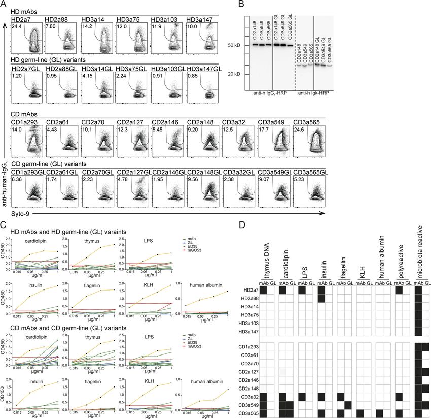
unaffected by somatic mutations in the Fab region of the anti-        endogenous SIgA are not captured in this approach. In total, we
body and rely on glycan moieties associated with the Fc part          generated and screened 280 human mAbs for their microbiota
of the antibody and the secretory component (Mathias and              reactivity (IgA, 105 from HD and 84 from CD donors; IgG, 57
Corthesy, 2011; Pabst and Slack, 2020). The relevance of such         from HD and 34 from CD donors).
noncanonical binding to members of the microbiota is not well            To avoid potential competition for epitope binding between
defined.                                                              these recombinant antibodies and endogenous IgA, fecal samples
    To investigate the specificity of IgA for the microbiota, a       from Rag2-deficient mice, which lack B cells and therefore sol-
recent study in mice showed that single mAbs bound to diverse         uble Ig, were used. Microbial cells were identified by flow cy-
but defined subsets of microbial populations (Bunker et al.,          tometry based on their scatter profiles and staining intensity
2017). This binding profile was associated with antibody poly-        with the DNA-binding dye Syto9. Material isolated from feces of
reactivity and was independent of somatic mutations (Bunker           germ-free mice and in vitro–cultured Escherichia coli was used to
et al., 2017). Yet, in the human gut, germline (GL)-encoded IgA is    establish precise gating of bacteria (Fig. 1 A).
virtually absent, and IgA-secreting plasma cells are character-          Bacterial staining with the control antibody (mG053;
ized by high numbers of somatic mutations (Barone et al., 2011;       Wardemann et al., 2003) and antibody-free cell culture super-
Benckert et al., 2011; Lindner et al., 2015). Indeed, human mAbs      natant showed variable background staining (0.52 ± 0.31, mean

                                                                                                                                                   Downloaded from http://rupress.org/jem/article-pdf/217/11/e20200275/1047083/jem_20200275.pdf by guest on 18 August 2020
directed against LPS O-antigens of Klebsiella pneumoniae recog-       ± SD, n = 36). Thus, to exclude the possibility of false positives,
nize intestinal microbes, and this binding required somatic           we considered binding of ≥1% total microbial cells as the
mutations (Rollenske et al., 2018). We suggest referring to this      threshold of antibody reactivity. This definition of antibody-
phenomenon of single mAbs binding to different bacterial spe-         positive populations does not exclude the possibility of true
cies as cross-species reactivity (Pabst and Slack, 2020). Mech-       binding of antibodies with lower binding capacity to bacteria.
anistically, cross-species reactivity is not fully understood.           Both IgA and IgG mAbs showed a wide spectrum of micro-
    Here, we characterized a panel of IgG and IgA mAbs derived        biota reactivity, including antibodies with no microbiota binding
from human small intestine of both healthy donors (HDs) and           and antibodies binding >5% of all fecal bacteria (Fig. 1, B and C).
Crohn’s disease (CD) patients. This approach allowed us to            We therefore categorized antibodies to have no/low (0–1% of
contrast the microbiota-binding capacity of IgA and IgG, but also     bacteria), intermediate (1–5%), and high (>5%) binding capacity
to compare antibody specificity for members of the gut micro-         for gut bacteria. Antibodies with high binding capacity for gut
biota under healthy and inflamed conditions. Our data demon-          bacteria were present among mAbs of both IgA and IgG origin
strate that microbiota binding is a frequent property of IgA and      (Fig. 1 B). Notably, high microbiota-binding capacity was ob-
IgG under both healthy and inflamed conditions. In most cases,        served in both collections of mAbs but clearly more pronounced
the binding capacity and spectrum of human IgA was crucially          in antibodies derived from CD patients compared with HD-
dependent on somatic mutations and did not correlate with             derived antibodies (Fig. 1 C). We therefore conclude that
polyreactivity. We therefore suggest that, in adult humans, a         microbiota-binding capacity is a characterizing property of a
system of affinity matured, microbiota cross-species–reactive         substantial proportion of human intestinal IgA and IgG anti-
IgA is a common aspect of SIgA–microbiota interactions in             bodies in both healthy and inflamed gut.
the gut.
                                                                      Intestinal IgA antibodies bind diverse but distinct groups of
                                                                      gut bacteria
Results                                                               To further characterize the binding specificity and mechanisms
Microbiota-reactive IgA and IgG antibodies are abundant in the        regulating the binding of human intestinal antibodies to mi-
adult human small intestine                                           crobiota, we selected 15 high-microbiota-reactive mAbs cloned
To investigate the microbiota reactivity and specificity of Fab-      from IgA plasmablasts/plasma cells (six from HDs, nine from CD
dependent canonical binding of human intestinal antibodies            patients; Fig. 1 C) for further analyses (Fig. S1 A). Two of the
under steady-state and inflammatory conditions, we generated          selected HD IgA mAbs are clonally related (HD3a14 and HD3a75),
two collections of recombinant mAbs. A first set of 162 anti-         suggesting that the two antibodies might show similar proper-
bodies was derived from the terminal ileum of three HDs, re-          ties and specificity.
ported previously (Benckert et al., 2011). A second collection of         All selected antibodies showed high microbiota-binding ca-
118 antibodies was obtained and generated from patients with          pacity across independent experiments performed with murine
IBD (CD; Table S1). For both HD and CD donors, IgA- and IgG-          donor feces obtained from different cages and collected at dif-
derived antibodies were cloned from single lamina propria             ferent times (Fig. S1 B). Notably, across the experiments, we
IgA+CD38+CD27+ and IgG+CD38+CD27+ plasmablasts/plasma                 observed marked variability in the percentage of antibody-
cells, respectively. To allow for unbiased comparison of gut          bound bacteria. Performing experiments on aliquots of pooled
microbiota binding of IgA- and IgG-derived antibodies, all mAbs       fecal material reduced variability (Fig. S1 B), suggesting that
were expressed as human IgG1 in mammalian cells (Benckert             microbial variations in the fecal material contributed to the
et al., 2011; Tiller et al., 2008). Thus, differences between indi-   observed variability in antibody binding.
vidual antibodies with respect to microbiota-binding capacity and         Major variability with respect to microbiota-binding profiles
specificity reflect differences in canonical Fab-dependent antigen    was also apparent when testing high-microbiota-reactive mAbs
binding, whereas potential noncanonical binding properties of         for binding to bacteria present in human feces. Screening of

Kabbert et al.                                                                                            Journal of Experimental Medicine    2 of 14
High microbiota reactivity of human intestinal IgA                                                     https://doi.org/10.1084/jem.20200275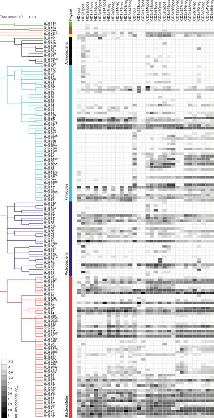
Downloaded from http://rupress.org/jem/article-pdf/217/11/e20200275/1047083/jem_20200275.pdf by guest on 18 August 2020
Figure 1. Microbiota-reactive IgA antibodies are abundant in the adult human small intestine. (A) IgA-free microbes were isolated from feces of SPF
Rag2-deficient mice. Gates were set according to in vitro–cultured GFP-expressing E. coli and fecal material isolated from germ-free mice (top row). GFP signal
or Syto9 nucleic acid dye were used to identify bacteria (bottom row). Anti-human IgG1 antibody was used to detect mAb-positive fecal bacteria. Repre-
sentative staining is depicted for previously described low (mGO53) and high (ED38) polyreactive control antibodies (Meffre et al., 2004; Wardemann et al.,
2003) and staining with supernatant from nontransfected HEK-cells (Med CTRL) on feces from SPF Rag2-deficient mice. (B and C) Two collections of an-
tibodies derived from IgA- or IgG-expressing plasmablasts/plasma cells from HDs (Benckert et al., 2011) or CD patients were screened for reactivity to microbes
isolated from SPF Rag2-deficient mice. (B) Symbols represent individual antibodies (162 HD mAbs and 118 CD mAbs) assayed in one representative experiment.
Binding to
Downloaded from http://rupress.org/jem/article-pdf/217/11/e20200275/1047083/jem_20200275.pdf by guest on 18 August 2020
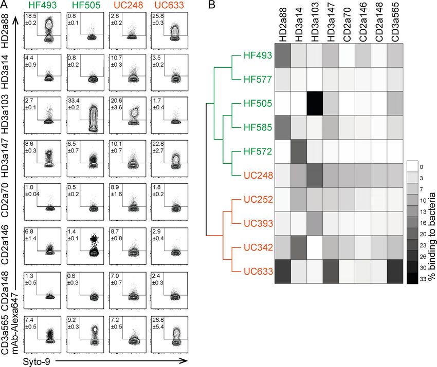
Figure 2. Binding capacity of intestinal IgA antibodies to human gut bacteria shows donor-dependent variability. Bacteria were isolated from fecal
samples of five healthy individuals (HF) and five ulcerative colitis (UC) patients and stained with directly conjugated mAbs. (A) Representative FACS plots of
microbiota reactivity of mAbs to human gut bacteria. Microbes were stained with Syto9 nucleic acid dye, and microbiota reactivity of AF647 directly conjugated
mAbs was assessed by flow cytometry. Data are representative of two independent experiments (mean ± SD). (B) Fecal samples were characterized by 16S
rRNA amplicon sequence analysis. Dendrogram clustering of individual fecal samples is based on their bacterial genomic sequence composition. Heat map
depicts binding capacity of single mAbs to bacteria isolated from respective HF and UC donors (organized as a dendrograms) displayed as mean of two in-
dependent experiments.
bound multiple bacterial taxa. Antibody-positive samples showed antibodies, i.e., healthy and inflamed intestine, revealed largely
a notable spread in Shannon effective counts (which integrates overlapping binding profiles to gut bacterial populations (Fig. 3 D,
the evenness of molecular species and can be considered as a left panel). This indicates that, although CD is often accompanied
proxy for numbers of dominant species), suggesting a po- by major shifts in the microbiota composition (Fadlallah et al.,
tential heterogeneity of antibody binding profiles. Notably, 2018), gut inflammation is not necessarily associated with an
α-diversity of antibody-positive samples did not correlate extensive shift in IgA specificity for particular members of the
with bacteria binding capacity (Fig. 3 C), i.e., samples sorted microbiota. In contrast, β-diversity analysis revealed major differ-
with antibodies binding to a comparably smaller percentage of ences between antibody-positive and antibody-negative
all gut bacteria showed a similarly high diversity as samples bacterial fractions across both sets of experiments (Fig. 3 D,
purified with antibodies binding to a very large proportion of middle and right panel). In conclusion, both α- and β-diversity
all gut bacteria. This further underlines that antibodies with analyses indicate that single monoclonal intestinal antibodies
high binding capacities to gut microbiota show distinct binding do not enrich for individual bacterial species but instead spe-
specificities. cifically bind broadly to distinct groups of gut bacteria.
To assess and visualize differences between samples
(β-diversity), we used principal coordinates analysis (PCoA) based Intestinal IgA shows cross-species reactivity
on generalized UniFrac distances (Chen et al., 2012). Comparison To identify bacterial taxa that explain the observed difference in
of antibody-positive samples of HD- and CD donor–derived β-diversity, we examined the relative abundance of individual
Kabbert et al. Journal of Experimental Medicine 4 of 14
High microbiota reactivity of human intestinal IgA https://doi.org/10.1084/jem.20200275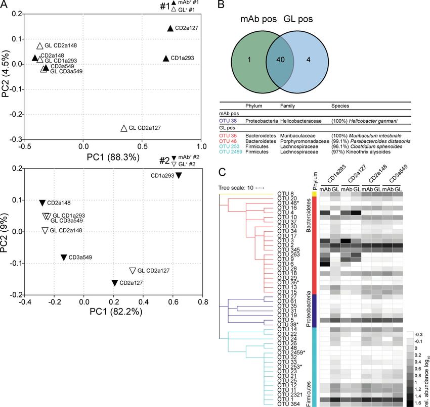
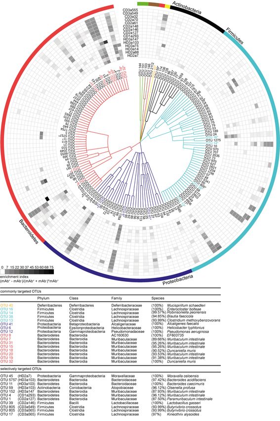
Downloaded from http://rupress.org/jem/article-pdf/217/11/e20200275/1047083/jem_20200275.pdf by guest on 18 August 2020
Figure 3. Intestinal IgA antibodies bind diverse groups of gut bacteria. (A) Representative illustration of antibody-positive (mAb pos bac) and antibody-
negative bacteria (mAb neg bac) obtained by cell sorting. The diversity and composition of sorted bacteria were characterized by 16S rRNA amplicon se-
quencing. (B) α-Diversity parameters (richness and Shannon effective count) for sorted antibody-positive (pos) and -negative (neg) bacteria. Two separate
experiments, #1 and #2, were performed. Groups were compared by one-way ANOVA and paired Wilcoxon test (*, P < 0.05; ns, not significant). (C) Correlation
analysis considering the Shannon effective count as displayed in B and bacteria binding in percent. Symbols represent the mean of two independent ex-
periments. (D) β-Diversity displayed as PCoA of generalized Unifrac distances. The plots depict comparison between antibody-positive samples from HDs and
CD patients (left plot) and between antibody-positive and -negative samples from HDs (middle) and CD patients (right). FSC, forward scatter; SSC, side scatter.

operational taxonomic units (OTUs) in antibody-positive and                      antibodies, as defined by a twofold enrichment in the antibody-
-negative samples. For all mAbs investigated, antibody-positive                  positive fraction for at least three individual mAbs tested, in-
samples contained multiple, phylogenetically distant, OTUs (Fig.                 dicating that these OTUs are preferential targets of intestinal
S2). However, OTUs highly abundant in the antibody-positive                      antibodies (Fig. 4 and Fig. S2). We will refer to these OTUs as
samples were typically still detectable in the antibody-negative                 commonly targeted OTUs.
samples (Fig. S2). Therefore, we expressed the capacity of in-                      Taxonomic assignment of these commonly targeted OTUs
dividual antibodies to enrich for distinct OTUs as an enrichment                 included microbial members of the main abundant phyla in the
index. The index considers the ratio of relative OTU abundances                  mouse gut, Firmicutes, Proteobacteria, Bacteroidetes, and De-
in the antibody-positive and -negative samples plus the OTU                      ferribacteres (Fig. 4 and Fig. S2) such as Enterocloster bolteae
abundance in the positive sample (see Materials and methods).                    (OTU 16), Mucispirillum schaedleri (OTU 40), Alcaligenes faecalis
Indeed, for all antibodies tested, the enrichment index high-                    (OTU 41), Helicobacter typhlonius (OTU 6), and several species that
lighted several OTUs (Fig. 4). 16 OTUs were targeted by multiple                 were identity matched to members of the family Muribaculaceae

Kabbert et al.                                                                                                            Journal of Experimental Medicine        5 of 14
High microbiota reactivity of human intestinal IgA                                                                     https://doi.org/10.1084/jem.20200275
Downloaded from http://rupress.org/jem/article-pdf/217/11/e20200275/1047083/jem_20200275.pdf by guest on 18 August 2020

Figure 4. High-microbiota-reactive intestinal IgA is cross-species reactive. Single OTUs are organized as a dendrogram (phylum to family, annotations
displayed in colors) based on the RDP taxonomic classification (Wang et al., 2007). Only OTUs occurring at a relative abundance of ≥0.5% in any positive

Kabbert et al.                                                                                                       Journal of Experimental Medicine      6 of 14
High microbiota reactivity of human intestinal IgA                                                                https://doi.org/10.1084/jem.20200275
fraction are displayed. Antibody specificity to a given OTU is displayed as enrichment index (as defined in the graph and determined as mean of two or more
replicate experiments). 16 commonly targeted OTUs enriched twofold by ≥3 mAbs are indicated in colors, marked by asterisks, and listed below the figure.
Selectively targeted OTUs (OTUs enriched by only a single mAb among the high-microbiota-reactive antibodies) are listed in black and marked by asterisks. The
lineage and taxonomic identity (closest species with a valid name and corresponding 16S rRNA gene sequence identity) of these denoted OTUs were obtained
using EZbiocloud (Yoon et al., 2017). Nonlisted phyla are as follows: Deinococcus-Thermus (green), Cyanobacteria (brown), and Deferribacteres (yellow).
Taxonomic classifications of OTUs are listed in Table S2.
(OTU 7, 15, 18, 19, 20, 31, and 32). These taxa have previously been antibodies with no/low, intermediate, and high microbiota
reported to be coated by endogenous IgA (Fadlallah et al., 2018; reactivity. In addition, polyreactive antibodies were not over-
Magri et al., 2017; Palm et al., 2014; Sterlin et al., 2020). In con- represented among antibodies with high binding capacity for
trast, some OTUs showed particularly strong enrichment by microbiota (Fig. 5 B and Fig. S3 D). Hence, correlation analysis
single antibodies. This pattern was observed for antibodies de- did not reveal any positive relationship between polyreactivity
rived from HDs as well as CD patients (Fig. 4 and Fig. S2). Notably, and percentage of antibody-bound bacteria (HD IgA, P = 0.97;
OTUs with an overall low relative abundance were targeted by CD IgA, P = 0.41). Because we did not find an overrepresenta-
Downloaded from http://rupress.org/jem/article-pdf/217/11/e20200275/1047083/jem_20200275.pdf by guest on 18 August 2020
specific mAbs, e.g., Moraxella osloensis (OTU 45; HD2a7), Bacte- tion of polyreactive antibodies among high-microbiota-binding
roides acidifaciens (OTU 5757; HD3a103), Muribaculum sp. (OTU 4; antibodies, we further investigated the possibility that the high
CD1a293), and Lactobacillus gasseri (OTU 39; CD2a146). Therefore, capacity for microbiota binding of human intestinal IgA might
IgA responses are not only directed against dominant members of rely on accumulated somatic mutations.
the microbiota but also target underrepresented populations of IgA- and IgG-derived antibodies from both HDs and CD patients
the overall gut microbial communities. Our data demonstrate that showed the typical pattern of highly mutated intestinal antibodies in
individual intestinal antibodies can bind a broad range of mi- both heavy and light chain V gene sequences (Barone et al., 2011;
crobial species while exhibiting unique binding profiles. Impor- Benckert et al., 2011; Berkowska et al., 2015; Lindner et al., 2015;
tantly, we observed phylogenetically unrelated taxa among Fig. 6 A and Fig. S3 E). No major difference was detected when
selectively and commonly targeted OTUs, suggesting that single comparing the number of mutations in the nonselected collection of
monoclonal human IgA antibodies can bind to unrelated groups antibodies to the set of antibodies with high microbiota-binding
of gut bacteria. We therefore conclude that cross-species reac-
tivity is one characterizing property of human intestinal IgA in
healthy and inflamed gut.
The mAbs tested here were expressed as human IgG1 anti-
bodies independently of their original isotype and subclass.
Therefore, it seems unlikely that the binding patterns described
herein rely on Fc-mediated interactions and glycan mod-
ifications. Nonetheless, glycan-mediated interactions between
mAbs and individual members of the gut microbiota have been
reported (Nakajima et al., 2018), and different mAbs may differ
in their precise glycan modification. To directly determine the
contribution of antibody glycan structures to microbiota bind-
ing, we deglycosylated a set of purified mAbs. Deglycosylation
did not affect the overall binding capacity of mAbs (Fig. S3, A
and B), suggesting that glycan modifications do not contribute to
the observed microbiota-binding patterns.
Somatic mutations but not polyreactivity confer cross-species
binding and high microbiota reactivity
To examine whether polyreactivity was an important determi-
nant of cross-species reactivity of human intestinal mAbs, we
screened the HD and CD collections of IgA- and IgG-derived
antibodies for binding to a panel of seven unrelated antigens
commonly used to test for polyreactivity. Antibodies that showed
binding to more than two antigens were classified as polyreactive. Figure 5. High microbiota reactivity does not correlate with poly-
Based on this criterion, 23% of IgA antibodies from HDs and 13% reactivity. (A) IgA mAbs were tested in at least two replicate experiments for
from CD patients were polyreactive (Fig. 5 A and Fig. S3 C; polyreactivity and classified as either polyreactive (black) or nonpolyreactive
(white). (B) Bar charts show the relative distribution of polyreactive and
Benckert et al., 2011). Among IgG-derived antibodies, we found
nonpolyreactive HD-derived and CD patient–derived IgA antibodies among
similar frequencies of polyreactive antibodies (23% for HD antibodies with no/low, intermediate, and high microbiota reactivity. Corre-
mAbs and 18% for CD; Fig. S3 D). Notably, polyreactive IgA and lation between polyreactivity and microbiota reactivity was determined by
IgG antibodies were observed at similar frequencies among linear regression (HD IgA, P = 0.97; CD IgA, P = 0.41).
Kabbert et al. Journal of Experimental Medicine 7 of 14
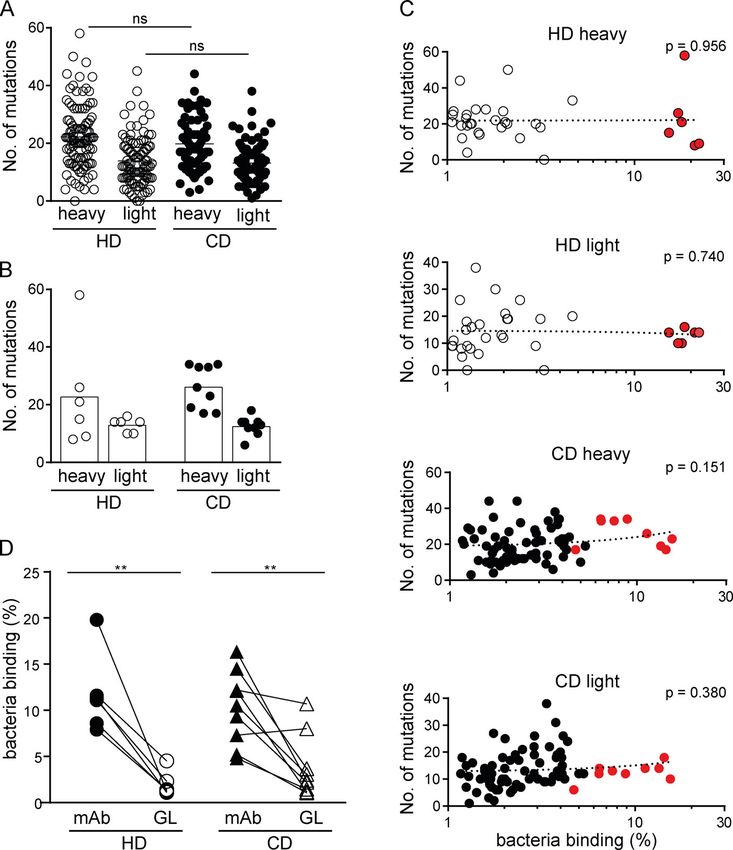
High microbiota reactivity of human intestinal IgA https://doi.org/10.1084/jem.20200275Figure 6. Somatic mutations confer high
microbiota-binding capacity. (A and B) Num-
bers of somatic mutations in the heavy and light
chain V genes displayed for all IgA antibodies
obtained from HDs and CD patients (A) and
for high-microbiota-reactive IgA antibodies (B).
(C) Microbiota-binding capacity, displayed as
percentage of bound microbiota determined as
mean of two or more replicate experiments (for
antibodies showing microbiota binding ≥1%),
was correlated to the number of somatic muta-
tions in heavy and light chain V genes for indi-
vidual IgA antibodies. Mean microbiota-binding
capacity of selected high-microbiota-reactive mAbs
are shown in red. P values were obtained by
linear regression analysis. (D) Pairwise com-
parison of microbiota-binding capacity of mAbs
and their respective, predicted GL variant. Symbols
Downloaded from http://rupress.org/jem/article-pdf/217/11/e20200275/1047083/jem_20200275.pdf by guest on 18 August 2020
represent the mean value of two independent
experiments. Significance was tested by paired
Mann–Whitney U test, **, P < 0.01; ns, not
significant.
capacity (Fig. 6, A and B). The overall number of mutations in heavy of bound microbes. Two of these antibodies maintained or even
or light chains of IgA-derived and IgG antibodies did not correlate showed increased binding capacity to bacteria (CD2a148 and
with their capacity to bind gut bacteria (Fig. 6 C), with the exception CD3a549), and two mAbs showed reduced overall binding but
of a positive correlation in HD-derived IgG light chains (Fig. S3 F). still recognized a sortable bacterial population (CD1a293 and
To directly address the contribution of somatic mutations to CD2a127) in their GL configuration (Fig. 6 D and Fig. S4 A).
microbiota-binding capacity as well as specificity, we generated Mutated and GL antibodies largely bound the same bacterial
the predicted GL variants of IgA antibodies with high microbiota- species (Fig. 7 B), although often with differing efficiencies
binding capacity (Fig. S4, A and B). For all reverted HD-derived (Fig. 7 C). β-Diversity analysis of populations bound by the
antibodies and for the majority of antibodies (seven of nine) de- mutated antibodies and their corresponding GL variants
rived from CD patients, the GL variant showed a significant loss of showed close clustering for CD2a148 and CD3a549 and similar
microbiota binding compared with the mutated variant (Fig. 6 D relative abundance of enriched OTUs (Fig. 7 A). There were
and Fig. S4 A). Our data therefore strongly suggest that in notable differences in the relative binding capacities of mutated
the human gut, somatic mutations significantly contribute to mAbs compared with their GL variants to multiple individual
microbiota-binding capacity and cross-species reactivity of IgA. OTUs (Fig. 7 C). Moreover, for two of the mAbs, CD1a293 and
Importantly, GL reversion of antibodies did not render antibodies CD2a127, the populations of bacteria bound by the mutated
polyreactive. In fact, the polyreactive antibodies HD2a7, CD3a32 version and GL variant were substantially different (Fig. 7 C).
and CD3a565 lost polyreactivity after GL reversion (Fig. S4, C and For these two antibodies, some of the OTUs enriched by the
D), indicating that these antibodies might have become poly- mutated antibodies were less abundant in the population of
reactive during somatic hyper mutation and cross-species reac- bacteria bound by the GL variants, indicating that even though
tive mAbs did not derive from originally polyreactive antibodies. GL reversion retained some microbiota-binding capacity, mu-
However, four CD-derived GL-variant mAbs retained sufficient tations in these two antibodies contribute to their microbiota
microbiota-binding capacity to allow for sort-based purification specificity and/or binding strength.
Kabbert et al. Journal of Experimental Medicine 8 of 14
High microbiota reactivity of human intestinal IgA https://doi.org/10.1084/jem.20200275Downloaded from http://rupress.org/jem/article-pdf/217/11/e20200275/1047083/jem_20200275.pdf by guest on 18 August 2020
Figure 7. Somatic mutations contribute to the gut microbiota specificity of human IgA. Fecal bacteria bound by mAbs carrying somatic mutations and
their corresponding GL variants were obtained by cell sorting and analyzed by 16S rRNA gene amplicon sequencing in two independent sets of experiments, #1
and #2. (A) β-Diversity PCoA plots based on generalized Unifrac distances depict comparison between sorted positive samples of mutated antibodies (filled
symbols) and corresponding GL variants (open symbols). (B) Venn diagram depicts shared and selectively enriched OTUs in mAb-positive and GL-positive
samples. Selectively enriched OTUs are marked by asterisks, EZbiocloud identified, and listed (Yoon et al., 2017). (C) Dendrogram clustering of OTUs organized
(phylum annotations displayed in colors) based on the RDP taxonomic classification (Wang et al., 2007). Only OTUs occurring at a relative abundance ≥0.5% in
any positive fraction are displayed. Heat map shows log10-transformed relative abundances of OTUs.
Collectively, these data show that fecal microbiota binding was binding capacity was observed for different microbiota config-
unrelated to polyreactivity, whereas in most cases, GL reversion urations, including murine samples as well as human samples
impaired microbiota-binding capacity or altered antibody specificity obtained from either HDs or patients with IBD. High microbiota-
for the microbiota. Therefore, our data demonstrate that a system of binding capacity was associated with binding to a broad range
affinity-matured IgA is one central feature of SIgA–microbiota in- of phylogenetically diverse bacteria. We refer to this mode of
teractions in the healthy and inflamed gut of adult humans. binding as cross-species reactivity (Pabst and Slack, 2020) and
speculate that Ig binding to bacteria may constitute an important
mechanism to regulate the gut microbiota and the integrity of
Discussion the intestinal mucosa. Cross-species reactivity does not make
Here we show that IgA and IgG antibodies derived from intes- any assumption about the mode of binding but points to inter-
tinal plasmablasts/plasma cells in adult humans can bind to esting functional consequences for intestinal antibody function
a large proportion of fecal microbes. Such high microbiota- in the complex gut microbiota environment.
Kabbert et al. Journal of Experimental Medicine 9 of 14
High microbiota reactivity of human intestinal IgA https://doi.org/10.1084/jem.20200275In the setting of a highly complex and dynamically changing 2017) and here in humans suggests at least some level of anti-
ecosystem such as the gut microbiota, it is difficult to picture body specificity. In contrast, hypermutated antibodies abundant
how antibodies selectively binding to single bacteria can be in the human gut are likely to be the result of ongoing selection
produced at functionally meaningful levels. How does the host for high-affinity binding. Cross-species reactivity of high-
generate sufficient amounts of secretory antibodies to affect the affinity antibodies might rely on epitopes shared between dif-
microbiota? A possible explanation is the concept of enchained ferent bacterial species such as conserved glycan structures or
growth, which refers to antibody-mediated cross-linking and ag- highly conserved peptides (Rollenske et al., 2018; Bunker et al.,
glutination of dividing bacteria (Moor et al., 2017). For instance, 2019; Diard et al., 2019 Preprint; Sterlin et al., 2020). The iden-
Salmonella-specific antibodies primarily cross-link dividing bacte- tification of epitopes targeted by IgA across multiple members of
ria to enable agglutination of the pathogen in a situation in which the microbiota is the crucial next step to further understand the
few targeted bacteria are diluted within a complex and numerous phenomenon of cross-species reactivity and to dissect pathways
endogenous microbiota (Moor et al., 2017). Cross-species–reactive of how cross-species reactivity is achieved.
antibodies might add to this concept by contributing to the ag- Notably, bacteria commonly targeted by antibodies isolated
glutination of planktonic members of the microbiota. from HDs were also bound by high-microbiota-reactive anti-
Cross-species reactivity appears to be a feature of both mouse bodies from CD patients. Therefore, selective broad binding to
Downloaded from http://rupress.org/jem/article-pdf/217/11/e20200275/1047083/jem_20200275.pdf by guest on 18 August 2020
(Bunker et al., 2017) and human intestinal antibodies. However, distinct OTUs seems to be a hallmark of intestinal IgA in both
although cross-species reactivity was proposed to be mainly healthy and inflamed intestine. Thus, in both situations, the
achieved by “innate-like” polyreactive antibodies in mice (Bunker profile of IgA-targeted bacteria comprised commonly targeted
et al., 2017), we demonstrate here that, in adult humans, IgA OTUs in addition to specific members of the microbiota. These
cross-species reactivity typically depends on the accumula- observations indicate that major changes in gut microbiota
tion of somatic mutations. It is possible that these two mecha- composition as observed in IBD patients do not necessarily
nisms of cross-species antibody reactivity may simply reflect a dominate the microbiota specificity of IgA and are consistent
fundamental difference between the two hosts. However, we with our previous observation that alterations of the microbiota
speculate that these seemingly contradictory findings may in fact do not rapidly result in major changes of the plasma cell rep-
reveal distinct facets of the intestinal immune response, which ertoire (Lindner et al., 2015).
may act in concert to protect against pathogens and create The presence of cross-species–reactive antibodies under
microbiota-reactive antibodies throughout life in both mice and healthy and inflamed conditions might hint at the functional
humans. At the outset, the newborn receives maternal anti- importance of such antibodies. Cross-species reactivity may
bodies that help shape the microbiota (Pabst et al., 2016), and contribute to efficient agglutination of opportunistic pathogens,
provide protection while the immune system initially develops. possibly including the endogenous microbiota as scaffolding. Yet
Subsequently, in young individuals with relatively few germinal proinflammatory functions can also be envisioned, and cross-
centers and little affinity maturation, polyreactive antibodies species reactivity might further exacerbate the inflammatory
may be the dominant mechanism of microbiota binding. Indeed, response to members of the microbiota. In particular, in sit-
such antibodies are prevalent in the gut of young mice (Bunker uations of an impaired intestinal barrier such as in IBD patients,
et al., 2017) and are present at low frequency in our collection of cells in the lamina propria will encounter IgA-coated intestinal
human antibodies. However, during aging, these may become bacteria. IgA coating of the bacteria has the potential to modu-
supplanted by affinity-matured antibodies that accumulate so- late the ensuing immune response. Depending on the precise
matic mutations, thereby enabling or even enhancing cross- context, interaction with the IgA Fc receptor FcαRI can initiate
species reactivity. Such antibodies are the distinguishing type proinflammatory or inhibitory responses (Bakema and van
of highly microbiota-reactive antibodies described here and are Egmond, 2011).
consistent with the well-documented finding of highly mutated An important step in understanding the generation of
plasma cell repertoires in the human gut exposed to a complex microbiota-targeting Ig responses and cross-species reactivity
and dynamic environment and microbiota (Barone et al., 2011; might come from a deeper understanding of how somatic hy-
Benckert et al., 2011; Lindner et al., 2015). permutation shapes these antibodies. Indeed, our data suggest
Intriguingly, cross-species reactivity does not extend to all that accumulating somatic mutations fundamentally contribute
members of the microbiota: some bacteria detected by se- to antibody cross-species reactivity. In adult humans, the con-
quencing did not show relevant binding by any of the tested tinuous exposure to varied but structurally similar bacterial
antibodies, whereas others were commonly targeted by differ- antigens may favor the proliferation and differentiation of
ent mAbs or specifically enriched by single antibodies. Thus, B cells with cross-species specificity. In this context, an impor-
intestinal antibodies display unique and specific binding profiles tant step forward may come from a deeper understanding of
for distinct members of the gut microbiota. But what determines which bacterial species contribute to the initial activation of the
which bacterial taxa are targeted? B cell response and subsequent affinity maturation. Indeed,
Polyreactive antibodies, per definition, bind unrelated mol- when testing antibodies selected for high microbiota reactivity
ecules. Thus, binding of polyreactive antibodies to different to murine fecal samples, these antibodies also showed high
bacterial species might function via unrelated antigens present microbiota reactivity to human fecal material from HDs and
on the different bacteria. The enrichment of defined bacterial patients with IBD. This suggests that either closely related bac-
taxa by intestinal antibodies as described in mice (Bunker et al., terial strains are present in all samples or that intestinal
Kabbert et al. Journal of Experimental Medicine 10 of 14
High microbiota reactivity of human intestinal IgA https://doi.org/10.1084/jem.20200275antibodies are indeed directed against structurally similar bac- CD38+CD27+IgG+ plasmablasts/plasma cells were sorted into 96-well
terial antigens shared among different members of the micro- PCR plates using a FACSVantage cell sorter with Diva configuration
biota, independent of the host. (BD Bioscience), snap frozen on dry ice, and stored at −80°C until
Collectively, our data highlight a new facet of IgA: high further processing.
microbiota and cross-bacterial species reactivity of human in-
testinal IgA rely on somatic mutations. We propose that cross- PCR amplification and expression vector cloning
species-specific antibodies provide a mechanism to efficiently Single-cell cDNA synthesis and nested PCR amplification of IGG
interact with multiple microbial members and represent one of or IGA and IGK or IGL genes was performed as described
the major mechanisms by which the intestinal immune system (Benckert et al., 2011). All PCR products were sequenced before
interacts with the intestinal microbiome. and after cloning into previously described eukaryotic expres-
sion vectors (Tiller et al., 2008). In brief, PCR products of IgH
V(D)J and IGK Igk and IGL Igl VJ variable regions were cloned
Materials and methods into separate expression vectors encoding for the constant re-
Mice gions of the human IgG1, IGK and IGL light chain to allow for
Rag2-deficient mice and C57BL/6J WT mice were bred and expression of all antibodies as fully human Fc-IgG1 antibodies. Ig
Downloaded from http://rupress.org/jem/article-pdf/217/11/e20200275/1047083/jem_20200275.pdf by guest on 18 August 2020
reared at RWTH Aachen University under specific pathogen– gene sequence analysis, including Ig gene usage, clonal rela-
free (SPF) conditions. Germ-free C57BL/6J mice were bred and tionships, IgG subclass, somatic mutations, IgH CDR3 length,
reared at the Laboratory for Animal Sciences at Hannover and positive charged amino acids, was performed using IgBlast
Medical School. Animal work was performed in compliance with (http://www.ncbi.nlm.nih.gov/igblast/).
ethics regulations of German Law for the Protection of Animal
Welfare (Tierschutzgesetz) and approved by North Rhein- Expression and purification of recombinant antibodies
Westphalia State Agency for Nature, Environment and Consumer 239T HEK cells were transiently transfected with equal amounts
Protection (Landesamt fur Natur, Umwelt und Verbraucherschutz of both corresponding IgH and IgL chain plasmids using a cal-
Nordrhein-Westfalen). cium phosphate kit (Invitrogen; K2780-01). Transfected HEK
cells were cultured in Pro 293a Media (Lonza) supplemented
Human fecal material with 1 mM sodium pyruvate, 2 mM L-glutamine, and 100 U/ml
The human biomaterials were provided by the centralized bio- penicillin G–streptomycin (all from Gibco). Supernatants were
material bank of the Medical Faculty of RWTH Aachen University collected 60 and 72 h after transfection and filtered through a
(RWTH cBMB) and were used in accordance with the regulations sterile filter (0.20 µm) and stored at 4°C. Igs were purified from
of the RWTH cBMB and the Ethics Vote 206/09 of the Ethics supernatants using Protein A–based affinity chromatography
Committee of the Medical Faculty of RWTH Aachen University. (Äkta Start; GE Healthcare Life Sciences). Protein A HiTrap
columns (GE Healthcare Life Sciences) were loaded with Ig-
Production of mAbs containing supernatant and washed with 20 mM sodium-
Generation of mAbs from three HDs undergoing right-sided phosphate buffer, and antibodies were eluted using 0.1 M
hemicolectomy has been described before (Benckert et al., citric acid (pH 3.0), pH neutralized with 1 M Tris-HCl (pH 9.0).
2011). None of the donors had a history of intestinal inflamma- Collected supernatants were concentrated and washed with
tion, and the samples showed no signs of inflammation as deter- PBS, and antibodies were collected in PBS. Concentrations of
mined by macroscopic evaluation and histopathologic examination purified antibodies were determined by ELISA.
of the adjacent mucosa (Benckert et al., 2011). A second collection
of antibodies was generated from surgical samples of three CD Deglycosylation of antibodies
patients undergoing ileocoecal resection (CD1, male, 25 yr old; To remove human IgG Fc N-glycan moieties by deglycosylation,
CD2, male, 47 yr; CD3, male, 22 yr) and were obtained after GLycINATOR (EndoS2) an endoglycosidase for hydroxylation of
signed informed consent in accordance with protocols reviewed β1,4 linkage of core GlcNAc residues, was used according to the
and approved by the Charité University Hospital institutional manufacturer’s instructions (GlycINATOR R; Genovis). Degly-
review board (EA1/257/12). cosylation was performed with 1 unit of GlycINATOR/1 µg IgG
and incubated for 30 min at 37°C. Deglycosylation was con-
Single B cell sorting firmed by mass spectrometry and Western blot. Samples were
Intestinal plasmablasts/plasma cells were isolated as previously reduced with Tris(2-carboxyethyl)phosphine before electro-
described (Benckert et al., 2011). In brief, lamina mucosa and spray ionization mass spectrometry analyses, desalted using a
propria were dissected from lamina muscularis mucosae by C4 ZipTip (Millipore), and analyzed in MeOH:2-PrOH:0.2% FA
blunt preparation, and 2–3-mm tissue pieces were digested us- (30:20:50). The solutions were infused through a fused silica
ing 0.1% DNase and 0.1% collagenase followed by discontinuous capillary (internal diameter, 75 µm) at a flow rate of 1 µl/min
Ficoll density gradient centrifugation (GE Healthcare). Purified and sprayed through a PicoTip (internal diameter 30 µm; both
lamina propria lymphocytes were stained on ice with fluorochrome- from New Objective). Nano-electrospray ionization mass spec-
coupled anti-human CD38 FITC, anti-human CD27 PE, anti-human trometry analyses of the samples was performed on a Synapt
CD19 PECy7, anti-human IgG APC (all from BD Bioscience) or anti- G2_Si mass spectrometer, and the data were recorded with
human IgA APC (Jackson Laboratory). Single CD38+CD27+IgA+ or MassLynx 4.2 software 2/2 (both from Waters). Mass spectra
Kabbert et al. Journal of Experimental Medicine 11 of 14
High microbiota reactivity of human intestinal IgA https://doi.org/10.1084/jem.20200275were acquired in the positive-ion mode by scanning an m/z weight of GL variants was validated by Western blot analysis for
range from 100 to 5,000 daltons with a scan duration of 1 s and heavy and light chains according to standard procedures.
an interscan delay of 0.1 s. The spray voltage was set to 3 kV,
source temperature 80°C, and cone voltage 50 V. The recorded Bacterial flow cytometry and bacterial sort purification
m/z data were then deconvoluted into mass spectra by applying by FACS
the maximum-entropy algorithm MaxEnt1 (MaxLynx) with a Fecal pellets from SPF Rag2-deficient and germ-free WT mice
resolution of the output mass 0.5 daltons/channel and Uniform were collected and human fecal material (from five HDs and five
Gaussian Damage Model at the half height of 0.7 daltons. ulcerative colitis patients) was obtained and stored at −80°C.
Deglycosylation efficiency of human IgG1 was independently Murine fecal pellets or aliquots from human feces were ho-
analyzed by Western blot. Heat-denatured samples were sepa- mogenized in 1.5-ml tubes in 1 ml of filtered (0.2 µm) HBSS.
rated on a 12% acrylamide gel (Bio-Rad) and transferred onto a Homogenized samples were centrifuged at 500 rpm for 1 min,
nitrocellulose membrane (0.45 µm, Bio-Rad; 1620115). For de- and the process was repeated until the supernatant was clear of
tection of IgG1 heavy chains, membranes were blocked with 5% debris. Clear supernatant was centrifuged at 8,000 rpm (VWR
milk powder in PBS/0.1% Tween-20 overnight at 4°C and in- Microstar 17R) for 8 min, and the supernatant was discarded.
cubated with HRP-conjugated anti-IgG1-HRP antibody (Jackson The bacterial pellet was resuspended in 100 µl of filtered HBSS/
Downloaded from http://rupress.org/jem/article-pdf/217/11/e20200275/1047083/jem_20200275.pdf by guest on 18 August 2020
ImmunoResearch; 109-035-098) for 1 h. To detect glycans, 2% normal goat serum (Sigma-Aldrich) and passed through
membranes were blocked with Hepes 2% Tween-20 buffer, a sterile cell strainer (40 µm). The OD600 was determined in a
washed twice with Hepes, and incubated with concanavalin- 1:100 dilution, and suspensions were adjusted to OD600 of 0.13 ±
A–HRP for 16 h at room temperature (0.2 µg/ml in Hepes, 0.01 or 0.2 ± 0.01 for human fecal suspensions (5 × 10⁷/ml
0.05% Tween-20, and 1 mM CaCl2, MgCl2, MnCl2, and conca- bacteria). Antibody concentrations were adjusted to 1 µg/ml in
navalin A [Sigma-Aldrich; L6397]). HRP activity was detected HBSS/2% normal goat serum. 2 µl of murine bacterial suspen-
with ECL substrate (Bio-Rad). sions were stained with 100 µl of diluted antibody, incubated on
ice for 20 min, and washed in 1 ml HBSS/2% goat serum at
ELISA 13,000 rpm for 8 min at 4°C. The supernatant was discarded,
The concentration of recombinant antibodies was determined and pellets were resuspended in 100 µl anti-human IgG1 AF647
by ELISA. ELISA 96 plates (NUNC Maxi Sorp) were coated with (1:200; 0.5 mg/ml, Alexa Fluor 647–conjugated AffiniPure Goat,
1.25 ng/ml of anti-human IgG Fc (AffiniPure goat anti-human anti-human IgG [H + L], Jackson ImmunoResearch) and incu-
IgG, Fcγ-specific; Jackson ImmunoResearch; 109-005-098) in bated for 20 min on ice. 50 µl of Syto9 dye (Syto 9 green fluo-
PBS. Plates were washed with PBS/0.05% Tween-20 and blocked rescent nucleic acid stain, 5 mM in DMSO; Thermo Fisher
with PBS/2% BSA for 1 h at 37°C. Human serum IgG1 standard Scientific) in HBSS was added and incubated for 10 min on ice.
(Sigma-Aldrich) and recombinant antibodies diluted in PBS/ For bacterial suspensions isolated from human fecal material,
0.05% Tween-20 at twofold dilution were incubated for 1.5 h at 2 µl of bacterial suspensions were stained with 5 µg/ml AF647-
37°C and detected with HRP-conjugated (Peroxidase AffiniPure directly conjugated mAbs for 20 min on ice followed by Syto9
goat anti-human IgG, Fcy-specific; Jackson ImmunoResearch, staining and washing as described for murine samples. A mini-
109–035-098) diluted in PBS/0.05% Tween-20 at a concentration mum of 100,000 Syto9-positive events were acquired for each
of 0.8 µg/ml (Jackson ImmunoResearch Laboratories) according sample. For FACS purification, samples were prepared as de-
to the manufacturer’s instructions. ODs were measured at 450 scribed above in replicates, pooled, and sorted on a FacsAria II
nm, and antibody concentrations were determined using the (BD). Sorted samples were centrifuged at 13,000 rpm for 8 min
SpectraMax microplate reader and software (Molecular Devices) at 4°C, and pellets were stored at −80°C.
and Excel 2010 (Microsoft).
16S rRNA gene amplicon sequencing and analysis
Generation of GL variants Bacterial DNA from human fecal samples was extracted via glass
Sequences were analyzed by IgBLAST comparison with Gen- bead preparation and further isolated using the QIAamp DNA
Bank, and GL segments with the highest probability were de- Stool Kit (Qiagen) according to the manufacturer’s protocol.
termined. The reverted sequences were generated as gBlock Metagenomic DNA of sort-purified murine bacterial samples of
double-stranded DNA fragments from IDT (Integrated Tech- mAb-positive and mAb-negative fractions and input material
nologies) with included flanking primer and restriction sites. GL was extracted using the Qiagen DNA isolation kit according to
(gBlock) fragments (100 ng/µl) and original vector plasmids the manufacturer’s instructions. OTU profiles obtained for fecal
containing IgH and IgL sequences were digested with respective material and isolated bacteria were highly comparable (not de-
restriction enzymes (all from NEB). Vector backbone was puri- picted). The V3/V4 regions of 16S rRNA genes were amplified
fied by gel electrophoresis in a 1.8% agarose gel, and gBlocks (35 cycles) using primers 515F and 816R. PCR amplicons were
were purified by PCR purification Kit (Qiagen). Ligation was purified by agarose gel electrophoresis (1.5%) followed by pu-
performed with T4 DNA ligase (NEB), and products were rification using a QIAquick Gel Extraction Kit (Qiagen). Empty
transformed into competent 5-α E. coli cells (NEB). For each samples were included in the overall sample processing (FACS
antibody, several clones were analyzed, and the GL and original buffer collected during cell sorting) and analyzed along with
insert sequences were confirmed with IgBLAST comparison H2O and buffer controls used during library preparation to
with GenBank and Gentle free software (GENtle). The molecular identify potential artifact sequences. DNA was quantified using
Kabbert et al. Journal of Experimental Medicine 12 of 14
High microbiota reactivity of human intestinal IgA https://doi.org/10.1084/jem.20200275the Quant-iT PicoGreen dsDNA Kit assay (Invitrogen), and PCR OTUs in HD and CD mAb-positive and -negative samples and
products were stored at −20°C until sequencing. The amplicon input material, with OTUs phylogenetically organized as a
libraries were sequenced with single barcodes in paired-end dendrogram. Fig. S3 shows representative FACS plots of binding
mode (2 × 300 nt) using MiSeq sequencer (Illumina Miseq; capacity of mAbs or their deglycosylated counterparts and
Uniklinik/RWTH Aachen). Raw reads were processed using the quantification of independent experiments; summary of poly-
IMNGS platform based on the UPARSE approach (Edgar, 2013). reactivity and microbiota reactivity for all tested IgA and IgG
OTUs were clustered at a sequence identity of 97%, and only mAbs derived from HDs and CD patients; overall distribution of
OTUs occurring at a relative abundance >0.25% in at least one polyreactive IgG mAbs among the HD and CD antibody collec-
sample were used for further analyses. Taxonomic assignment tions tested; relative distribution of polyreactive and non-
was based on the Ribosomal Database Project (RDP) classifier polyreactive IgG mAbs among no/low, intermediate, or high
v2.11 (Wang et al., 2007) with a confidence cutoff of 80%, and microbiota-reactive mAbs; absolute number of mutations in
dendrogram clustering of OTUs (phylum to family) was based on IgG H and L V genes; and correlation of number of mutations in
genomic sequence similarity obtained by the RDP taxonomic Ig genes to microbiota-binding capacity for HD- and CD-
classification (Wang et al., 2007). The lineage and taxonomic derived IgG mAbs. Fig. S4 shows representative FACS plots of
identity (closest species with a valid name and corresponding microbiota-binding capacity of selected high-microbiota-reac-
Downloaded from http://rupress.org/jem/article-pdf/217/11/e20200275/1047083/jem_20200275.pdf by guest on 18 August 2020
16S rRNA gene sequence identity) of relevant OTUs were ob- tive HD- and CD-derived IgA mAbs before and after GL rever-
tained using EZbiocloud (Yoon et al., 2017). Diversity and sample sion; representative Western blot analysis to validate the molecular
composition analyses were performed using Rhea v2.0 in R weight of heavy and light chains; polyreactivity ELISA repre-
(Lagkouvardos et al., 2017). All 16S rRNA data are freely avail- sentations for selected high-microbiota-reactive HD- and CD-
able under BioProject accession no. PRJNA622437. derived IgA mAbs before and after GL reversion tested
against seven unrelated antigens; and a summary heat map
Polyreactivity ELISA of polyreactivity and microbiota reactivity of selected high-
Polyreactivity assays were performed as described previously microbiota-reactive HD- and CD-derived IgA mAbs before
(Benckert et al., 2011). In brief, ELISA plates were coated with and after GL reversion. Table S1 contains Ig sequence analysis
50 µl of the respective antigens (10 µg/ml calf thymus DNA, of CD-derived IgA+ and IgG+ intestinal plasmablasts/plasma
5 µg/ml human recombinant insulin, 10 µg/ml LPS, 5 µg/ml cells and microbiota reactivity of tested mAbs. Table S2 lists
flagellin, 10 µg/ml cardiolipin, 10 µg/ml human albumin, and taxonomic classification of all identified OTUs in HD and CD
10 µg/ml KLH) in PBS, overnight at 4°C. ELISA plates were mAb–positive and –negative samples and input material.
washed three times with PBS and incubated with 50 µl of an-
tibody at 1, 0.25, 0.06, and 0.015 µg/ml in PBS for 1 h. Recom-
binant human mAbs mGO53 (nonpolyreactive; Wardemann Acknowledgments
et al., 2003) and ED38 (high polyreactive; Meffre et al., 2004) We thank Christian Busse for help with Ig sequence analysis;
were used for comparison on each plate. ELISA plates were Toralf Kaiser for single-cell sorting; and Sabine von Oy, Chris-
developed with HRP-labeled goat anti-human IgG Fcy specific tina Petrick, Lena Küsgens, Matthias F. Müllenbeck, and Cor-
antibody (Jackson ImmunoResearch; 109-035-098) at a con- nelia Kreschel for experimental support. We thank Emma Slack
centration of 0.8 µg/ml with 2 mM EDTA and 0.05% Tween-20 for helpful discussion and support of mass spectrometry analysis.
and HRP chromogenic substrate (ABTS; Pierce). OD450 was The work was supported by the Flow Cytometry Facility, a
measured using a microplate reader (Molecular Devices) and core unit of the Interdisciplinary Center for Clinical Research
Soft-Max software (Molecular Devices). The cutoff value for Aachen within the Faculty of Medicine at RWTH Aachen Uni-
each plate was determined based on the highest OD450 value at a versity. Funds were obtained from the Behrens Weise Founda-
concentration of 1 µg/ml of the nonpolyreactive control mAb tion to H. Wardemann and from the German Research Foundation:
(mGO53). Antibodies showing binding to at least two different grant DFG 921/4-1 to O. Pabst, Project-ID 403224013 – SFB 1382
antigens were considered polyreactive. (B06 to O. Pabst and Q02 to T. Clavel), and grant B3988 to
J. Benckert.
Statistical analysis Author contributions: H. Wardemann, J. Benckert, and T.
Statistical analysis for FACS-generated data was performed with Rollenske generated the collection of antibodies. J. Kabbert per-
GraphPad Prism 7. P values ≤0.05 were considered significant. formed experiments, analyzed data, and prepared figures. T.C.A.
Statistics for 16S rRNA gene amplicon analyses are described in Hitch and T. Clavel contributed to 16S rRNA gene amplicon
detail in Rhea (https://lagkouvardos.github.io/Rhea/; Lagkouvardos analyses and drafted or reviewed corresponding figures and text.
et al., 2017). O. Pabst conceived the study and supervised the work. J. Kabbert,
V. Cerovic, and O. Pabst analyzed data and wrote the manuscript.
Online supplemental material
Fig. S1 shows representative FACS plots of bacterial staining Disclosures: The authors declare no competing interests exist.
with selected high-microbiota-reactive mAbs from HDs and CD
patients and mAb staining quantification of bacterial material Submitted: 14 February 2020
obtained from unrelated feces or aliquots of pooled fecal mate- Revised: 25 May 2020
rial. Fig. S2 shows log10-transformed relative abundance of all Accepted: 22 June 2020
Kabbert et al. Journal of Experimental Medicine 13 of 14
High microbiota reactivity of human intestinal IgA https://doi.org/10.1084/jem.20200275You can also read